Abstract
Preserving patients' native tissues has posed many challenges for surgeons. Increased life expectancy is leading to a proportionately older surgical population with weaker tissues. The growing population of morbidly obese patients in addition to those with multiple comorbidities which influence the native strength and perfusion of tissues compounds the surgeon's challenge. Certainly, there is a rising demand for materials to replace or augment a patient's native tissue when it has been compromised. Over time, the number of products available has increased substantially. The ideal substitute, however, is debatable. The manufacturing and processing of these materials has become more complex and this has resulted in a significant increase in cost. The composition of the mesh, clinical scenario, and operative technique all interact to impact the long-term results. Surgeons require a thorough understanding of these products to guide proper selection and use, to ensure optimal outcomes for patients, and to properly steward financial resources. This review will outline the properties of commonly used materials, highlighting the strength and weakness of each. It will then discuss recommendations regarding mesh selection, coding, and reimbursement. While general principles and trends can be highlighted, further studies of biologic versus synthetic meshes are clearly necessary.
Keywords: biological mesh, synthetic mesh, coding
CME objectives: Upon reviewing this manuscript, the reader should be able to
Outline the properties of commonly used materials, highlighting the strength and weakness of each.
Address mesh complications such as shrinkage, erosion, tissue–mesh interface failure, eventration, and hernia recurrence.
Discuss issues relating to coding, reimbursement, and cost.
Colorectal surgeons encounter numerous clinical scenarios that require supplemental material to augment or replace a patient's native tissues. Tissue destruction from infection and loss of abdominal domain from damage control procedures can lead to large hernia defects. Parastomal hernias and pelvic prolapse repair frequently require mesh to strengthen a patient's attenuated fascia. Each of these circumstances has unique factors which must be taken into account in planning surgical interventions. A large number of products have been developed to meet these needs. Each of these materials has unique properties that have implications for their use in clinical practice.
While there have been innumerable studies involving the use of mesh, the variety of applications, nuances of surgical technique, and differences in study design make comparisons difficult. This review will outline the properties of commonly used materials, highlighting the strength and weakness of each. It will address mesh complications such as shrinkage, erosion, tissue–mesh interface failure, eventration, and hernia recurrence. Finally, it will discuss issues relating to coding, reimbursement, and cost.
For the purpose of discussion, these products are divided into two groups: synthetic and biologic mesh.
Synthetic Mesh
In considering synthetic mesh, several mechanical factors must be taken into account: tensile strength, porosity, elasticity, and method of fabrication. The tensile strength of most synthetic materials generally far exceeds the physiologic demand. However, excessive strength can lead to increased inflammation and loss of elasticity. The porosity of a mesh affects its incorporation into surrounding tissues. In general, small pores generate a strong inflammatory response that can reduce tissue ingrowth. Although larger pores allow more ingrowth and may preserve elasticity, it comes at the expense of creating an adequate scaffold for fibrous tissue growth. Finally, the material can be constructed by knitting or weaving. Knitted mesh is generally more porous and flexible than woven mesh. Woven mesh, because of the increased fiber density, is generally stronger, but serves as a poor scaffold for fibrous ingrowth.
Synthetic meshes can be either permanent or absorbable. Permanent materials are generally composed of polypropylene, polyester, or expanded polytetrafluoroethylene (ePTFE). Each of these materials has benefits and limitations. They are often combined with each other or additional material to create “composite” meshes designed to take advantage of their strengths while combating their deficiencies. A wide variety of these composite meshes have been approved for clinical use. Absorbable meshes generally contain Dexon or Vicryl, and are designed to be completely degraded over time.
Polypropylene
Polypropylene has been extensively used in a wide variety of surgical procedures and is relatively inexpensive. Experimental studies have shown that polypropylene mesh is well incorporated into the anterior abdominal wall within 2 weeks of implantation. However, the inflammatory reaction may predispose to adhesion formation and result in contraction of the mesh and surrounding tissues.1 This vigorous inflammatory reaction is thought to contribute to postoperative pain and loss of elasticity. As a result, polypropylene is available in multiple thicknesses and pore sizes. The lightweight material is designed to decrease the volume of polypropylene, and hence the inflammatory reaction resulting in improved abdominal wall compliance, less contraction of the mesh, and better tissue incorporation.2
While the inflammatory response generated by polypropylene contributes to its durability, it also increases adhesion formation when the mesh is used adjacent to the bowel. As a result, polypropylene is rarely used alone in the peritoneal cavity. Polypropylene may be combined with either a temporary or permanent material to reduce adhesion formation or isolate it from contact with the bowel. Temporarily lining the mesh with poliglecaprone, carboxy-methylcellulose, titanium, and omega-3 fatty acid is designed to isolate the polypropylene from the bowel during the immediate postoperative period when adhesion formation is at its peak. ePTFE (discussed later) has also been used in conjunction with polypropylene to create a permanent barrier to protect the bowel.
The inflammatory response to polypropylene also causes the material to contract by 30 to 50%. In addition to causing separation with the native tissue, the contraction can lead to rolling of composite meshes, exposing the polypropylene component to the bowel surface.
Polyester
Polyester is a carbon-based polymer frequently used in fabrics. Early studies raised concerns about higher infection, small bowel obstruction, recurrence, and fistula rates compared with other synthetic materials.3 While subsequent data have not supported this report, the stigma has limited its popularity. Polyester meshes continue to be clinically available, with the caveat that they should be separated from the surface of the bowel.4 Polyester may offer some advantages over polypropylene. In an animal model of ventral hernia repair, a polyester mesh coated with a collagen hydrogel matrix (Parietex) showed superior incorporation into tissue than a composite mesh of polypropylene and sodium hyaluronate/carboxymethylcellulose (Sepramesh, Bard, Davol, Inc., Warwick, RI).5
Expanded Polytetrafluoroethylene
ePTFE is a microporous woven mesh that was originally used in vascular grafts. The material used in abdominal cases generally has two sides—one side is smooth with small (3 µm) pores, the other has larger pores (> 100 µm) with ridges and groves. The material is designed to place the smooth side toward the bowel to minimize adhesions, and the rough side toward the fascia to allow for tissue ingrowth.6 However, experimental studies have shown limited ingrowth of fibers and minimal inflammatory changes surrounding ePTFE grafts. This may be a result of small pore size, hydrophobicity, or the electronegative charge of the mesh.1
In an animal model of ventral hernias, grafts constructed with ePTFE were compared with those of polypropylene. While the ePTFE grafts showed less evidence of adhesions, there was no ingrowth of fibrocollagenous tissue into the ePTFE graft. The polypropylene mesh was completely incorporated. In addition, hernia recurrence was 60% in the ePTFE group, compared with 0% in the polypropylene group. All of the recurrent hernias were at the junction of the mesh and the native tissue, suggesting that the lack of ingrowth into the ePTFE resulted in insufficient anchorage of the mesh to the fascia.7 An experimental model in rabbits compared ePTFE mesh (Dualmesh, W.L. Gore & Associates, Inc. Newark, DE) with a composite mesh of polypropylene and sodium hyaluronate/carboxymethylcellulose (Sepramesh). There was no significant difference in adhesion formation or strength of incorporation. However, the ePTFE mesh had significantly more shrinkage in size (50.8 vs. 32.6%).8
Absorbable Material
The development of absorbable mesh using Dexon or Vicryl was triggered by the complications of using permanent mesh in contaminated fields. The material is completely absorbed between 90 and 180 days and generally results in a hernia where the mesh was placed. They do not have to be removed in the setting of infection, and therefore are often used as a temporary barrier in contaminated fields.
Newer biosynthetic prostheses are being developed. The BIO-A mesh [W. L. Gore & Associates, Inc., Newark, DE] is a copolymer of polyglycolic acid and trimethylene carbonate in a three-dimensional matrix. It is designed to maintain its structure long enough for tissue ingrowth, but completely degrade in approximately 6 to 7 months. It is available as a fistula plug, inguinal plug, and mesh.
Biological Mesh
Biological grafts are derived from human, bovine, and porcine tissue that has been decellularized to leave a collagen matrix. This structure acts as a regenerative framework that supports remodeling and new collagen deposition. The characteristics of each material are unique and dependent on the tissue source and the specific methods used to remove the cells and sterilize the graft. The subtle biochemical alterations in the collagen structure that take place as a result of this processing influence the biocompatibility, foreign body response, and immunogenic potential of the graft.
To create a durable and permanent repair, the mesh must integrate into the host tissue. This process begins with an inflammatory response, followed by cellular and vascular infiltration and finally matrix remodeling. Each of these steps is critical to the long-term success of the graft and is dependent upon the biochemical properties of the mesh. Host macrophages at the junction of the mesh control the inflammatory response. If this response is too vigorous, it can lead to excessive scaring, graft encapsulation, and degradation. The inflammatory response signals fibroblasts resulting in new collagen deposition. Angiogenesis must also occur to allow for tissue remodeling, otherwise the graft will be replaced by scar tissue. Finally, graft integration occurs with new collagen deposition and potentially resorption of the graft.9 Because they are revascularized and incorporated into the host tissue, biological meshes theoretically generate less of a foreign body response and are more resistant to infection.
The grafts are exposed to various enzymes that degrade them over time. To result in a successful repair, they must maintain their structure long enough for them to be integrated into the host tissue. Collagenases are enzymes that are commonly found in healing wounds and are involved in the breakdown of collagen. The collagen matrix can be chemically cross-linked to resist degradation by these enzymes. Non–cross-linked mesh is typically degraded in 2 to 3 months, whereas the cross-linked material can last several years. Theoretically, this allows the mesh to maintain its structure with slower incorporation into the native tissue. In addition to cross-linking, other elements of the tissue processing affect the rate of degradation. The rate of degradation and the ability to withstand mechanical stress is unique for each material.10
The article “Biomaterials: So Many Choices, So Little Time. What Are the Differences?” in this series on biomaterials detailed specifics of dermis-based versus non–dermis-based homografts versus xenografts (pp. 132–137). In the next section, we will discuss two aspects of specific grafts to set the stage for a comparison of pros and cons of various meshes as they relate to mesh complications.
Homograft
Human acellular dermal matrix was the first biological mesh available, and gained widespread popularity early in its history. Initial reports were promising, with good tissue incorporation and low infection rates. The majority of infections were managed with local wound care, and graft removal was necessary only in 4%. However, follow-up studies showed a high incidence of laxity, eventration, and recurrent herniation.9 11 Eventration appears to be a significant issue with this biomaterial, and the amount of stretch increases over time. In a study of trauma patients, laxity occurred in 67% of patients at 60 days, and 100% at 1 year.12
Xenografts
Small intestinal submucosa (SIS) tissue repair products are biologic grafts created from porcine SIS. Biodesign (Cook Medical, Inc., Bloomington, IN) is available in multiple thicknesses. It has been used in contaminated fields, and seems to hold up well when the degree of contamination is minimal. However, it does not perform as well with gross contamination or when the fascia cannot be reapproximated (i.e., when it is used as a “bridge”).10
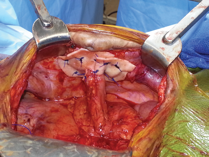
Strattice (LifeCell Corporation, Bridgewater, NJ) is a non–cross-linked porcine dermal product. It too has been used in contaminated fields and recurrences are higher when it is used as a bridge. Unlike polypropylene, its adherence and potential erosion into bowel is minimal, allowing it to be placed in direct contact with bowel, as seen in Fig. 1, used in a parastomal hernia repair reinforcement.
Fig. 1.

Biologic mesh comes directly in contact with the colon without impunity in this parastomal hernia repair reinforcement. Photo credit: Dr. Jennifer Ayscue.
Hybrid Mesh
Because both biologic and synthetic materials come with their own unique set of advantages and disadvantages, it is possible that they could be combined in a manner that would exploit the advantages of both, while minimizing the disadvantages. Recently, a hybrid made up of lightweight macroporous polypropylene encased in 8-ply porcine SIS has been released (Fig. 2). While data supporting the use of this product are lacking, it may, in fact, be helpful in situations where the advantages of each type of mesh were desirable. Developers of this product theorize that the biologic component will shield the synthetic component from potential infection while allowing the host to invade and replace the SIS with native tissue over time. Once the biologic component is replaced, the synthetic would be incorporated into the surrounding tissue. This could potentially allow for placement against the viscera with diminished risk of fistulization, or it may allow the use of the product as a bridge in a contaminated environment without the associated high risk of incisional hernia.
Fig. 2.

Image of Zenapro (Cook Medical, Inc., Bloomington, IN), a hybrid made up of porcine small intestinal submucosa encasing a lightweight macroporous polypropylene mesh.
Mesh Complications, Surgical Environment, and Technical Factors
Table 1 summarizes the complications of biologic and synthetic mesh as they relate to shrinkage, erosion, interface failure, eventration, and hernia recurrence.
Table 1. Shortcomings of mesh repairs by mesh type prone to complication.
| Shrinkage | Erosion | Interface failure | Eventration | Hernia recurrence | |
|---|---|---|---|---|---|
| Synthetic mesh | |||||
| Generalizations | Woven mesh shrinks less | Permanent mesh more susceptible | Small pore mesh more prone to interface failure than large pore mesh | ||
| Specific examples | Polypropylene mesh contracts 30–50% Sutures lead to less shrinkage than tacks |
Polypropylene and polyester mesh, when not lined, can erode into bowel | ePTFE interface failure due to small pore size, hydrophobicity, electronegative charge of the mesh | Dexon, Vicryl absorbed in 3– 6 mo with hernia recurrence near 100% | |
| Biologic mesh | |||||
| Generalizations | Biologics in general are less prone to erosion into bowel | Homografts more prone to eventration | Xenografts lead to recurrence when used as a bridge | ||
Patients undergoing reconstructive procedures that require supplemental material are generally complex, challenging, and unique cases. Balancing the patient's needs with the limitations of products available can be a daunting task. In selecting the type of graft material used, the degree of contamination and the proximity of the bowel need to be taken into account.
A retrospective cohort study in 200 patients undergoing open repair of incisional hernia compared four types of synthetic mesh: polypropylene, ePTFE, polyester, and double filament. A variety of surgical approaches were used. The recurrence rate was significantly higher in the polyester group. In addition, the fistula rate was 15.6% for polyester, 1.7% for polypropylene, and 0% for ePTFE and double-filament.3 While there have been isolated incidents of erosion into the bowel with ePTFE, it is highly unusual.13
We know from animal models that tissue matrix processing impacts upon the organism's reaction to a given material. Factors intrinsic to the matrix, patient physiologic factors, or surgical environment can impair the ability of the mesh to integrate into host tissue and can compromise revascularization. Fibroblast infiltration has been shown to increase the healing strength of an incised wound reinforced with mesh, while exaggerated inflammatory response by lymphocytes and neutrophils may promote rapid mesh degradation with resultant weakening or failure of the mesh material.14 15 16 17
Surgical Environment
Using National Surgical Quality Improvement Program data, a large study of more than 33,000 cases of ventral hernia repair looked at complication rates for clean, clean-contaminated, and contaminated cases. They compared patients undergoing repair with mesh to those having repair without mesh. The type of mesh included synthetics and biological mesh. The deep incisional surgical site infection rate was 1% in clean cases, 3% in clean-contaminated cases, and 4.5% in contaminated cases.18 There is experimental evidence that infection interferes with the integration of mesh into the host tissue.17 19
Permanent synthetic meshes are susceptible to infection, limiting their use in contaminated fields. A recent meta-analysis showed that the overall infection rate was 5%. Risk factors for infection included smoking, American Society of Anesthesiologists score > 3, and emergency operation. A variety of synthetic meshes and surgical techniques were employed. There was no difference in infection rate between microporous and macroporous mesh, but the authors cautioned that there were multiple confounding factors that precluded any solid conclusion on this issue. Mesh removal was performed in 70% overall, and 100% of the ePTFE grafts.20
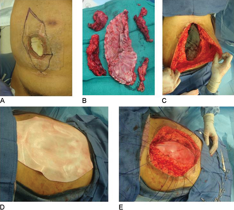
The management of infected mesh depends on the type of material involved. In general, infections involving polypropylene mesh can be drained, with excision of exposed, unincorporated mesh (Fig. 3). Grafts using ePTFE usually need to be excised.21 Absorbable materials can be used in an infected field; however, they often result in fascial defects once the material has dissolved. As they are degraded, they can generate dense adhesions that may complicate subsequent repair.
Fig. 3.

Repair of infected synthetic mesh with biologic mesh. (A) Synthetic mesh seen eroding though skin. Outline shows mesh extension. (B) Specimen photo of excised mesh and mesh-fascial scar. (C) Facial defect prepared for biologic mesh underlay. (D) Biologic mesh measured and cut to size over defect allowing >3 cm overlap with fascia. (E) Mesh underlay with suture fixation. Fascial edges were then approximated over mesh (not pictured). Photo credit: Dr. Praful Ramineni.
Biological mesh has been extensively used in clean-contaminated and contaminated fields, and short-term outcomes appear promising.22 While (as expected) wound infection rates are high, graft removal is unusual.23 Fig. 4 demonstrates an exposed biologic mesh that is likely contaminated with skin flora, and possibly enteric flora because the patient also had a colostomy. Mesh removal was not undertaken. Granulation tissue can be seen growing through the pores.
Fig. 4.

Exposed biologic mesh in a patient with Crohn disease who has an ileostomy with a leaking appliance in close proximity to the wound resulting in likely contamination with enteric flora. Photo credit: Dr. Neil Mauskar.
It is worth mentioning that experimental studies have demonstrated that the degree of contamination may adversely affect subsequent repair strength.24 In addition, long-term follow-up of patients, such as the one highlighted in Fig. 3, reveals a hernia recurrence rate of over 50% at 3 years.25
A meta-analysis of bioprosthetics for incisional hernia repair found that when combining mesh product by source, the recurrence rate was 23.2% for human dermis and 7.4% for porcine SIS. They reported a mesh disintegration rate of 0.5%. However, they concluded that there was an insufficient level of high-quality evidence on the use of biological mesh in ventral hernia repair.26
Because they generate an extensive adhesive response, simple polypropylene and polyester meshes generally should not be placed adjacent to the bowel. The use of a composite material, ePTFE, or biological mesh should be considered.27 28
Technical Factors
Subtle details of surgical technique can have a significant impact on long-term results. The operative approach must take into account not only patient factors but also variables related to the mesh. As each case can present its own unique challenges, the surgeon must be creative and be able to adapt, but adhere to certain basic principles.
With synthetic mesh, the method of mesh fixation impacts the amount of mesh contraction. Suture fixation results in less contraction than the use of tacks.29 To combat changes in the geometry of the mesh as a result of contraction, a 5-cm overlap is generally recommended. In addition, placing the omentum between the bowel and the mesh may protect the bowel from the inflammatory properties of the mesh and thereby limit adhesions as well as the potential for fistula formation.30
When considering a biological repair, the position of the mesh has a major impact on recurrence rates. When biological mesh is sewn to the edge of the fascia and used as a “bridge,” recurrence rates are as high as 80%. When the fascia can be reapproximated and the mesh used to reenforce the repair, the recurrence rate drops to approximately 20%. In addition, the type of suture material used to secure the graft has been shown to be important. Permanent suture material reduced the recurrence rate from 25 to 10%.31 It is also important to establish excellent contact between the biologic prosthesis and host tissues. Mesh placed with large buckles or wrinkles will impair host cell migration into the matrix and may have a negative impact on integration into surrounding tissues.
Component Separation Procedures: An Alternative to “Bridging” Procedures
When fascia cannot be primarily reapproximated, rather than bridging a defect with mesh alone and covering this repair with subcutaneous tissue and skin, modified flap procedures called “components separation/releases” allow for primary fascial closure and restoration of the midline. These can be performed alone or with mesh reinforcement (either biologic or synthetic). Reinforcement in this manner reduced hernia recurrence from 80% in bridged procedures using acellular dermal matrix to 20% in reinforcement procedures.32 Some examples of specific techniques include external oblique release, internal and external oblique release, “sliding door” release, “lateral” release, anterior rectus fascia release, and transversus abdominus release.
Fig. 5 provides an example of a classic Ramirez anterior component separation. Flaps are created anteriorly and the external oblique muscle is released. This allows for medial mobilization of the fascia, followed by closure. A small incision in the external oblique fascia can be made just 1 cm lateral to the lateral aspect of the rectus abdominus muscle. Fig. 5A shows the resultant defect. Surgeons will reinforce with a mesh in an overlay, underlay, or sublay fashion (Table 2). Overlay has been advocated because it allows reinforcing of the midline and the lateral edges. This method is typically used by plastic surgeons, while general and colorectal surgeons tend to prefer underlays or retrorectus placements. Running a suture along the lateral cut edges of the external oblique fascia will provide the necessary tension along the graft overlay to keep it in place (Fig. 5B).
Fig. 5.

Component separation with biologic mesh reinforcement (onlay). Photo credit: Dr. Tung Tran.
Table 2. Component separation procedures vary in what they expose mesh to contact.
| Location of mesh insertion | Mesh in contact with |
|---|---|
| Onlay | Subcutaneous fat Fascia |
| Underlay | Intestines Air/Peritoneal fluid Peritoneum |
| Sublay | Fascia Musculature |
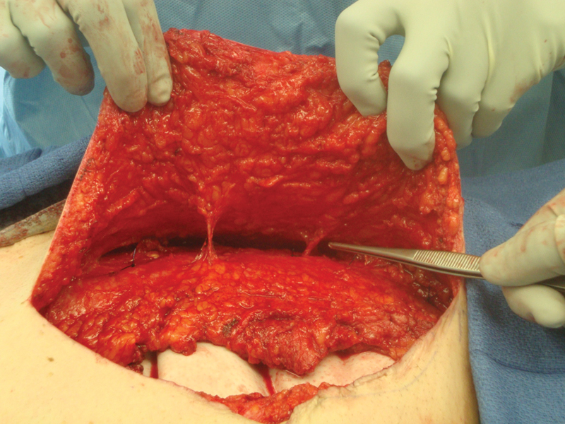
The creation of large flaps runs the risk of wound contamination because it leaves a large space for potential hematomas and seromas to form; therefore, wide closed suction drainage and extended antibiosis are advocated. In addition, preserving blood supply or perfusion to the large skin flaps through perforators is key in flap survival and decreasing infection rates (Fig. 6). Sublay procedures allow the preservation of these perforators.
Fig. 6.

Preservation of perforators in a sublay biologic mesh procedure. Photo credit: Dr. Praful Ramineni.
Sublay procedures studied in comparison to primary repair procedures and onlay procedures in a prospective randomized trial involving 161 patients by Venclauskas et al resulted in less wound complications (49 vs. 24%, total; 45 vs. 24%, seroma; 14 vs. 2%; 10.5 vs. 2% in recurrent hernia for onlay vs. sublay, respectively), concluding that reinforced sublay is superior in reduction of wound complications and recurrence.33
Cost, Reimbursement, and Coding
In the United States, coding varies by location of care with outpatient procedures performed at ambulatory surgery centers (ASCs) consistently costing less in U.S. Dollars (USD or $) than those performed in hospital settings. Furthermore, those performed as an outpatient are consistently less costly than those performed on inpatients. For example, in 2009,33 CPT 49561 (repair initial incisional or ventral hernia: incarcerated or strangulated) reimbursed at a rate of approximately $1.2K at ASCs, $2.1K in the hospital outpatient setting, and $5.3K in the hospital inpatient setting. All of these charges were associated with physician professional reimbursement rates of $838. When synthetic mesh is used, an “add-on” code +49658 is appended for an additional reimbursement potential of $562 in the ASC setting and $1K in the hospital outpatient setting. The physician garners an additional $250 as professional fees. Use of biologics in the setting of component separation can be reimbursed only in the hospital inpatient setting. In 2009, CPT 15734 (cutting and preparation of pedicle grafts or flaps) with add-on code +15430 (acellular xenograft implant, graft first 100 cm2 or less) provides physician professional fees of approximately $1.7K with hospital reimbursement of $21K.34 35 Although these rates represent only a snapshot in time, and these numbers constantly fluctuate, it serves to demonstrate that the costs of primary repair with synthetic mesh are roughly a quarter of that of component separation with biologic mesh.
Table 3 details the add-on codes necessary to bill for use of mesh, while Table 4 provides some coding combinations for common procedures performed by colorectal surgeons using mesh.36
Table 3. CPT “add-on” codes commonly used in mesh procedures.
| CPT code | CPT code descriptor |
|---|---|
| +49568 | Implantation of mesh or other prosthesis for open incisional or ventral hernia repair or mesh for closure of debridement for necrotizing soft tissue infection (List separately in addition to code for the incisional or ventral hernia repair) |
| +15777 | Implantation of biologic implant (e.g., acellular dermal matrix for soft tissue reinforcement (e.g., breast, trunk) (list separately in addition to code for primary procedure) |
| +57267 | Insertion of mesh for pelvic floor defect |
Table 4. Example of coding combinations for procedures.
| Repair initial incisional hernia with tissue matrix/mesh and component separation of muscle parts | 15734 Musculofascial flaps trunk right (i.e., component separation) |
| 15734–59 Musculofascial flaps trunk left (i.e., component separation) | |
| • Modifier “59” distinct procedural service 49560 Repair initial incisional hernia | |
| • Modifier “51” for multiple procedure same site +49568 Add-on implantation of mesh | |
| Parastomal repair with biologic | 44346 Revision of colostomy, simple; with repair paracolostomy hernia (separate procedure) |
| +15777 Implantation of biologic implant (e.g., acellular dermal matrix for soft tissue reinforcement (e.g., breast, trunk) (list separately in addition to code for primary procedure) |
Critical to understanding the cost–benefit of these, more complex, costly, but perhaps more durable repairs, are the costs of recurrence and readmissions. This is an area where further inquiry and analysis is much needed.
Attempting to account for success rates, an industry-sponsored cost analysis (and therefore potentially inherently biased) found that the average cost of a hernia repair using a 587-cm2 piece of mesh (which accounts for 5 cm of overlap, circumferentially) was approximately $20K to $26K when biologics were employed, versus $13K when synthetics (resorbable and coated polypropylene) were employed. They undertook a cost analysis of the biologics based on a systematic review of success rates of SIS ($23K) and acellular human dermis ($26K).36 At 88% success for SIS meshes and 78% for acellular human dermis meshes, the authors compared it to a roughly 81% success rate of non–cross-linked porcine dermis meshes ($26K)37 to conclude that SIS mesh repairs were the most cost-effective, when accounting for hernia repair success rates.38
In a difficult economy, rising health-care costs are under consistent scrutiny by both governmental health-care agencies which set standards for reimbursement as well as hospitals trying desperately to function without sustaining significant financial losses. Makers and suppliers of biologic and synthetic meshes frequently coach surgeons on how best to code. Recent studies have shown that hernia repair procedures, especially those using biologics, usually cost hospitals more than they can gain in reimbursement, not accounting for readmissions.
A study featured in General Surgery News which focused a spotlight on this issue was championed by Reynolds et al at University of Kentucky.39 They analyzed cost data on 415 consecutive open ventral hernia repairs (CPT codes 49560, 49561, 49565, and 49566) performed over a 3-year period at a tertiary care referral center.
Among inpatients undergoing the primary procedure of a ventral hernia repair, 46 were repaired without mesh, 79 were repaired with synthetic mesh, and 48 with biologic mesh. Median direct costs for cases performed without mesh were $5,432; median direct costs for those using synthetic and biologic mesh were $7,590 and 16,970, respectively (p < 0.01). Median net losses for repairs without mesh were $500. Median net profit of $60 was observed for synthetic mesh-based repairs. The median contribution margin for cases using biologic mesh was $4,560, and the median net financial loss was $8,370. Outpatient ventral hernia repairs, with and without synthetic mesh, resulted in median net losses of $1,560 and $230, respectively.39 The author, however, was quoted to admit that the limitation of the study was a lack of a link to readmission and reoperation data, which may account for the added cost of use of biologics having a financial advantage.40
Conclusion
Synthetic and biological meshes are widely used in surgical practice and the number of new products continues to grow. To optimize surgical outcomes, the practicing surgeon must have a thorough understanding of these products to guide proper selection and use. The heterogeneous patient population, variety of techniques employed, and large number of products available make comparisons between existing studies difficult. Randomized clinical trials of various meshes used in technically standardized manners in similar patient populations would help lend Level 1 evidence to the growing body of scientific literature on this subject.
Acknowledgments
The authors gratefully acknowledge Ms. Haripriya Ayyala, Dr. Kirthi Kolli, and Dr. Shola Cole for editorial assistance. The authors also acknowledge Drs. Jennifer Ayscue, Praful Ramenini, Tung Tran, and Neil Mauskar for providing photos for the figures.
References
- 1.Morris-Stiff G J, Hughes L E. The outcomes of nonabsorbable mesh placed within the abdominal cavity: literature review and clinical experience. J Am Coll Surg. 1998;186(3):352–367. doi: 10.1016/s1072-7515(98)00002-7. [DOI] [PubMed] [Google Scholar]
- 2.Cobb W S, Kercher K W, Heniford B T. The argument for lightweight polypropylene mesh in hernia repair. Surg Innov. 2005;12(1):63–69. doi: 10.1177/155335060501200109. [DOI] [PubMed] [Google Scholar]
- 3.Leber G E, Garb J L, Alexander A I, Reed W P. Long-term complications associated with prosthetic repair of incisional hernias. Arch Surg. 1998;133(4):378–382. doi: 10.1001/archsurg.133.4.378. [DOI] [PubMed] [Google Scholar]
- 4.Rosen M J. Polyester-based mesh for ventral hernia repair: is it safe? Am J Surg. 2009;197(3):353–359. doi: 10.1016/j.amjsurg.2008.11.003. [DOI] [PubMed] [Google Scholar]
- 5.Judge T W, Parker D M, Dinsmore R C. Abdominal wall hernia repair: a comparison of sepramesh and parietex composite mesh in a rabbit hernia model. J Am Coll Surg. 2007;204(2):276–281. doi: 10.1016/j.jamcollsurg.2006.11.003. [DOI] [PubMed] [Google Scholar]
- 6.Bachman S Ramshaw B Prosthetic material in ventral hernia repair: how do I choose? Surg Clin North Am 2008881101–112., ix [DOI] [PubMed] [Google Scholar]
- 7.Simmermacher R KJ, Schakenraad J M, Bleichrodt R P. Reherniation after repair of the abdominal wall with expanded polytetrafluoroethylene. J Am Coll Surg. 1994;178(6):613–616. [PubMed] [Google Scholar]
- 8.Johnson E K, Hoyt C H, Dinsmore R C. Abdominal wall hernia repair: a long-term comparison of Sepramesh and Dualmesh in a rabbit hernia model. Am Surg. 2004;70(8):657–661. [PubMed] [Google Scholar]
- 9.Novitsky Y W, Rosen M J. The biology of biologics: basic science and clinical concepts. Plast Reconstr Surg. 2012;130(5) 02:9S–17S. doi: 10.1097/PRS.0b013e31825f395b. [DOI] [PubMed] [Google Scholar]
- 10.Annor A H, Tang M E, Pui C L. et al. Effect of enzymatic degradation on the mechanical properties of biological scaffold materials. Surg Endosc. 2012;26(10):2767–2778. doi: 10.1007/s00464-012-2277-5. [DOI] [PMC free article] [PubMed] [Google Scholar]
- 11.Patel K M, Bhanot P. Complications of acellular dermal matrices in abdominal wall reconstruction. Plast Reconstr Surg. 2012;130(5) 02:216S–224S. doi: 10.1097/PRS.0b013e318262e186. [DOI] [PubMed] [Google Scholar]
- 12.de Moya M A, Dunham M, Inaba K. et al. Long-term outcome of acellular dermal matrix when used for large traumatic open abdomen. J Trauma. 2008;65(2):349–353. doi: 10.1097/TA.0b013e31817fb782. [DOI] [PubMed] [Google Scholar]
- 13.Foda M, Carlson M A. Enterocutaneous fistula associated with ePTFE mesh: case report and review of the literature. Hernia. 2009;13(3):323–326. doi: 10.1007/s10029-008-0441-6. [DOI] [PubMed] [Google Scholar]
- 14.Roessner E D, Thier S, Hohenberger P. et al. Acellular dermal matrix seeded with autologous fibroblasts improves wound breaking strength in a rodent soft tissue damage model in neoadjuvant settings. J Biomater Appl. 2011;25(5):413–427. doi: 10.1177/0885328209347961. [DOI] [PubMed] [Google Scholar]
- 15.Ono I. The effects of basic fibroblast growth factor (bFGF) on the breaking strength of acute incisional wounds. J Dermatol Sci. 2002;29(2):104–113. doi: 10.1016/s0923-1811(02)00019-1. [DOI] [PubMed] [Google Scholar]
- 16.Chang P J, Chen M Y, Huang Y S. et al. Morphine enhances tissue content of collagen and increases wound tensile strength. J Anesth. 2010;24(2):240–246. doi: 10.1007/s00540-009-0845-1. [DOI] [PubMed] [Google Scholar]
- 17.Orenstein S B, Qiao Y, Kaur M, Klueh U, Kreutzer D L, Novitsky Y W. Human monocyte activation by biologic and biodegradable meshes in vitro. Surg Endosc. 2010;24(4):805–811. doi: 10.1007/s00464-009-0664-3. [DOI] [PubMed] [Google Scholar]
- 18.Choi J J, Palaniappa N C, Dallas K B, Rudich T B, Colon M J, Divino C M. Use of mesh during ventral hernia repair in clean-contaminated and contaminated cases: outcomes of 33,832 cases. Ann Surg. 2012;255(1):176–180. doi: 10.1097/SLA.0b013e31822518e6. [DOI] [PubMed] [Google Scholar]
- 19.Bellón J M, García-Carranza A, García-Honduvilla N, Carrera-San Martín A, Buján J. Tissue integration and biomechanical behaviour of contaminated experimental polypropylene and expanded polytetrafluoroethylene implants. Br J Surg. 2004;91(4):489–494. doi: 10.1002/bjs.4451. [DOI] [PubMed] [Google Scholar]
- 20.Mavros M N, Athanasiou S, Alexiou V G, Mitsikostas P K, Peppas G, Falagas M E. Risk factors for mesh-related infections after hernia repair surgery: a meta-analysis of cohort studies. World J Surg. 2011;35(11):2389–2398. doi: 10.1007/s00268-011-1266-5. [DOI] [PubMed] [Google Scholar]
- 21.Cobb W S Kercher K W Heniford B T Laparoscopic repair of incisional hernias Surg Clin North Am 200585191–103., ix [DOI] [PubMed] [Google Scholar]
- 22.Janfaza M, Martin M, Skinner R. A preliminary comparison study of two noncrosslinked biologic meshes used in complex ventral hernia repairs. World J Surg. 2012;36(8):1760–1764. doi: 10.1007/s00268-012-1576-2. [DOI] [PubMed] [Google Scholar]
- 23.Kim H, Bruen K, Vargo D. Acellular dermal matrix in the management of high-risk abdominal wall defects. Am J Surg. 2006;192(6):705–709. doi: 10.1016/j.amjsurg.2006.09.003. [DOI] [PubMed] [Google Scholar]
- 24.Harth K C, Blatnik J A, Anderson J M, Jacobs M R, Zeinali F, Rosen M J. Effect of surgical wound classification on biologic graft performance in complex hernia repair: an experimental study. Surgery. 2013;153(4):481–492. doi: 10.1016/j.surg.2012.08.064. [DOI] [PubMed] [Google Scholar]
- 25.Rosen M J, Krpata D M, Ermlich B, Blatnik J A. A 5-year clinical experience with single-staged repairs of infected and contaminated abdominal wall defects utilizing biologic mesh. Ann Surg. 2013;257(6):991–996. doi: 10.1097/SLA.0b013e3182849871. [DOI] [PubMed] [Google Scholar]
- 26.Bellows C F, Smith A, Malsbury J, Helton W S. Repair of incisional hernias with biological prosthesis: a systematic review of current evidence. Am J Surg. 2013;205(1):85–101. doi: 10.1016/j.amjsurg.2012.02.019. [DOI] [PubMed] [Google Scholar]
- 27.Shankaran V, Weber D J, Reed R L II, Luchette F A. A review of available prosthetics for ventral hernia repair. Ann Surg. 2011;253(1):16–26. doi: 10.1097/SLA.0b013e3181f9b6e6. [DOI] [PubMed] [Google Scholar]
- 28.Vrijland W W, Bonthuis F, Steyerberg E W, Marquet R L, Jeekel J, Bonjer H J. Peritoneal adhesions to prosthetic materials: choice of mesh for incisional hernia repair. Surg Endosc. 2000;14(10):960–963. doi: 10.1007/s004640000180. [DOI] [PubMed] [Google Scholar]
- 29.Beldi G, Wagner M, Bruegger L E, Kurmann A, Candinas D. Mesh shrinkage and pain in laparoscopic ventral hernia repair: a randomized clinical trial comparing suture versus tack mesh fixation. Surg Endosc. 2011;25(3):749–755. doi: 10.1007/s00464-010-1246-0. [DOI] [PubMed] [Google Scholar]
- 30.Karabulut B, Sönmez K, Türkyilmaz Z. et al. Omentum prevents intestinal adhesions to mesh graft in abdominal infections and serosal defects. Surg Endosc. 2006;20(6):978–982. doi: 10.1007/s00464-005-0473-2. [DOI] [PubMed] [Google Scholar]
- 31.Janis J E, O'Neill A C, Ahmad J, Zhong T, Hofer S O. Acellular dermal matrices in abdominal wall reconstruction: a systematic review of the current evidence. Plast Reconstr Surg. 2012;130(5) 02:183S–193S. doi: 10.1097/PRS.0b013e3182605cfc. [DOI] [PubMed] [Google Scholar]
- 32.Jin J, Rosen M J, Blatnik J. et al. Use of acellular dermal matrix for complicated ventral hernia repair: does technique affect outcomes? J Am Coll Surg. 2007;205(5):654–660. doi: 10.1016/j.jamcollsurg.2007.06.012. [DOI] [PubMed] [Google Scholar]
- 33.Venclauskas L, Maleckas A, Kiudelis M. One-year follow-up after incisional hernia treatment: results of a prospective randomized study. Hernia. 2010;14(6):575–582. doi: 10.1007/s10029-010-0686-8. [DOI] [PubMed] [Google Scholar]
- 34.Sample Reimbursement Cases Available at: http://www.covidien.com/imageServer.aspx/doc187708.pdf?contentID=15216&contenttype=application/pdf
- 35.Ritter C Optimizing Reimbursement for Biological Implants Presented at: Michigan Ambulatory Surgery Association, 2013; Acme, Michigan [Google Scholar]
- 36.Hiles M, Record Ritchie R D, Altizer A M. Are biologic grafts effective for hernia repair?: a systematic review of the literature. Surg Innov. 2009;16(1):26–37. doi: 10.1177/1553350609331397. [DOI] [PubMed] [Google Scholar]
- 37.Itani K Samir A Baumann D et al. Prospective multicenter clinical study of single-stage repair of infected or contaminated abdominal incisional hernias using StratticeTM Reconstructive Tissue Matrix Poster presented at: American College of Surgeons 96th Clinical Congress; October 3–7, 2010; Washington, DC
- 38.Hiles M Briggs C M The overall cost of complex ventral hernia repair with biologic grafts General Surgery News 2010. Available at: http://www.cookbiodesign.com/library/Cost_DispellingMyths.pdf
- 39.Reynolds D Davenport D L Korosec R L Roth J S Financial implications of ventral hernia repair: a hospital cost analysis J Gastrointest Surg 2013171159–166., discussion 166–167 [DOI] [PubMed] [Google Scholar]
- 40.Frangou C Ventral hernia repairs a financial bust for hospitals? Money lost on most procedures at one facility General Surgery News2012 [Google Scholar]


