Chronic overnutrition and lack of physical activity result in excess deposition of adipose tissue and insulin resistance, which play key roles in the pathophysiology of type 2 diabetes and associated cardiovascular disease (1). The expansion of adipose tissue mass in dietary-induced obesity involves both hyperplasia and hypertrophy of adipocytes, and these processes are associated with extensive neovascularization mediated by the sprouting of endothelial cells (ECs) from preexisting blood vessels (2). Normal angiogenesis depends on the intricate balance between angiogenic factors, such as vascular endothelial growth factor (VEGF), fibroblast growth factors, and transforming growth factor-β, as well as angiostatic factors, such as angiostatin, endostatin, and the thrombospondins (3). Under chronic overnutrition and obesity, adipocytes produce EC-specific mitogens and angiogenic factors that favor vessel formation. Furthermore, ECs produce growth factors and cytokines that increase adipogenesis and fat deposition (4,5). Thus, adipocytes and ECs interact to regulate both adipogenesis and angiogenesis, and this interaction prompts the expansion of the capillary bed in conjunction with expanding regional adipose depots.
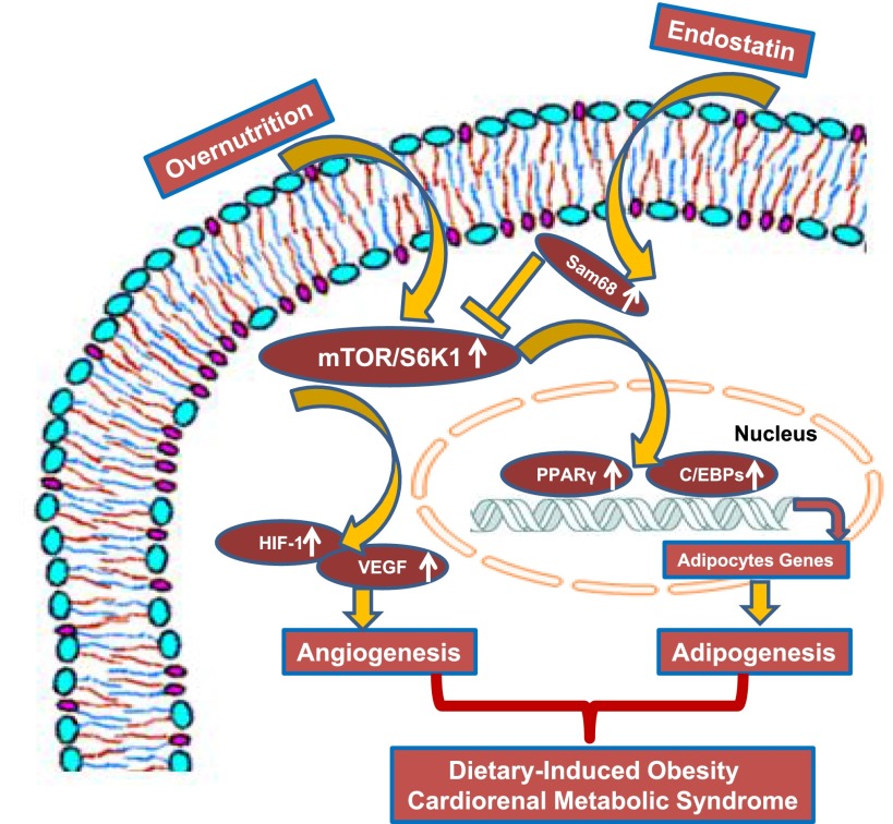
Adipogenesis is promoted by a network of transcription factors, including the CCAAT/enhancer–binding proteins (C/EBP)β, C/EBPδ, C/EBPα, and peroxisome proliferator–activated receptor γ (PPARγ) (6) (Fig. 1). Research from our laboratory has confirmed that mammalian target of rapamycin (mTOR)/ribosomal S6 kinase (S6K1) activation is enhanced under conditions of overnutrition (1) and impacts on PPARγ-C/EBPα signals to regulate adipogenic differentiation (6). To this point, mTOR/S6K1 signaling modulates cellular growth, energy metabolism, nutrient availability, and stress responses by assembling distinct signaling complexes and pathways mediated by two different protein complexes, mTOR complex 1 (mTORC1) and mTORC2 (1). Indeed, mTORC1 and its complexes have been shown to positively regulate adipogenesis and lipogenesis by activating sterol regulatory element–binding proteins. This activation is mediated in both an mTOR/S6K1-dependent and mTOR/S6K1-independent manner (7). Further, mTORC2 plays a key role in adipogenesis by controlling protein kinase B (Akt) activity in the early stage of adipogenesis, but this process is dispensable in the regulation of the function of the mature adipocyte (6,7). In these processes, mTOR function is regulated by Sam68 RNA–binding protein through mTOR alternative splicing (8). One study reported that mice with ablated Sam68 RNA–binding protein exhibit a lean phenotype as a result of increased energy expenditure, decreased commitment to early adipocyte progenitors, and defects in adipogenic differentiation, suggesting that an error in mTOR transcript impaired by Sam68 decreases activities of the mTORC1 pathway, resulting in a defect in adipogenesis (8).
Figure 1.
Chronic overnutrition activates mTOR/S6K1 signals to prompt adipogenesis and angiogenesis, resulting in obesity, insulin resistance, and the cardiorenal metabolic syndrome.
There is also an important connection between the Akt/mTOR signaling pathway and angiogenesis. For example, activation of the Akt/mTOR pathway in tumor adipose tissue can increase VEGF secretion through hypoxia-inducible factor 1 (HIF-1)-dependent and -independent mechanisms (5,9) (Fig. 1). Proinflammation cytokines and reactive oxygen species are upregulated in obesity and insulin resistance in part by the activation of mTOR signaling and contribute in VEGF-mediated vascular permeability, thereby promoting ocular neovascularization and proliferative diabetic retinopathy (10). These studies provide a new light on the molecular interplay between angiogenesis and adipogenesis in the pathogenesis of obesity and the cardiorenal metabolic syndrome (1).
Endostatin, a cleavage product of the COOH-terminal of collagen, is a potent inhibitor of EC proliferation and migration and angiogenesis mediated by interaction with integrins on ECs (11). It has been shown that the systemic administration of endostatin increases apoptotic activity of tumor cells, decreases microvessel density, and regresses primary tumors (11), suggesting that the antiangiogenic effects of endostatin prevent neovascularization and support tumor regression. In this issue of Diabetes, Wang et al. (12) investigate the effect of endostatin in dietary-induced adipogenesis, angiogenesis, and obesity. This study convincingly demonstrates that endostatin inhibited adipogenesis, angiogenesis, and dietary-induced obesity, in part, through mechanisms independent of the endostatin role as an integrin ligand. This investigation discloses that the antiadipogenic mechanism of endostatin lies in its interaction with Sam68 RNA–binding protein in the nuclei of preadipocytes. This interaction competitively impairs the binding of Sam68 to intron 5 of mTOR, causing an error in mTOR transcription. This consequently decreases mTOR expression and results in decreased activities of the mTORC1 pathway, which in turn leads to defects in adipogenesis. Therefore, these data suggest that endostatin may prevent dietary-induced obesity and its related metabolic disorders, including insulin resistance, glucose intolerance, and hepatic steatosis, by inhibition of angiogenesis and adipogenesis through pathways that modulate mTOR signaling and are integrin independent.
The observation that endostatin has both antiobesity and antiangiogenic functions is translationally relevant as endostatin becomes a potential application for antiobesity therapy and the prevention of the cardiorenal metabolic syndrome (1). It should be remembered that angiogenesis may be viewed as “good” or “bad” in patients with obesity and diabetes. For example, hyperglycemia and insulin resistance prompt the generation of reactive oxygen species and oxidative stress and inhibit endothelial nitric oxide synthase and HIF-1 activity, which result in the impairment of myocardial angiogenesis and an inadequate development of collateral blood vessels in patients with diabetes and coronary artery disease (3,13). Conversely, vasculopathy or excessive angiogenesis can participate in various human diseases, including cancer, diabetes complications, and impaired wound healing. Thus, it remains to be determined how endostatin may work in patients with dietary-induced obesity and the metabolic syndrome as endostatin may have both positive and negative roles in controlling obesity, diabetes, and associated cardiovascular disease at different stages of weight gain and the syndrome. In addition, rapamycin, an FDA-approved drug that inhibits mTOR/S6K1 signaling, has been used for coating coronary stents, treating renal cancer, and suppressing the immune response (3). Indeed, rapamycin may have potential benefit roles in dietary-induced obesity by inhibition of angiogenesis and adipose tissue expansion via effects on mTOR/S6K1 signaling (1,12). Future studies need to explore the impact of rapamycin on endostatin production in the context of dietary-induced obesity.
In summary, the study by Wang et al. (12) presents important new evidence that endostatin effectively protects mice against dietary-induced obesity and related metabolic disorders by the inhibition of adipogenesis and angiogenesis. Further animal and potentially clinical studies are needed to garner additional evidence that targeting of endostatin has therapeutic utility in the attenuation of dietary-induced obesity and the development of the cardiorenal metabolic syndrome.
Article Information
Acknowledgments. The authors would like to thank Brenda Hunter for her editorial assistance.
Funding. J.R.S. receives funding from the National Institutes of Health (R01-HL-73101-01A and R01-HL-107910-01) and the Veterans Affairs Merit System (0018).
Duality of Interest. No potential conflicts of interest relevant to this article were reported.
Footnotes
See accompanying article, p. 2442.
References
- 1.Jia G, Aroor AR, Martinez-Lemus LA, Sowers JR. Overnutrition, mTOR signaling, and cardiovascular diseases. Am J Physiol Regul Integr Comp Physiol 2014;307:R1198–R1206 [DOI] [PMC free article] [PubMed] [Google Scholar]
- 2.Manrique C, DeMarco VG, Aroor AR, et al. Obesity and insulin resistance induce early development of diastolic dysfunction in young female mice fed a Western diet. Endocrinology 2013;154:3632–3642 [DOI] [PMC free article] [PubMed] [Google Scholar]
- 3.Kim JA, Jang HJ, Martinez-Lemus LA, Sowers JR. Activation of mTOR/p70S6 kinase by ANG II inhibits insulin-stimulated endothelial nitric oxide synthase and vasodilation. Am J Physiol Endocrinol Metab 2012;302:E201–E208 [DOI] [PMC free article] [PubMed] [Google Scholar]
- 4.Tahergorabi Z, Khazaei M. Imbalance of angiogenesis in diabetic complications: the mechanisms. Int J Prev Med 2012;3:827–838 [DOI] [PMC free article] [PubMed] [Google Scholar]
- 5.Soumya SJ, Binu S, Helen A, Reddanna P, Sudhakaran PR. 15(S)-HETE-induced angiogenesis in adipose tissue is mediated through activation of PI3K/Akt/mTOR signaling pathway. Biochem Cell Biol 2013;91:498–505 [DOI] [PubMed] [Google Scholar]
- 6.Yoon MS, Zhang C, Sun Y, Schoenherr CJ, Chen J. Mechanistic target of rapamycin controls homeostasis of adipogenesis. J Lipid Res 2013;54:2166–2173 [DOI] [PMC free article] [PubMed] [Google Scholar]
- 7.Haissaguerre M, Saucisse N, Cota D. Influence of mTOR in energy and metabolic homeostasis. Mol Cell Endocrinol 2014;397:67–77 [DOI] [PubMed] [Google Scholar]
- 8.Huot MÉ, Vogel G, Zabarauskas A, et al. The Sam68 STAR RNA-binding protein regulates mTOR alternative splicing during adipogenesis. Mol Cell 2012;46:187–199 [DOI] [PubMed] [Google Scholar]
- 9.Karar J, Maity A. PI3K/AKT/mTOR pathway in angiogenesis. Front Mol Neurosci 2011;4:51. [DOI] [PMC free article] [PubMed] [Google Scholar]
- 10.Sasore T, Reynolds AL, Kennedy BN. Targeting the PI3K/Akt/mTOR pathway in ocular neovascularization. Adv Exp Med Biol 2014;801:805–811 [DOI] [PubMed] [Google Scholar]
- 11.Mucci LA, Stark JR, Figg WD, et al. Polymorphism in endostatin, an angiogenesis inhibitor, and prostate cancer risk and survival: A prospective study. Int J Cancer 2009;125:1143–1146 [DOI] [PMC free article] [PubMed] [Google Scholar]
- 12.Wang H, Chen Y, Lu X-a, Liu G, Fu Y, Luo Y. Endostatin prevents dietary-induced obesity by inhibiting adipogenesis and angiogenesis. Diabetes 2015;64:2442–2456 [DOI] [PubMed] [Google Scholar]
- 13.Hayden MR, Karuparthi PR, Habibi J, et al. Ultrastructure of islet microcirculation, pericytes and the islet exocrine interface in the HIP rat model of diabetes. Exp Biol Med (Maywood) 2008;233:1109–1123 [DOI] [PMC free article] [PubMed] [Google Scholar]


