

Abstract
Being capable of characterizing DNA local bending is essential to understand thoroughly many biological processes because they involve a local bending of the double helix axis, either intrinsic to the sequence or induced by the binding of proteins. Developing a method to measure DNA bend angles that does not perturb the conformation of the DNA itself or the DNA-protein complex is a challenging task. Here, we propose a joint theory-experiment high-throughput approach to rigorously measure such bend angles using the Tethered Particle Motion (TPM) technique. By carefully modeling the TPM geometry, we propose a simple formula based on a kinked Worm-Like Chain model to extract the bend angle from TPM measurements. Using constructs made of 575 base-pair DNAs with in-phase assemblies of one to seven 6A-tracts, we find that the sequence CA6CGG induces a bend angle of 19° ± 4°. Our method is successfully compared to more theoretically complex or experimentally invasive ones such as cyclization, NMR, FRET or AFM. We further apply our procedure to TPM measurements from the literature and demonstrate that the angles of bends induced by proteins, such as Integration Host Factor (IHF) can be reliably evaluated as well.
INTRODUCTION
DNA bending was first revealed in the mid-80s on the mitochondrial DNA of trypanosomatid parasites, the kinetoplast DNA (kDNA) (1) and attributed to the intrinsic bending property of the A-tracts sequences present in kDNA (2,3). These A-tracts were not only abundantly found in other prokaryotic and eukaryotic organisms but they were also shown to have a biological role, for example, by participating in the regulation of transcription (4–13). The binding of protein to DNA, that occurs in most of DNA-related biological processes, was also observed to induce the local bending of DNA (3,14–16).
As a result, for both intrinsic, sequence-dependent, or protein-induced bending of DNA, a large amount of work has been carried out to characterize it on structural and thermodynamical grounds (15,16). It is now commonly accepted that the bendability of specific DNA sequences relates to their capacity to be bent under the action of DNA-binding proteins. The bendability of these sequences may stem from their intrinsic bend, their low bending modulus or some specific breathing behavior of the DNA duplex structure that would facilitate its interactions with proteins. A DNA-analysis server based on the bending propensities of tri-nucleotides, that were deduced from DNase I digestion data (17), can be used to predict DNA structure from sequence and get an estimation of the foreseen DNA bend angles (18). However, characterizing rigorously the local bending of DNA molecules is a crucial issue that remains highly challenging (13,19,20).
To probe the DNA bendability experimentally, the most popular but complex technique remains the DNA cyclization method which provides a measure of the efficiency of cyclization of DNA fragments in presence of DNA ligase (21,22). However, this method does not permit to distinguish between changes in bending modulus that could be due to either permanent or transient structural defects and the presence of a local bend, a question for which other experimental strategies are needed. Another indirect approach consists in carrying out gel shift electrophoresis experiments using DNA molecules with several intrinsically bent sequences in-phase and in opposition of phase or circularly permuted DNA fragments (23–25). Though easy to handle, this technique can only provide rough estimates of bending angle and may be difficult to employ in all cases. Both Nuclear Magnetic Resonance (NMR) and X-ray require expensive equipment and tedious sample preparation and analysis procedures. They are therefore not routinely employed to detect and quantify the angle of an unknown DNA local bend but to bring details at the base-pair (bp) scale of a DNA structure already known to exhibit a local bend (26,27). Bend angles have also been deduced from more indirect techniques based on distance-dependent processes such as Fluorescence Resonance Energy Transfer (FRET) (28,29) or Plasmon resonance coupling of nanoparticles (30). Note that for these four last methods, the investigations are restricted to DNA molecules much shorter than the persistence length. Bending angles can also be extracted from single-molecule force-extension curves at large forces, as first proposed in (31). In this approach, the bending angle is inferred from the apparent persistence length, itself being a parameter used to fit experimental force-extension curves. However, extracting accurate values of the persistence length in this context presents several inherent difficulties, notably the fact that the fitting equation is supposed to be valid in the large-force regime where non-linear stretching should also be taken into account (32). More recently direct visualizations by Atomic Force Microscopy (AFM) (33,34) and cryo-electron microscopy (35) have given quantitative measurements of the bend angles. However these methods have limitations. Though AFM apparently gives the most direct access to the bend angles, the measurements are potentially biased by the sample preparation (36,37). By contrast, the technically demanding cryo-EM is supposed to preserve close-to-native state of the DNA complex, but it may also induce biases due especially to the sample confinement into a 50-nm-thick layer (35).
In the present work, we propose a physical method for the measure of DNA bend angles in a single DNA molecule and DNA-protein complex, which leaves intact the DNA conformation by ensuring minimal interaction with surfaces or tagged particles. This method combines our recently developed High Throughput Tethered Particle Motion (HT-TPM) technique and analytical modeling. HT-TPM enables the tracking of the conformational dynamics of hundreds of single DNA molecules in parallel, free to fluctuate in solution (38) (Figure 1A, Supplementary Information video). To extract the bend angles from HT-TPM data, we developed a simple analytical formula based on a kinked Worm-Like Chain (WLC) model that we validated on simulated data. Applied to constructs made of 575 bp DNAs with in-phase assemblies of 1 to 7 6A-tracts, we find that the sequence CA6CGG has an intrinsic bend angle of 19° ± 4°. In addition, the slight difference between our experimental data and our analytical model for a regular DNA suggests that even a DNA molecule with a randomly chosen sequence may contain a global curvature. We further apply our procedure to TPM measurements from the literature and demonstrate that the angles of bends induced by proteins, such as Integration Host Factor (IHF) can be reliably evaluated as well.
Figure 1.

(A) Schematic HT-TPM setup to measure the apparent length of a DNA molecule. The amplitude of motion, Rexp∥, of a particle tethered to a surface by a DNA with a contour length L depends on its effective length which varies with the angle θ of a bend located at distance l from one end chosen nearby the center of the molecule. For high-throughput measurements, individual DNA-particle complexes are immobilized on an array of functionalized sites. See ‘Materials and Methods’ section for details. (B) Representation of a typical 575-bp-long DNA fragment used in this study with the variable 88 bp central region (sizes are indicated in bp, details in Supplementary Information). The central region contains n copies of a CA6CGG sequences (in red), which are predicted to be bent (see text). The sequences of the 88-bp-long insert of the eight DNA molecules studied here are represented with the CA6CGG sequences. Each sequence is named as a function of the number of A contained in the CA6CGG (6A), followed by the number of repeats (nX) and the phasing (in exponent, P: in-phase, O: in opposition of phase).
MATERIALS AND METHODS
DNA constructs
DNA molecules were produced by PCR (oligonucleotides (Sigma-Aldrich): Biot-F575 ATAAGGGCGACACGGAAATG and Dig-R575 CGTGCAAGATTCCGAATACC) on pTOC plasmids, derived from pBR322 (39). Synthetic (GeneScript) DNA molecules (88 bp) containing increasing number of A6-tracts (0 to 7) were synthesized (Figure 1B) and were inserted between the HindIII and SalI restriction sites of pBR322. PCR products were purified as described in Diagne et al. (40).
HT-TPM setup and procedure
HT-TPM on chip assembly and the experimental setup are such as described in (38). The DNA/particle complexes were visualized in TPM buffer with an ionic strength of 165 mM (1.06 mM KH2PO4, 3.00 mM Na2HPO4, 154 mM NaCl, 1 mg.ml−1 Pluronic F127, 0.1 mg.ml−1 BSA) at 21 ± 1°C using a dark-field microscope (Axiovert 200, Zeiss). Acquisitions of 5 min were performed at a recording rate of 25 Hz with acquisition time of 40 ms on a CMOS camera Dalsa Falcon 1.4M100. The field of observation covers an area of  × 160 μm. Details can be found in Supplementary Information text.
The software Nanomultiplex co-developed with Magellium Toulouse (request should be addressed at info@magellium.fr) tracks in real time the positions of all the particles using the centroid method, averages these absolute positions on a 5 s window giving access to the anchoring point of the DNA molecule, calculates the 2D-vector positions of the bead  relative to the anchoring point of the DNA which corrects for experimental drift, calculates the asymmetry factor of the particle trajectories (41), and the amplitude of motion of the particle defined as
  and noted Rexp∥raw. In a general manner, we will use indifferently
  and R in the following. The averages, performed in the calculation of the asymmetry factors and amplitudes of motion, are taken over a sliding window of 5 s along the time trace. We invite the reader to refer to (42) for the detailed calculations of Rexp∥raw.
Finite exposure time of detectors,  , equal here to 40 ms, can lead to a blurring effect in single molecule (or particle) tracking experiments, as investigated for example in (42). The correlation time of the positions
  , of about 20 ms, was calculated for each DNA-particle complex and then injected in the following equation:
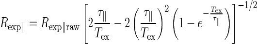 |
(1) |
to correct the amplitudes of motion recorded on each trace from blurring. In order to quantify the small differences expected on Rexp∥, we set up a two-step procedure that is described in detail in the Supplementary Information text.
TPM simulations
We performed Kinetic Monte Carlo simulations on the particle-DNA complex to predict the particle to anchor 2D-distance. We invite the reader to refer to (42) for the details of these Kinetic Monte Carlo simulations.
The bent sequences used in the experiments are simulated by setting a fixed angle between three successive monomers located at the center of the DNA molecule. A full range of angles were studied in successive simulations: 0, 18, 30, 45, 50, 60, 72, 90, 120 and 180°. The 2D-vector of the particle position  is measured throughout simulations and utilized to estimate the amplitude of motion defined as
  , the average being taken along the trajectory.
Influence of the particle size on the angle measurements
The particle size cannot be known exactly. Indeed, on the one hand, there is a limited control of their radius by the manufacturer leading to an uncertainty of 3 nm. On the other hand, the subsequent functionalization of the particles confer them a slightly larger radius Rp (Figure 1A). Indeed, the layer of antibody used to connect the DNA molecule to the particle is expected to be a few nanometers wide. Since in Equation (2), Rp precisely refers to the distance between the particle center and the extremity of the DNA molecule, and being not able to exactly infer it, we chose to explore two cases: Rp = 150 nm, previously used, and Rp = 155 nm, the real values probably dwelling between both values.
For Rp = 155 nm, θ1 was found to be equal to 17° ± 2°, while for Rp = 150 nm, θ1 = 15°± 2°. The bend angle values obtained considering or not a 5 nm increase of the effective radius of the particles in this precise case cannot be distinguished. Though particles with a well-defined size would lead to a more precise bend angle, the uncertainty on the exact value of Rp does not appear to be a critical issue in a typical case such as the one described here.
RESULTS
A new experimental strategy
The presence of a localized bent structure within a DNA molecule is expected to induce the reduction of the apparent end-to-end distance of the entire DNA molecule. Such an effect will be much easier to detect on short DNA molecules. Such an effect will be much easier to detect on short DNA molecules. To measure it, we chose 575-bp-long DNA molecules that were immobilized on a functionalized coverslip by one end, and attached to a 150 nm radius particle at their other end permitting their video tracking (Figure 1A). This experimental design represents a good compromise for an easy particle detection and a nearly force-free measurement, as the effective force exerted by the particle on the DNA is only a fraction of pN (43). In that way, the 2D projection of the particle displacement relative to the anchoring point of the DNA molecule gives access to its root-mean-squared end-to-end distance projected on the grafting surface, noted Rexp∥raw, which depends on the length and the conformational state of the monitored molecule. Rexp∥raw was corrected for the blurring effect, caused by the acquisition system, to obtain Rexp∥, using Equation (1) of ‘Materials and Methods’ section. TPM is capable of revealing changes in tether length equivalent to an apparent contour length as small as 100 bp (42,44). To obtain a good precision, inferior to 1 nm, a large amount of experimental data (see Table 1) is required because of the intrinsic dispersion of data due to an unavoidable variability of the DNA-particle and DNA-substrate links. To do so, we take advantage of our recently developed biochip that permits us to accumulate acquisitions on several hundreds of single DNA molecules in parallel by HT-TPM with a typical error on Rexp∥ equal to 0.4 nm (38).
Table 1. Amplitude of motion,  , corrected from the blurring effect and end-to-end distance,
  , extracted with a particle radius
  nm for the set of constructs.
| DNA samples | Number of trajectories |
 (nm) (nm) |
Uncertainty (nm) | RDNA(nm) |
|---|---|---|---|---|
| 6An0 | 3496 | 149.7 | 0.2 | 105.4 |
| 6An1P | 2728 | 149.1 | 0.2 | 104.1 |
| 6An2P | 2354 | 149.7 | 0.2 | 105.4 |
| 6An3P | 2904 | 149.6 | 0.2 | 105.2 |
| 6An4P | 2604 | 146.5 | 0.2 | 98.5 |
| 6An6P | 348 | 145.2 | 0.4 | 95.5 |
| 6An7P | 431 | 140.4 | 0.4 | 84.1 |
| 6An4O | 2990 | 151.2 | 0.2 | 108.6 |
In order to evaluate the capacity of this technique to detect and quantify local bending angles, we produced a series of DNA molecules based on a unique plasmid series. It incorporates a central 88 bp region, smaller than the DNA persistence length ( bp), containing one to seven CA6CGG sequences in-phase or in opposition of phase (Figure 1B and Materials and Methods section). We chose the CA6CGG sequence, known to be a sequence inducing a large bend (45). The in-phase A-tracts are located every integer number of helix turns, whereas those in opposition of phase are located every half-integer number of helix turns (Figure 1B).
By HT-TPM, we measured Rexp∥raw and calculated Rexp∥, the amplitude of motion, for this series of 575-bp-long DNA molecule (Table 1).
A kinked Worm-Like Chain model to analyze TPM data
Obtaining the bend angle from  requires the calculation of the root-mean-square end-to-end distance of the DNA molecule,
  , and an appropriate theoretical model giving the variation of
  as a function of a local bend angle, denoted by
  . To validate this analytical tool, we simulated a DNA-particle complex in a TPM setup (see Materials and Methods section and Supplementary Information text) where a bend of fixed angle
  varying from 0 to 180° was incorporated in the middle of the DNA molecule. The corresponding projection of the mean-square end-to-end distance
  was computed accordingly, as sketched in Figure 1A.
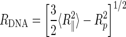
R
DNA was extracted from these numerical results by correcting for the effects of the particle and of the glass substrate (42,43). To achieve it, we explored two strategies. The first one is a minimal model in which the particle and the DNA molecule are considered statistically independent and the effect of the substrate is ignored. This last assumption is correct when the particle is very small, 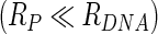 , and the DNA molecule is either very long
  or very short
  , leading to
 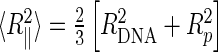 and thus to:
 |
(2) |
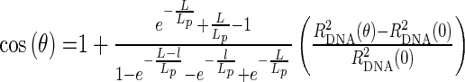
However, it has been shown that the effect of the particle can matter, because the hard-core interaction between the particle and the substrate reduces the number of degrees of freedom accessible to the molecule. The second strategy relies on a more sophisticated protocol proposed by Segall et al. in (43) to correct for the effects of the particle and of the glass substrate for very long DNA. We refer to this approach as Segall's method. It consists in solving the equation:
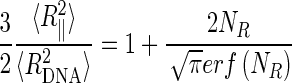 |
(3) |
where erf is the error function and  . To use this Equation (3) in the present context, we crudely extrapolated the results of (43), obtained for the Gaussian chain case
  , to the semi-flexible regime
  , by replacing
  by
by  . Note that Equation (2) is nothing but the expansion at order 1 in
  of Equation (3).
To obtain the bending angle, we fit the corrected data using a WLC model (46) on a polymer of length L with persistence length Lp, with a bend located at distance l from one end which locally induces a curvature with an angle θ (see Figure 1A). The mean-squared end-to-end distance is given by (Supplementary Information text, note that a similar formula has been obtained in 2 dimensions in (33)):
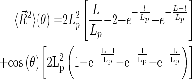 |
(4) |
For L = 575 bp and l = L/2, one derives from Equation (4) the fitting formula,  . In Figure 2, both methods were applied to the numerical data. For example, for
  = 0, the true WLC value of
  is
is 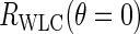 = 121.9 nm (see Materials and Methods section), in-between the values obtained with Segall's model and the minimal one. Whereas the minimal method underestimates
  , Segall's one overestimates it, both methods leading to comparable relative errors of about 10%. We thus choose D as a free fitting parameter in order to account for an offset at θ = 0. One observes in Figure 2 that whereas the minimal method gives a very satisfying fit, Segall's one leads to a worse one. Hence extrapolating Segall's calculation to the case of semi-flexible DNA molecules including a local bend appears to be less adapted than the minimal model. We thus use the minimal model in the remainder of this paper.
Figure 2.

Simulated HT-TPM amplitudes of motion Rsim∥ (•), together with the corrected values RDNA obtained through Segall's method (♦) and the minimal one (▪); see text for details. The DNA molecule length is L = 575 bp and the particle radius is Rp = 150 nm, as in the experiments. Fits of the numerical data, corrected by both the Segall's method and the minimal one, are shown as dotted lines using Equation (4). The curve corresponding to the WLC model, with no adjusted parameters, is shown as a solid line.
Bending angle measurements on a regular DNA
For the set of DNA molecules containing series of in-phase 6A-tracts (Table 1), we found by HT-TPM that Rexp∥ diminishes from 149.7 to 140.4 nm. When the initial 88 bp internal fragment of the DNA molecules (6An0) was replaced by a fragment containing only one (6An1P), two (6An2P) or three 6A-tracts (6An3P) in-phase, Rexp∥ remained in a range of 0.6 nm from its initial value (no 6A-tract), which appears to be very close to the 0.4 nm incertitude range we estimated for our HT-TPM measurements according to the method described in the Materials and Methods section. Rexp∥ decreased down to 146.5 nm for DNA molecules with four 6A-tracts in-phase (6An4P), 145.2 nm with six 6A-tracts in-phase (6An6P) and 140.4 nm with seven 6A-tracts in-phase (6An7P). These decreases in Rexp∥ might stem from an intrinsic bend of the 6A-tract or a decrease in bending modulus of this sequence. In this latter case, one would expect the insertion of four 6A-tracts (6An4O) in opposition of phase to lead to a decrease in Rexp∥ similar to the one measured on 6An4P. In fact, 6An4O showed no decrease in Rexp∥ as it was found equal to 151.2 nm. Taken together, these results prove that, beyond four 6A-tracts, Rexp∥ decreased when the number of 6A-tracts in-phase increases due to an intrinsic bend of the 6A-tract.
In order to calculate  , we injected these Rexp∥ values in Equation (2). We assume then that each of the n successive 6A-tracts inserts in-phase imposes the same bending angle θ1 and postulate as a first order simplification hypothesis that θ = nθ1. With L = 575 bp, l = 318 bp, we now obtain the following equation:
 |
(5) |
The fits of  shown in Figure 3 (black symbols and fits for Rp = 150 nm) are reasonably good leading to D = 92 ± 2 nm and θ1 = 15° ± 2° for the value of the bend angle of each CA6CGG sequence. In the Materials and Methods section, we studied the influence of small variations of the particle size on this result and found comparable results within error bar.
Figure 3.

Experimental data after correction of the particle effects for the labeling particle Rp = 150 nm (•) using the minimal model, and two series of fits with D and θ1 (straight line), or D, θ1 and θ0 (dotted line) as free parameters. Error bars are smaller than the symbol size. Using Equation (5), ones finds: D = 92.0 nm, θ1 = 15° for Rp = 150 nm. Using Equation (6), one finds D = 91 nm, θ1 = 19°, θ0 = 25° (see Table S3 for details).
Influence of a global DNA curvature on the angle measurements
The experimental values of Figure 3 suggested that RDNA might be non-monotonous when n increases, contrary to what is expected from Equation (5). One reason for this observation could be the existence of an intrinsic curvature of the DNA molecule. To account for it, we have chosen to refine our analysis and use the more general fitting form:
 |
(6) |
where  mimics the whole intrinsic bend of the
  molecule. For sake of simplicity, we implicitly assume that it is accumulated at the same position as the bends when
  (17,23). The resulting fits, displayed in Figure 3 (dotted lines), are slightly better than before.
To sum up, the proposed refinement suggests that the true bending angle  is 19° ± 4° per bend, while the global bend of the DNA molecule would have an angle
  between 25° ± 20°. Although the fit including a global curvature of the DNA molecule seems better when looking at Figure 3, this new parameter
  can only be determined with a poor precision. This is due to the fact that this angle is actually delocalized on the whole 575 bp sequence. A more sophisticated model is needed to take this point better into account.
Influence of the bending modulus of the bent sequence on the angle measurements
Alternatively, it was in principle possible that the increase in the DNA end-to-end distance observed at small values of  was associated with an increase of the molecular rigidity of the inserts. We analytically derived the corrections to
  due to an insert of finite length and of increased rigidity using the WLC model (Supplementary Information text). At small insert length, the increase is linear with the insert length (Supplementary Figure S1). At first order, this effect is additive with the effect of a bend. The corresponding fits are displayed in Supplementary Figure S2 and Table S3. The obtained values of
  were close to the previous ones, thus showing that this alternative approach does not differ significantly from the previous one. This could be anticipated since a linear expansion of Equation (6) adds a linear term into Equation (5) if one assumes that
  . As a result, the combined HT-TPM/kinked-WLC approach might not easily discriminate between an increase of rigidity of the inserts and an intrinsic bend. However, when fitting the data of the DNA molecules, containing 0 to 4 6A-tracts inserts, with this additional linear term, we found a linear increase of the amplitude of motion of 1.9 nm per 10 bp 6A-tract. It would correspond to a persistence length of about 89 nm for this bent sequence. There exists another way to evaluate the rigidity contribution of the 6A-tracts. It consists in considering the data obtained for 6An0 (no 6A-tracts) and 6An4O (four 6A-tracts in opposition of phase). An increase in RDNA of 3.2 nm (105.4 versus 108.6 nm; Table 1) was then measured and gave access to a persistence length equal to 129 nm for this 55 bp sequence by using the formula in Supplementary Information text. These persistence lengths are estimated with a low precision due to the poor sensitivity of RDNA to an increase of Lp above 150 bp (Supplementary Figure S4). In any case, whatever the way used to estimate the persistence lengths of these 6A-tract sequences, values were very high compared to the 50 nm measured for random double strand DNA in the present salt conditions (47). As a result, an increase in rigidity of the 6A-tracts could not account for our data, and introducing a global bend
  is probably more adapted to this case.
DISCUSSION
DNA bending can be measured using HT-TPM
In this paper, we develop a procedure to measure a bend angle localized inside long DNA molecules with a good accuracy. For that, we built a platform plasmid based on pBR322 that enabled us to generate several 575-bp-long DNA molecules with various 88-bp-long DNA constructs close to their center. We used 6A-tracts as a model of bent sequences and showed that, by assembling them every 10.5 bp, we could amplify the observed decrease in Rexp∥ as a function of the number of bent sequences. Furthermore, making use of the HT-TPM, we are able to reliably detect variations of Rexp∥ down to 2%. Using Equations (1) and (2), we corrected experimental raw data for the effects of both camera averaging and particle radius and obtain  , which was correctly fitted using a kinked WLC model (Equation (4)). We thus got access to the bend angle of the chosen sequence θ1 = 19° ± 4.
To sum up, this entire procedure can be readily applied to quantify the bend related to a specific DNA structure together with an evaluation of the intrinsic curvature of the entire DNA molecule. To do so, our platform plasmid can be used to build an ad hoc series of DNA molecules with increasing numbers of the DNA sequences under study assembled in-phase. HT-TPM measurements have first to be corrected for the camera blurring effect using Equation (1) before being injected in Equation (2) to get the DNA end-to-end distance. The resulting series of data, DNA end-to-end distance as a function of the number of assembled sequences, are adjusted with Equation (4) where an intrinsic bend angle is incorporated if necessary.
Comparison with other methods for the measurements of CA6CGG bend angles
Crothers et al. combined numerical simulations and cyclization experiments on DNA fragments of 105 and 210 bp lengths to get the relative bend induced by this sequence (48). They found a bend angle equal to 19° ± 2°. It should be stressed that cyclization is an indirect method which (i) enforces the looping of short DNAs ( ) that can also be due to kinks or small denaturation bubbles (22), (ii) requires numerical simulations of the specific sequence with a large number of unknown parameters for the fitting procedure. In our case, we need at most three free parameters, namely D, θ0 and θ1.
More precise results were obtained by MacDonald et al. who used NMR spectroscopy on partially aligned DNA molecules (26). In particular, they found that the overall helix axis of the DNA dodecamer GGCA6CGG exhibits a bend of 19°± 1° toward the minor groove of the A-tract. In addition, NMR results confirmed the major role played by the joint located between the 6A-tract and the next CG sequence that was found to be responsible for 14° of the 19° of the global bend angle of the dodecamer. NMR is limited to small DNA fragments and cannot be easily extended to DNA-protein complexes.
X-ray crystallography showed A-tracts structures with various conformations due to the crystal constraints (49). As it is complicated to determine the most representative one, we did not try to compare X-ray crystallography results to ours.
Experiments based on FRET also gave access to the bend angle value of the CA6CGG-CGA6CGG-CA6CGG sequence prepared in solutions with a broad range of salt concentration (28). The bend angle was deduced from the average dye-to-dye distance between two fluorophores located at the ends of the 31 bp DNA fragment. It was shown to vary from 23° ± 4° to 41° ± 4° with NaCl concentration increasing from 10 to 500 mM. At the ionic strength of 165 mM we carried out the experiments, Tóth et al. found a bend angle ranging between 36° ± 4° and 41° ± 4° which is in slight disagreement with the 3 × (19° ± 4°) = 57° ± 7° that we obtain. This discrepancy could be due to the sequence located on the middle which is slightly different with the flanking C being replaced by G. According to Koo et al. (45), the middle sequence should induce a bend smaller, up to 10%, than the two other ones. As a result, the total bend angle of the Toth's sequences is necessarily smaller than the one obtained by a 3-fold assembly of our sequence. Another source of discrepancy is that distance measurements by FRET are highly sensitive to the precise geometry of the attachment of the dyes at the extremity of the DNA molecules which is difficult to determine accurately. Moreover, the dyes are very close to the studied sequence and may interfere with it.
Finally, direct imaging of two to eight 6A-tracts assembled in-phase has been made by AFM (33). By measuring the planar end-to-end distance of these molecules, it was deduced a mean bend angle for each 6A-tract of about 13.5° with a precision of about 1°. The high precision that was obtained here does not take into account the possible bias induced by the mechanical constraints exerted onto the DNA molecule deposited on mica and visualized in air. Indeed the divalent ions that stick the DNA to the surface are likely to modify significantly the electrostatic interactions and therefore the DNA elastic properties (37).
Using HT-TPM to characterize the 6A-tracts enabled us to obtain values in reasonable agreement with those measured in these preceding studies by other methods. Besides, our measurements were carried out on DNA molecules containing the bent inserts far from the surface and the labeling particle, which thus do not interfere with the insert under examination. This is not possible with the previously mentioned techniques. Though very informative, most of them remain limited to the study of short DNA fragments. Furthermore, our underlying model is simple, appealing to elementary and robust polymer physics arguments, whereas interpreting data incoming from NMR, X-ray scattering, FRET or cyclization experiments is far more indirect and tedious. Finally, parallelizing single molecule experiments is a pre-requisite here, the very small variations of the amplitude of motion that we intend to monitor requiring intensive sampling.
DNA heterogeneity and mechanical characterization
As expected the end-to-end distance of 6An0, calculated as  ), is found notably smaller, 107.7 ± 7.1 nm, than the expected WLC value, namely 121.9 nm. It suggests that the WLC model is not perfectly adequate to the DNA sample studied here. More importantly, whatever the equation we used to fit our HT-TPM data, the fits do not match exactly the experimental data within error bars. This can be due to an under-estimation of our confidence intervals. Even though increasing our sample sizes, we did not manage to reduce this discrepancy. We propose that it could be due to the intrinsic bend accumulated along the molecule, the additional effect of which would reduce the overall apparent end-to-end distance. Notably, when deleting some random DNA regions and replacing them by bent inserts, we conjecture that the deleted regions already bore some small but non-zero curvature, the effect of which can be either compensated or accentuated when replaced by an insert. This leads to small corrections of the end-to-end distance around the average trend described by Equation (4). This work thus highlights that a given sequence generically involves a small but non-vanishing intrinsic bend to DNA molecules, the amplitude of which typically grows like the square root of the molecule length, owing to the central-limit theorem, because the small bending angles related to individual base pairs add up independently along the chain. From a statistical physics perspective, our results point to the need to adapt statistical models to incorporate the fact that typical DNA molecules with a ‘random’ sequence adopt a curved shape rather than a straight one in the rigid regime L < Lp (13).
Quantification of protein-induced DNA bends by HT-TPM
TPM is one of the very few methods permitting the monitoring of DNA looping. In addition, relying on the theoretical framework proposed here, we claim that HT-TPM can also reveal the protein-induced bending of the DNA interaction site and give access to the protein-induced bend angle.
When studying the interaction of a protein with a DNA molecule, a decrease in  may be observed after the addition of the protein on DNA molecules containing only one site of interaction (40,44,50–52). In this case, we can still evaluate the bend angle θ from the two DNA end-to-end distance values obtained before and after addition of the protein by using Equation (7), derived from Equation (4) assuming that the global intrinsic bend
  is negligible,
 |
(7) |
Note that if an intrinsic angle θ0 was present before insertion of the sequence of interest, this formula gives access to the angle variation θ –θ0 instead of θ.
As a test of its accuracy, we applied this simplified method to 6An4P, 6An6P and 6An7P and obtained a mean value of one 6A-tract insert equal to 15° ± 2°, the uncertainty is here calculated as the standard deviation of the data obtained for the three DNA molecules (Supplementary Table S5). This value is in good agreement with the value 15° ± 2° obtained from the fit performed on the entire set of data with  as the only free angle parameter (
 . In addition, using Equation (7) and considering equal to 2% the minimal variation in Rexp∥ that can be detected with HT-TPM on this 575 bp DNA molecule, one can only measure bend angles larger than 33°. To measure smaller bend angles, the construction of a series of repeated sequence is required.
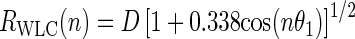
On the ground of this simple formula (Equation (7)), we analyzed the TPM results obtained by Mumm et al. (52) concerning the bend induced by the Integration Host Factor (IHF) that is known to bind to some specific binding sites and play thus a major role as architectural protein in prokaryotes. Upon binding of the protein to a single site of a L = 1943-bp-long DNA, the molecule behaved as a 1659-bp-long DNA which corresponds to  decreasing from
  249 to 229 nm. The binding site being located at l = 301 bp away from one extremity, we evaluate the IHF induced-angle to be about 180° using Equation (7). This value is in very good agreement with the crystallography measurements that show that IHF provokes a bend with an angle of at least 160° (53), especially as it should be considered that the end-to-end distance tends to saturate for such high angles.
Our analytical models consider separately the cases of a bend with a fixed angle and of a local flexible hinge, while a great number of DNA-binding proteins are now considered to induce both effects. To account for these two mechanical changes occurring simultaneously as well as the extended deformation of the protein-DNA interaction site, a more precise theory is needed. Nevertheless, we believe that our method could still be applied to those cases and give access to some valuable though less precise information.
We can therefore conclude that the combined HT-TPM/kinked-WLC approach provides an efficient method to estimate the angle of fixed local DNA bend either intrinsic to a sequence or induced by the binding of proteins. With many advantages over the existing methods, we believe that our approach will permit the refined characterization of DNA geometry in various contexts and to shed new light on DNA–protein complexes.
SUPPLEMENTARY DATA
Supplementary Data are available at NAR Online.
Acknowledgments
We thank François CORNET for his helpful discussions on the DNA construction. We belong to the GDR CellTiss.
FUNDING
CNRS, University of Toulouse 3 and ANR-11-NANO-010 ‘TPM On a Chip’. Funding for open access charge: ANR-11-NANO-010 ‘TPM On a Chip’.
Conflict of interest statement. None declared.
REFERENCES
- 1.Borst P. Why kinetoplast DNA networks. Trends Genet. 1991;7:139–141. doi: 10.1016/0168-9525(91)90374-y. [DOI] [PubMed] [Google Scholar]
- 2.Marini J.C., Levene S.D., Crothers D.M., Englund P.T. Bent helical structure in kinetoplast DNA. Proc. Natl. Acad. Sci. U.S.A. 1982;79:7664–7668. doi: 10.1073/pnas.79.24.7664. [DOI] [PMC free article] [PubMed] [Google Scholar]
- 3.Wu H.-M., Crothers D.M. The locus of sequence-directed and protein-induced DNA bending. Nature. 1984;308:509–513. doi: 10.1038/308509a0. [DOI] [PubMed] [Google Scholar]
- 4.Horn G.T., Wells R.D. The leftward promoter of bacteriophage lambda. Structure, biological activity, and influence by adjacent regions. J. Biol. Chem. 1981;256:2003–2009. [PubMed] [Google Scholar]
- 5.Struhl K. Naturally occurring poly(dA-dT) sequences are upstream promoter elements for constitutive transcription in yeast. Proc. Natl. Acad. Sci. U.S.A. 1985;82:8419–8423. doi: 10.1073/pnas.82.24.8419. [DOI] [PMC free article] [PubMed] [Google Scholar]
- 6.Mizuno T. Static bend of DNA helix at the activator recognition site of the ompF promoter in Escherichia coli. Gene. 1987;54:57–64. doi: 10.1016/0378-1119(87)90347-7. [DOI] [PubMed] [Google Scholar]
- 7.Ross W., Gosink K.K., Salomon J., Igarashi K., Zou C., Ishihama A., Severinov K., Gourse R.L. A third recognition element in bacterial promoters: DNA binding by the alpha subunit of RNA polymerase. Science. 1993;262:1407–1413. doi: 10.1126/science.8248780. [DOI] [PubMed] [Google Scholar]
- 8.Gaal T., Rao L., Estrem S.T., Yang J., Wartell R.M., Gourse R.L. Localization of the intrinsically bent DNA region upstream of the E. coli rrnB P1 promoter. Nucleic Acids Res. 1994;22:2344–2350. doi: 10.1093/nar/22.12.2344. [DOI] [PMC free article] [PubMed] [Google Scholar]
- 9.Tippner D., Afflerbach H., Bradaczek C., Wagner R. Evidence for a regulatory function of the histone-like Escherichia coli protein H-NS in ribosomal RNA synthesis. Mol. Microbiol. 1994;11:589–604. doi: 10.1111/j.1365-2958.1994.tb00339.x. [DOI] [PubMed] [Google Scholar]
- 10.Iyer V., Struhl K. Poly(dA:dT), a ubiquitous promoter element that stimulates transcription via its intrinsic DNA structure. EMBO J. 1995;14:2570–2579. doi: 10.1002/j.1460-2075.1995.tb07255.x. [DOI] [PMC free article] [PubMed] [Google Scholar]
- 11.Hillebrand A., Wurm R., Menzel A., Wagner R. The seven E. coli ribosomal RNA operon upstream regulatory regions differ in structure and transcription factor binding efficiencies. Biol. Chem. 2005;386:523–534. doi: 10.1515/BC.2005.062. [DOI] [PubMed] [Google Scholar]
- 12.Segal E., Widom J. Poly(dA:dT) tracts: major determinants of nucleosome organization. Curr. Opin. Struct. Biol. 2009;19:65–71. doi: 10.1016/j.sbi.2009.01.004. [DOI] [PMC free article] [PubMed] [Google Scholar]
- 13.Freeman G.S., Lequieu J.P., Hinckley D.M., Whitmer J.K., de Pablo J.J. DNA Shape Dominates Sequence Affinity in Nucleosome Formation. Phys. Rev. Lett. 2014;113:168101. doi: 10.1103/PhysRevLett.113.168101. [DOI] [PubMed] [Google Scholar]
- 14.Swinger K.K., Rice P.A. IHF and HU: flexible architects of bent DNA. Curr. Opin. Struct. Biol. 2004;14:28–35. doi: 10.1016/j.sbi.2003.12.003. [DOI] [PubMed] [Google Scholar]
- 15.Maher L.J., III Mechanisms of DNA bending. Curr. Opin. Chem. Biol. 1998;2:688–694. doi: 10.1016/s1367-5931(98)80104-x. [DOI] [PubMed] [Google Scholar]
- 16.Privalov P.L., Dragan A.I., Crane-Robinson C. The cost of DNA bending. Trends Biochem. Sci. 2009;34:464–470. doi: 10.1016/j.tibs.2009.05.005. [DOI] [PubMed] [Google Scholar]
- 17.Brukner I., Sánchez R., Suck D., Pongor S. Sequence-dependent bending propensity of DNA as revealed by DNase I: parameters for trinucleotides. EMBO J. 1995;14:1812–1818. doi: 10.1002/j.1460-2075.1995.tb07169.x. [DOI] [PMC free article] [PubMed] [Google Scholar]
- 18.Vlahovicek K., Kajan L., Pongor S. DNA analysis servers: plot.it, bend.it, model.it and IS. Nucleic Acids Res. 2003;31:3686–3687. doi: 10.1093/nar/gkg559. [DOI] [PMC free article] [PubMed] [Google Scholar]
- 19.Gietl A., Grohmann D. Modern biophysical approaches probe transcription-factor-induced DNA bending and looping. Biochem. Soc. Trans. 2013;41:368–373. doi: 10.1042/BST20120301. [DOI] [PubMed] [Google Scholar]
- 20.Harteis S., Schneider S. Making the bend: DNA tertiary structure and protein-DNA interactions. Int. J. Mol. Sci. 2014;15:12335–12363. doi: 10.3390/ijms150712335. [DOI] [PMC free article] [PubMed] [Google Scholar]
- 21.Zhang Y., Xi Z., Hegde R.S., Shakked Z., Crothers D.M. Predicting indirect readout effects in protein–DNA interactions. PNAS. 2004;101:8337–8341. doi: 10.1073/pnas.0402319101. [DOI] [PMC free article] [PubMed] [Google Scholar]
- 22.Vafabakhsh R., Ha T. Extreme bendability of DNA less than 100 base pairs long revealed by single-molecule cyclization. Science. 2012;337:1097–1101. doi: 10.1126/science.1224139. [DOI] [PMC free article] [PubMed] [Google Scholar]
- 23.Zinkel S.S., Crothers D.M. DNA bend direction by phase sensitive detection. Nature. 1987;328:178–181. doi: 10.1038/328178a0. [DOI] [PubMed] [Google Scholar]
- 24.Lane D., Prentki P., Chandler M. Use of gel retardation to analyze protein-nucleic acid interactions. Microbiol. Rev. 1992;56:509–528. doi: 10.1128/mr.56.4.509-528.1992. [DOI] [PMC free article] [PubMed] [Google Scholar]
- 25.Wang Q., Calvo J.M. Lrp, a major regulatory protein in Escherichia coli, bends DNA and can organize the assembly of a higher-order nucleoprotein structure. EMBO J. 1993;12:2495–2501. doi: 10.1002/j.1460-2075.1993.tb05904.x. [DOI] [PMC free article] [PubMed] [Google Scholar]
- 26.MacDonald D., Herbert K., Zhang X., Pologruto T., Lu P., Polgruto T. Solution structure of an A-tract DNA bend. J. Mol. Biol. 2001;306:1081–1098. doi: 10.1006/jmbi.2001.4447. [DOI] [PubMed] [Google Scholar]
- 27.Nelson H.C., Finch J.T., Luisi B.F., Klug A. The structure of an oligo(dA).oligo(dT) tract and its biological implications. Nature. 1987;330:221–226. doi: 10.1038/330221a0. [DOI] [PubMed] [Google Scholar]
- 28.Tóth K., Sauermann V., Langowski J. DNA curvature in solution measured by fluorescence resonance energy transfer. Biochemistry. 1998;37:8173–8179. doi: 10.1021/bi973135z. [DOI] [PubMed] [Google Scholar]
- 29.Wozniak A.K., Schröder G.F., Grubmüller H., Seidel C.A.M., Oesterhelt F. Single-molecule FRET measures bends and kinks in DNA. Proc. Natl. Acad. Sci. U.S.A. 2008;105:18337–18342. doi: 10.1073/pnas.0800977105. [DOI] [PMC free article] [PubMed] [Google Scholar]
- 30.Reinhard B.M., Sheikholeslami S., Mastroianni A., Alivisatos A.P., Liphardt J. Use of plasmon coupling to reveal the dynamics of DNA bending and cleavage by single EcoRV restriction enzymes. Proc. Natl. Acad. Sci. 2007;104:2667–2672. doi: 10.1073/pnas.0607826104. [DOI] [PMC free article] [PubMed] [Google Scholar]
- 31.Kulić I.M., Mohrbach H., Lobaskin V., Thaokar R., Schiessel H. Apparent persistence length renormalization of bent DNA. Phys. Rev. E. 2005;72:041905. doi: 10.1103/PhysRevE.72.041905. [DOI] [PubMed] [Google Scholar]
- 32.Manghi M., Destainville N., Palmeri J. Mesoscopic models for DNA stretching under force: New results and comparison with experiments. Eur. Phys. J. E. 2012;35:110. doi: 10.1140/epje/i2012-12110-2. [DOI] [PubMed] [Google Scholar]
- 33.Rivetti C., Walker C., Bustamante C. Polymer chain statistics and conformational analysis of DNA molecules with bends or sections of different flexibility. J. Mol. Biol. 1998;280:41–59. doi: 10.1006/jmbi.1998.1830. [DOI] [PubMed] [Google Scholar]
- 34.Buzio R., Repetto L., Giacopelli F., Ravazzolo R., Valbusa U. Label-free, atomic force microscopy-based mapping of DNA intrinsic curvature for the nanoscale comparative analysis of bent duplexes. Nucleic Acids Res. 2012;40:e84. doi: 10.1093/nar/gks210. [DOI] [PMC free article] [PubMed] [Google Scholar]
- 35.Bednar J., Furrer P., Katritch V., Stasiak A., Dubochet J., Stasiak A. Determination of DNA persistence length by cryo-electron microscopy. Separation of the static and dynamic contributions to the apparent persistence length of DNA. J. Mol. Biol. 1995;254:579–594. doi: 10.1006/jmbi.1995.0640. [DOI] [PubMed] [Google Scholar]
- 36.Sorel I., Piétrement O., Hamon L., Baconnais S., Le Cam E., Pastré D. The EcoRI−DNA complex as a model for investigating protein−DNA interactions by atomic force microscopy. Biochemistry. 2006;45:14675–14682. doi: 10.1021/bi060293u. [DOI] [PubMed] [Google Scholar]
- 37.Destainville N., Manghi M., Palmeri J. Microscopic mechanism for experimentally observed anomalous elasticity of DNA in two dimensions. Biophys. J. 2009;96:4464–4469. doi: 10.1016/j.bpj.2009.03.035. [DOI] [PMC free article] [PubMed] [Google Scholar]
- 38.Plenat T., Tardin C., Rousseau P., Salome L. High-throughput single-molecule analysis of DNA-protein interactions by tethered particle motion. Nucleic Acids Res. 2012;40:e89. doi: 10.1093/nar/gks250. [DOI] [PMC free article] [PubMed] [Google Scholar]
- 39.Watson N. A new revision of the sequence of plasmid pBR322. Gene. 1988;70:399–403. doi: 10.1016/0378-1119(88)90212-0. [DOI] [PubMed] [Google Scholar]
- 40.Diagne C.T., Salhi M., Crozat E., Salomé L., Cornet F., Rousseau P., Tardin C. TPM analyses reveal that FtsK contributes both to the assembly and the activation of the XerCD-dif recombination synapse. Nucl. Acids Res. 2014;42:1721–1732. doi: 10.1093/nar/gkt1024. [DOI] [PMC free article] [PubMed] [Google Scholar]
- 41.Blumberg S., Gajraj A., Pennington M.W., Meiners J.-C. Three-dimensional characterization of tethered microspheres by total internal reflection fluorescence microscopy. Biophys. J. 2005;89:1272–1281. doi: 10.1529/biophysj.105.061242. [DOI] [PMC free article] [PubMed] [Google Scholar]
- 42.Manghi M., Tardin C., Baglio J., Rousseau P., Salomé L., Destainville N. Probing DNA conformational changes with high temporal resolution by tethered particle motion. Phys. Biol. 2010;7:046003. doi: 10.1088/1478-3975/7/4/046003. [DOI] [PubMed] [Google Scholar]
- 43.Segall D.E., Nelson P.C., Phillips R. Volume-exclusion effects in tethered-particle experiments: bead size matters. Phys. Rev. Lett. 2006;96:088306. doi: 10.1103/PhysRevLett.96.088306. [DOI] [PMC free article] [PubMed] [Google Scholar]
- 44.Pouget N., Turlan C., Destainville N., Salomé L., Chandler M. IS911 transpososome assembly as analysed by tethered particle motion. Nucleic Acids Res. 2006;34:4313–4323. doi: 10.1093/nar/gkl420. [DOI] [PMC free article] [PubMed] [Google Scholar]
- 45.Koo H.S., Wu H.M., Crothers D.M. DNA bending at adenine. thymine tracts. Nature. 1986;320:501–506. doi: 10.1038/320501a0. [DOI] [PubMed] [Google Scholar]
- 46.Kratky O., Porod G. Röntgenuntersuchung gelöster Fadenmoleküle. Recl. Trav. Chim. Pays Bas. 1949;68:1106–1122. [Google Scholar]
- 47.Baumann C.G., Smith S.B., Bloomfield V.A., Bustamante C. Ionic effects on the elasticity of single DNA molecules. PNAS. 1997;94:6185–6190. doi: 10.1073/pnas.94.12.6185. [DOI] [PMC free article] [PubMed] [Google Scholar]
- 48.Koo H.S., Drak J., Rice J.A., Crothers D.M. Determination of the extent of DNA bending by an adenine-thymine tract. Biochemistry. 1990;29:4227–4234. doi: 10.1021/bi00469a027. [DOI] [PubMed] [Google Scholar]
- 49.DiGabriele A.D., Sanderson M.R., Steitz T.A. Crystal lattice packing is important in determining the bend of a DNA dodecamer containing an adenine tract. PNAS. 1989;86:1816–1820. doi: 10.1073/pnas.86.6.1816. [DOI] [PMC free article] [PubMed] [Google Scholar]
- 50.Tolić-Nørrelykke S.F., Rasmussen M.B., Pavone F.S., Berg-Sørensen K., Oddershede L.B. Stepwise Bending of DNA by a Single TATA-Box Binding Protein. Biophys. J. 2006;90:3694–3703. doi: 10.1529/biophysj.105.074856. [DOI] [PMC free article] [PubMed] [Google Scholar]
- 51.Dixit S., Singh-Zocchi M., Hanne J., Zocchi G. Mechanics of binding of a single integration-host-factor protein to DNA. Phys. Rev. Lett. 2005;94:118101. doi: 10.1103/PhysRevLett.94.118101. [DOI] [PubMed] [Google Scholar]
- 52.Mumm J.P., Landy A., Gelles J. Viewing single λ site-specific recombination events from start to finish. EMBO J. 2006;25:4586–4595. doi: 10.1038/sj.emboj.7601325. [DOI] [PMC free article] [PubMed] [Google Scholar]
- 53.Rice P.A., Yang S., Mizuuchi K., Nash H.A. Crystal structure of an IHF-DNA complex: a protein-induced DNA U-turn. Cell. 1996;87:1295–1306. doi: 10.1016/s0092-8674(00)81824-3. [DOI] [PubMed] [Google Scholar]
Associated Data
This section collects any data citations, data availability statements, or supplementary materials included in this article.