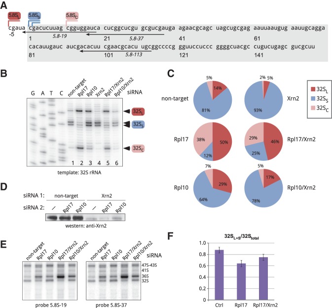
FIGURE 2.
Analysis of the 5′ end formation in mouse 32S pre-rRNA. (A) Nucleotide sequence of the mature short 5.8SS rRNA in the mouse (boxed) and positions of 5′ ends of the long, short, and cropped forms (5.8SL, 5.8SS, and 5.8SC). Positions of oligonucleotides used in hybridizations and primer extensions are indicated. (B) Primer extension analysis of the 5′ ends of gel-purified 32S pre-rRNA using primer 5.8S-113. Cells were analyzed 48 h after transfections with the indicated siRNAs. (C) Intensities of primer extension stops corresponding to 32SL, 32SS, and 32SC pre-rRNAs shown in B were quantified by phosphorimaging analysis and expressed as a percentage of the total (32SL+S+C). (D) Immunoblotting analysis of Xrn2 levels in cells transfected with the indicated siRNA combinations. (E) Northern hybridizations of the RNA samples used for primer extensions in B. RNA was separated on a formaldehyde–agarose gel, blotted and hybridized with the indicated probes. Probe 5.8S-19 overlaps with site C (see A) and does not hybridize with 32SC, while probe 5.8S-37 detects all three 32S forms (32SL, 32SS, and 32SC). (F) Quantification of 32SL+S forms relative to total 32S pre-rRNA from hybridizations with two 5.8S probes, as shown in E. Data are mean values from three independent transfections with the indicated siRNAs. Error bars, SEM; see Materials and Methods for details.

