

Abstract
Background:
A two year seroepidemiological study was designed to find out the seroprevalence and risk factors of canine visceral leishmaniasis (CVL) among stray and owned dogs in Tehran and Alborz Provinces of Iran.
Methods:
Blood samples of 602 dogs living in 11 counties of Tehran and Alborz Provinces were taken by venipuncture in 2008–2010. After separation of blood sera, anti-leishmanial antibodies were detected by direct agglutination test (DAT).
Results:
Overall, of the 408 and 194 serum samples collected randomly from dogs in 11 localities in Tehran and Alborz Provinces, 18/408 (4.41%) and 12/194 (6.18%) respectively were found positive. Among the localities, Shemiran in Tehran Province and Karaj In Alborz Province had the highest prevalence rates. No statistically significant differences were found between sex and living place but there was significant difference between living status (owned or stray) and CVL infection of dogs in Alborz Province (P= 0.018). The highest seroprevalence (7.5%) was found in dogs aged 3 to 5 years old. Only 20% of the seropositive dogs were symptomatic.
Conclusion:
Concerning possible human infections in Tehran and Alborz Provinces, both symptomatic and asymptomatic seropositive dogs should be considered as a risk.
Keywords: Visceral leishmaniasis, Direct Agglutination Test, Dog, Iran
Introduction
Visceral leishmaniasis is one of the most important parasitic diseases affecting humans, domestic and wild canines worldwide. The infection is transmitted by sandflies. Reservoir hosts vary within different geographic areas and can include domestic or wild canines ( Greene 2006). Domestic dogs (Canis familiaris) are principal reservoir hosts for Mediterranean type of visceral leishmaniasis caused by Leishmania infantum (Mohebali et al. 2005). The clinical manifestation of disease vary from asymptomatic, self-limiting infections to fatal visceral leishmaniasis.
Seroepidemiologic studies of canine leishmaniasis have shown a large number of asymptomatic seropositive animals in Meshkinshahr district of Ardabil Province (Moshfe et al. 2009). Canine leishmaniasis is not only a veterinary problem but also a serious public health concern. As the high proportion of infected dogs are asymptomatic, detection of specific antibodies remains the method of choice for mass screening of dogs in epidemiological surveys (Moshfe et al. 2008). Several diagnostic tests are available to detect anti-Leishmania antibodies in canine sera. In the present study, direct agglutination test (DAT) was used as it is a simple as well as a valid test and does not require specialized equipments (Moshfe et al. 2008).
Up to now, Ardabil, East Azerbaijan, Fars, Bushehr and Qom Provinces have been recognized as endemic foci of canine visceral leishmaniasis in Iran (Mohebali et al. 2001, Mohebali et al. 2005, Fakhar et al. 2005, Habibzadeh et al. 2007, Farzam et al. 2007, Moshfe et al. 2008, Khanmohammadi et al. 2010a, Khanmohammadi et al. 2010b).
The objective of this first ever study in Tehran and Alborz Provinces of Iran was to determine the seroprevalence and distribution of canine visceral leishmaniasis among dogs living in the urban and suburban areas of these highly populated regions with their new developing cities.
Materials and Methods
Study area
This survey was conducted over a period of 2 years (from July 2008 to october 2010) in 11 counties from two provinces of Tehran and Alborz located across the north of the central plateau of Iran at 35° 40′ 19″ N, 51° 25′ 28″ E and 34° 43′ 42″ N, 50° 58′ 19″ E, respectively (Fig. 1). Samples were collected from 8 counties (Damavand, Eslam Shahr, Rey, Robat karim, Shahriar, Shemiran, Tehran, Varamin) of Tehran and 3 counties (Hashtgerd, Karaj, Nazar Abad) of Alborz Provinces.The study population was selected by Convenience sampling. The geographical locations, where this study was carried out are shown in Fig. 1.
Fig. 1.
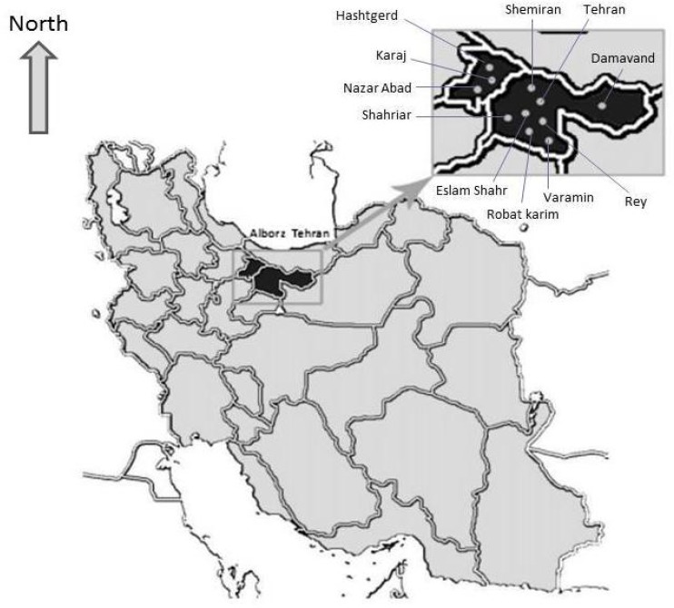
Map of Iran shows 2 provinces and 11 localities (Damavand, Eslam Shahr, Rey, Robat karim, Shahriar, Shemiran, Tehran, Varamin, Hashtgerd, Karaj, Nazar Abad) where this survey was conducted
Sampling
Totally, 602 dogs living in Tehran (n= 408) and Alborz (n= 194) Provinces were selected for this study. Ages of dogs were determined either by observing their teeth or asking from the owners, all dogs based on their ages put into three classes (0–3, 3–5 and >5 years). After the clinical examination of dogs, blood samples (2.5ml) were taken by venipuncture and put into 10ml polypropylene tubes and centrifuged at 800 × g for 5–10 minutes. Sera were separated and stored at −20 °C. All serum samples were tested by direct agglutination test (DAT) in leishmaniasis laboratory of the School of Public Health, Tehran University of Medical Sciences.
Direct Agglutination Test (DAT)
The Direct Agglutination Test antigens were prepared from indigenous L. infantum [MCAN/IR/07/Moheb-gh. (GenBank accession number FJ555210)] with the methods described by Harith et al. (1989) and Mohebali et al. (2005). Specific anti- Leishmania antibodies at a titer of ≥1: 320 in sera of indigenous dogs were considered as positive based on previous studies (Mohebali et al. 2011).
Data analysis
Chi-square (x 2) analysis was used to compare seroprevalence values relative to gender, age, geographical zone, clinical signs and living status of dogs. Data regarding gender and age of some dogs from total population (30/602 and 36/602 respectively) were missing, thus we did not consider those data in the analysis. Analyses were conducted using SPSS software version 17 (SPSS Inc, Chicago, Illinois, USA), with a probability (P) value of <0.05 as statistically significant.
Results
Eighteen out of 408 (4.4%) of the canine blood samples collected from Tehran Province, and twelve out of 194 (6.1%) from Alborz Province, were seropositive (≥1: 320) by DAT. In general, no statistical significant differences were observed between seroprevalence values found in Tehran and Alborz Provinces (P= 0.35). Sero-prevalence (Titer ≥1: 320) of canine Leishmania infection by gender in Tehran and Alborz Provinces are shown in Table 1. There was no significant difference among two sexes in both Tehran and Alborz Provinces (P= 0.933 and P= 0.349 respectively). Referring to three age groups, the highest seroprevalence (7.5%) was found in dogs aged 3 to 5 years old compared with other groups (P= 0.023), however there was no statistically significant difference among three age classes in each province (P= 0.134 and P= 0.082 in Alborz and Tehran Provinces respectively). Table 2 shows the seroprevalence (Titer ≥1: 320) of canine Leish-mania infection regarding age classes in Tehran and Alborz Provinces. Only 6/30 (20%) of the seropositive dogs showed clinical signs of disease including lethargy, cachexia, skin lesions, alopecia, epistaxis and myopathy and the remaining 80% were asymptomatic. As shown in Table 3, 11/226 (4.6%) and 1/79 (1.2%) of stray dogs and 7/182 (5.7%) and 11/115 (8.7%) of owned dogs from Tehran and Alborz Provinces were seropositive respectively. There was statistically significant difference between living status and canine VL infection of dogs in Alborz Province (P= 0.018 ). As shown in Table 4, Shemiran and Karaj counties in Tehran and Alborz Provinces showed the highest seroprevalence (14.2% and 10%, respectively).
Table 1.
Sero-prevalence (Titer ≥1: 320) of canine Leishmania infection by gender in Tehran and Alborz Provinces. Percentages were calculated in each group (male or female) totally and in each region
| sex | Male | Female | |||
|---|---|---|---|---|---|
|
| |||||
| No. of sampled dogs (%) | No. of infected dogs (%)(Titer ≥1: 320) | No. of sampled dogs (%) | No. of infected dogs (Titer ≥1: 320) (%) | P values | |
| Tehran | 232 (96) | 10 (4) | 146 (95.4) | 7 (4.6) | 0.933 |
| Alborz | 104 (93) | 8 (7) | 90 (96) | 4 (4) | 0.349 |
| Total | 336 (94.3) | 18 (5.7) | 236 (95.5) | 11 (4.5) | 0.852 |
Table 2.
Sero-prevalence (Titer ≥1: 320) of canine Leishmania infection regarding age groups in Tehran and Alborz Provinces of Iran. Percentages were calculated in each age group totally and in each region
| Age (yr) | 0–3 | 3–5 | >5 | ||||
|---|---|---|---|---|---|---|---|
|
| |||||||
| No. of sampled dogs (%) | No.of infected dogs (%) (Titer ≥1: 320) | No. of sampled dogs (%) | No. of infected dogs (%) (Titer ≥1: 320) | No. of sampled dogs (%) | No.of infected dogs (%)(Titer ≥1: 320) | P values | |
| Tehran | 179 (98.4) | 3 (1.6) | 138 (93.2) | 10 (6.8) | 59 (93.6) | 4 (6.4) | 0.082 |
| Alborz | 83 (99.6) | 2 (0.4) | 88 (92.3) | 7 (7.4) | 19 (90.5) | 2 (9.5) | 0.134 |
| Total | 262 (98) | 5 (2) | 226 (93) | 17 (7) | 78 (93) | 6 (7) | 0.013 |
Table 3.
Sero-prevalence (Titer ≥1: 320) of canine Leishmania infection regarding living status in Tehran and Alborz Provinces of Iran. Percentages were calculated in each group (stray or owned) totally and in each region
| Living status | Stray | Owned | |||
|---|---|---|---|---|---|
|
| |||||
| No. of sampled dogs (%) | No. of infected dogs (%) (Titer ≥1: 320) | No. of sampled dogs (%) | No. of infected dogs (%) (Titer ≥1: 320) | P values | |
| Tehran | 226 (95.4) | 11 (4.6) | 182 (96.3) | 7 (5.7) | 0.618 |
| Alborz | 79 (98.8) | 1 (1.2) | 115 (91.3) | 11 (8.7) | 0.018 |
| Total | 305 (96.2) | 12 (3.8) | 297 (94.3) | 18 (5.7) | 0.82 |
Table 4.
Distribution and seroprevalence of L.infantum infection (Titer ≥1: 320) among 602 dogs from 11 counties in Tehran and Alborz Provinces of Iran
| Province /Locality | No. of sampled dogs | No. / % of infected dogs(Titer ≥1: 320) |
|---|---|---|
| Tehran Province | 408 | 18 (4. 4) |
| Damavand | 62 | 4(6.4) |
| Eslam Shahr | 9 | 0(0) |
| Rey | 12 | 0(0) |
| Robat karim | 23 | 1(4.3) |
| Shahriar | 198 | 6(3) |
| Shemiran | 14 | 2(14.2) |
| Tehran | 79 | 5(6.3) |
| Varamin | 11 | 0(0) |
| Alborz Province | 194 | 12 (6.18) |
| Hashtgerd | 115 | 6(5.2) |
| Karaj | 60 | 6(10) |
| Nazar Abad | 19 | 0(0) |
| Total | 602 | 30(4.98) |
Discussion
Dogs can remain infected by L. infantum without displaying apparent clinical signs of VL for years even for their entire life (Moreno and Alvar 2002, Moshfe et al. 2009). Determination of the prevalence of canine Leishmania infection is necessary to define control measures for zoonotic visceral leishmaniasis (Mohebali et al. 2005). Reports from neighboring Turkey have shown the seroprevalences of 2.58% and 3.6% (Ozensoy et al. 1998, Aslantas et al. 2005), Jaffe showed that the disease was highly prevalent in central Israel and encroaching on urban areas (Jaffe et al. 2004), seroprevalence of 18% and 26.6% with DAT and ELISA, respectively have been reported from Pakistan (Rab et al. 1995). Based on our results, no statistically difference was found between genders. This finding is in consistent with other studies (Mohebali et al. 2001, Francasilva et al. 2003, Mohebali et al. 2005, Farzam et al. 2007, Moshfe et al. 2008, Khanmohammadi et al. 2010a) and in conflict with some others (Dantas-Torres et al. 2006, Khanmohammadi et al. 2010b, Zaffaroni et al. 1999). In the present study, the infection was more prevalent in dogs aged 3 to 5 years old. Simillarly, Bokai et al. (1998) showed the highest infection in middle-aged dogs. In the study of Mohebali et al. (2005) dogs aged 8 years or more, showed the highest seroprevalence. The results of the study of Dantas-Torres et al. (2006) showed that the seroprevalence rate was higher among juvenile (≤1 year) dogs than adults (>1 years).
In our study, only 20% of 30 seropositive dogs (titer ≥1: 320) showed the clinical features of disease and the remaining 80% were asymptomatic. Fourteen (46.6%) and ten (33.3 %) seropositive dogs in Tehran and alborz Provinces respectively were found asymptomatic which is in agreement with other studies reporting similar results (Mohebali et al. 2005, Moshfe et al. 2008, Dantas-Torres et al. 2006). There was statistically significant difference between living status and CVL infection of dogs in Alborz Province (P= 0.018). It seems that the owned dogs in this province, were much more likely to be exposed to Leish-mania infection than stray dogs. Owned dogs in Tehran Province also showed higher prevalence than stray dogs for CVL, but their difference was not significant (Table 3). Despite lower prevalnce for CVL in owned dogs population of Tehran Province, difference between two provinces in the same category was not significant (Table 3). Seroprevalence values of 6.1% and 4.4% for Leishmania infection among dogs in Alborz and Tehran Provinces, respectively, reflect the significant risk of exposure for both healthy dogs and human population in these regions. If lower cut-off value was considered (<1: 320) the prevalence should have been more than the current findings. Only 20% of the seropositive dogs were symptomatic. Since asymptomatic Leishmania-infected dogs as well as symptomatic ones, have a potential role in the maintenance of L. infantum infection (Moshfe et al. 2009), finding and reporting such number of infected asymptomatic dogs in these provinces of Iran for the first time should be considered as a risk from both veterinary and public health aspects. Among the localities, Shemiran (14.2 %) in Tehran Province and Karaj (10%) in Alborz Province had the highest prevalence rates.
Conclusion
Based on our survey these provinces especially those localities and suburbs with higher seroprevalence should be regarded as new endemic foci for CVL in Iran. The huge population living inside these areas particularly in new developing cities around both provinces, should be considered as at risk population.
Acknowledgements
The authors would like to express their gratitude to all those who helped them running this survey including A Aliari for his technical assistance in Small Animal Hospital, Faculty of Veterinary Medicine, University of Tehran, P Nobakht the local private veterinarian in Hashtgerd county, S Tehranian for helping in statistical review of the manuscript and S Charehdar for her technical assistance in Parasitology laboratory, School of Public Health, Tehran University of Medical Sciences. The authors declare that there is no conflict of interest.
References
- 1. Bokai S, Mobedi I, Edrissian GhH, Nadim A. ( 1998) Seroepidemiological study of canine visceral leishmaniasis in Meshkin-Shahr, northwest of Iran. Arch Razi Inst. 41– 46, 48– 49. [Google Scholar]
- 2. Clarisa B, Palatnik-de-Sousa ( 2008) Vaccines for leishmaniasis in the fore coming 25 years. Vaccine. 26: 1709– 1724. [DOI] [PubMed] [Google Scholar]
- 3. Dantas-Torres F, Edileuza Felinto de Brito M, Pinto Brandao-Filho S. ( 2006) Seroepidemiological survey on canine leishmaniasis among dogs from an urban area of Brazil, Vet Parasitol. 140: 54– 60. [DOI] [PubMed] [Google Scholar]
- 4. Fakhar M, Motazedian MH, Asgari Gh, Mohebali M, Mehrbani D. ( 2006) Introduction of a new endemic foci of kalaazar in south of Iran. Armaghan Danesh J. 11( 2): 103– 113. [Google Scholar]
- 5. Farzam M, Changizi E, Mohebali M, Akhoundi B, Salimi Bajestani MR. ( 2007) Seroepidemilogical survey of visceral leishmaniasis in shepherd dogs of Jahrom county, Fars Province. Vet J Iran. 4( 3): 58– 67. [Google Scholar]
- 6. França-Silva J, T da Costa R, Siqueira A, LL Machado-Coelho G, A da Costa C, Mayrink W, P Vieira E, S Costa J, Genaro O, Nascimento E. ( 2003) Epidemiology of canine visceral leishmaniosis in the endemic area of Montes Claros Municipality, Minas Gerais State, Brazil. Vet Parasitol. 111: 161– 173. [DOI] [PubMed] [Google Scholar]
- 7. Greene Craig E. ( 2006) Infectious diseases of the dog and cat, third edition, WB Saunders Co, Chapter 73, pp: 685– 698 [Google Scholar]
- 8. Habibzadeh S, Arshi S, Sadeghi-Bazargani H, Mohebali M. ( 2007) Rk39 Dipstick Test as a practical solution for canine leishmaniasis diagnosis among nomadic populations. J Anim Vet Adv. 6( 2): 175– 178. [Google Scholar]
- 9. Harith A, Salappendel RJ, Reiter I, Knapen F, Korte P, Huigen E, Kolk RHG. ( 1989) Application of a direct agglutination test for detection of specific anti- Leishmania antibodies in the canine reservoir. J Clin Microbiol. 27: 2252– 2257. [DOI] [PMC free article] [PubMed] [Google Scholar]
- 10. Jaffe ChL, Baneth G, Abdeen ZA, Schlein Y, Warburg A. ( 2004) Leishmaniasis in Israel and the Palestinian Authority. Trends Parasitol. 20( 7): 328– 332. [DOI] [PubMed] [Google Scholar]
- 11. Khanmohammadi M, Fallah E, Rahbari S, Sohrabi I, Farshchian M, Hamzavi F, Mohammadpour Asl A. ( 2010a) Seroepidemiological survey of canine Visceral leishemanisis in Sarab District, East Azerbaijan Province, Northwest of Iran. Afr J Microbiol Res. 4( 19): 2022– 2028. [Google Scholar]
- 12. Khanmohammadi M, Fallah E, Rahbari S, Sohrabi I, Farshchian M, Hamzavi F, Mohammadpour Asl A. ( 2010b) Study on seroprevalence of canine visceral leishmaniasis (CVL) in ownership dogs of Sarab, East Azerbaijan Province, Northwest of Iran with indirect immuno fluorescence antibody test (IFAT) and its health importance. J Anim Vet Adv. 9( 1): 139– 143. [Google Scholar]
- 13. Mohebali M, Hamzavi Y, Fallah E, Zarei Z. ( 2001) Study of canine visceral leishmaniasis in some parts of Iran and its health importance. J Vet Res. 56: 55– 59. [Google Scholar]
- 14. Mohebali M, Khamesipour A, Mobedi I, Zarei Z, Hashemi-Fesharki R. ( 2004) Double blind randomized efficacy trial of alum precipitated autoclaved Leishmania major vaccine mixed with BCG against canine visceral leishmaniasis in Mesh-kin-shahr district I.R. of Iran. Vaccine. 22: 4097– 4100. [DOI] [PubMed] [Google Scholar]
- 15. Mohebali M, Hajjaran H, Hazavi Y, Mobedi I, Arshi Sh, Zarei Z, Akhoundi B, Manouchehri Naeini K, Avizeh R, Fakhar M. ( 2005) Epidemiological aspects of canine visceral leishmaniosis in the Islamic Republic of Iran. Vet Parasitol. 129: 243– 251. [DOI] [PubMed] [Google Scholar]
- 16. Mohebali M, Edrissian GhH, Shirzadi MR, Akhoundi B, Hajjaran H, Zarei Z, Molaei S, Sharifi I, Mamishi S, Mahmoud-vand H, Torabi V, Moshfe A, Malmasi A, Motazedian MH, Fakhar M. ( 2011) An observational study on the current distribution of visceral leishmaniasis in different geographical zones of Iran and implication to health policy. Travel Med Infect Dis. 9( 2): 67– 74. [DOI] [PubMed] [Google Scholar]
- 17. Moreno J, Alvar J. ( 2002) Canine leishmaniasis: epidemiological risk and the experimental model. Trends Parasitol. 18: 399– 405. [DOI] [PubMed] [Google Scholar]
- 18. Moshfe A, Mohebali M, Edrissian Gh, Zarei Z, Akhoundi B, Kazemi B, Jamshidi Sh, Mahmoodi M. ( 2008) Seroepidemiological study on canine visceral leishmaniasis in Meshkin-Shahr district, Ardabil Province, Northwest of Iran. Iran J Parasitol. 3( 3): 1– 10. [Google Scholar]
- 19. Moshfe A, Mohebali M, Edrissian Gh, Zarei Z, Akhoundi B, Kazemi B, Jamshidi Sh, Mahmoodi M. ( 2009) Canine visceral leishmaniasis: Asymptomatic infected dogs as a source of L. infantum infection. Acta Trop. 112: 101– 105. [DOI] [PubMed] [Google Scholar]
- 20. Ozensoy S, Ozbel Y, Turgay N, Alkan MZ, Gul K, Gilman-Sachs A, Chang KP, Reed SG, Ali Ozcel M. ( 1998) Serodiagnosis and epidemiology of visceral leishmaniasis in Turkey. Am J Trop Med Hyg. 59( 3): 363– 369. [DOI] [PubMed] [Google Scholar]
- 21. Rab MA, Frame IA, Evans DA. ( 1995) The role of dogs in the epidemiology of human visceral leishmaniasis in northern Pakistan. T Roy Soc Trop Med H. 89: 612– 615. [DOI] [PubMed] [Google Scholar]
- 22. Zaffaroni E, Rubaudo L, Lanfranchi P, Mignone W. ( 1999) Epidemiological patterns of canine leishmaniosis in Western Liguria (Italy). Vet Parasitol. 81: 11– 19. [DOI] [PubMed] [Google Scholar]