Abstract
The Exaltation of Newcastle disease virus (END) phenomenon is induced by the inhibition of type I interferon in pestivirus-infected cells in vitro, via proteasomal degradation of cellular interferon regulatory factor (IRF)-3 with the property of the viral autoprotease protein Npro. Reportedly, the amino acid residues in the zinc-binding TRASH motif of Npro determine the difference in characteristics between END-phenomenon-positive (END+) and END-phenomenon-negative (END−) classical swine fever viruses (CSFVs). However, the basic mechanism underlying this function in bovine viral diarrhea virus (BVDV) has not been elucidated from the genomic differences between END+ and END− viruses using reverse genetics till date. In the present study, comparison of complete genome sequences of a pair of END+ and END− viruses isolated from the same virus stock revealed that there were only four amino acid substitutions (D136G, I2623V, D3148G and D3502Y) between two viruses. Based on these differences, viruses with and without mutations at these positions were generated using reverse genetics. The END assay, measurements of induced type I interferon and IRF-3 detection in cells infected with these viruses revealed that the aspartic acid at position 136 in the zinc-binding TRASH motif of Npro was required to inhibit the production of type I interferon via the degradation of cellular IRF-3, consistently with CSFV.
Keywords: bovine viral diarrhea virus, END phenomenon, interferon regulatory factor-3, Npro, type I interferon
The genus Pestivirus of the family Flaviviridae comprises 4 recognized species that are economically important pathogens in the veterinary field: bovine viral diarrhea viruses (BVDVs) 1 and 2, classical swine fever virus (CSFV) and border disease virus of sheep [30]. Pestiviruses are quasispecies; a single strain consists of different populations showing various characteristics. Two biotypes of pestiviruses, cytopathogenic (cp) and noncytopathogenic (ncp) viruses, are distinguished by their ability to induce a cytopathic effect (CPE) in tissue cultures [16]. Pestiviruses have been reported to inhibit the production of type I interferon in infected cells in vitro in coordination with the property of viral protein Npro [1, 4, 8, 28]. Subsequently, Newcastle disease virus (NDV) and some orbiviruses replicate efficiently and induce distinguishable CPE in cells infected with pestiviruses [15, 20]. The exaltation of NDV (END) phenomenon is a well-known property of BVDV and CSFV that has been used as a biological marker of vaccine virus and for the titration purpose. Ncp pestiviruses are further divided into two types by their ability to induce END phenomenon: END-phenomenon-positive (END+) and END-phenomenon-negative (END−) viruses. A pair of END+ and END− viruses can be cloned from the same virus stock using reverse plaque formation techniques [5, 19]. END− viruses do not enhance NDV, but induce intrinsic interference against Western equine encephalitis virus and vesicular stomatitis virus (VSV) [5, 19]. One of the END− viruses, the GPE− strain of CSFV, attenuated and cloned from the virulent ALD strain [29], is a Japanese live vaccine strain against classical swine fever (CSF) that contributed to the eradication of CSF in Japan.
It is well documented that END+ pestiviruses subvert host innate immune defenses by preventing the production of type I interferon. This occurs via the proteasomal degradation of cellular interferon regulatory factor (IRF)-3 in infected cells through its interaction with viral autoprotease Npro [1, 4, 8, 28]. Npro, the first functional unit of the pestivirus polyprotein, is followed by structural proteins (C, Erns, E1 and E2) and nonstructural proteins (p7, NS2, NS3, NS4A, NS4B, NS5A and NS5B) [3, 16, 25, 31, 35]. Npro is a unique protein in pestiviruses that autocatalytically cleaves itself from the nascent polyprotein to generate the N terminus of the viral capsid (C) protein [26]. Npro has two domains: a catalytic N-terminal domain and a C-terminal domain containing a zinc-binding TRASH motif. The zinc-binding TRASH motif, which includes the zinc-binding residues Cys112-Cys134-Asp136-Cys138, is required for IRF-3 binding and for the prevention of type I interferon production [7, 32].
Recent studies have shown that Npro of CSFV interacts with IRF-7 and dampens the production of type I interferon in plasmacytoid dendritic cells [4]. It has also been reported for CSFV that deletion of up to 19 amino acids from the N terminus of Npro does not abolish its capacity to inhibit the production of type I interferon [7, 24] and that amino acid substitutions C112A/R, C134A, D136N or C138A in Npro result in the disappearance of this function [24, 32]. In contrast, Ruggli et al. and Tamura et al. succeeded in restoring the Npro functions of wild-type END− viruses, the Ames-END− and GPE− strains of CSFV, by mutating a single amino acid residue (R112C and N136D, respectively) using reverse genetics techniques [24, 33].
Npro of both cp BVDV (e.g., the NADL strain) and ncp BVDV (e.g., the pe515 strain) blocks type I interferon production in vitro via proteasomal degradation of cellular IRF-3 [2, 8]. It has been reported for BVDV that nearly the entire Npro is required for the prevention of type I interferon production because removal of 30 residues from the N terminus or the removal of 24 or 88 residues from the C terminus abolishes this function [2, 8]. Deletion mutants expressing residues 1−69 or 70−168 of Npro also lack the ability to inhibit the production of type I interferon [2]. In addition, the previous reports revealed that substitutions of amino acid residue L8P, E22V or H49L of Npro abolish its capacity to function as an interferon antagonist [2, 6]. Till date, however, the basic mechanism by which BVDV inhibits type I interferon production has not been approached through engineering in vitro rescued viruses with mutations on the basis of the differences between a pair of END+ and END− viruses.
In the present study, to take advantage of the differences between a wild-type END− virus and its END+ virus pair, we cloned a pair of END+ and END− viruses from a viral stock of the BVDV2 GBK strain. Following this, we identified the amino acid residue determinants involved in the inhibition of type I interferon production by determining and comparing the complete genome sequence of both viruses as well as investigating mutant viruses generated from the END+ virus using reverse genetics techniques.
MATERIALS AND METHODS
Cells and viruses: Bovine testicle (BT) cells were grown in Eagle’s minimum essential medium (EMEM) (Nissui Pharmaceutical, Tokyo, Japan) supplemented with 0.295% tryptose phosphate broth (TPB) (Becton Dickinson, San Jose, CA, U.S.A.) and 5% fetal bovine serum (FBS) (Mitsubishi Chemical, Tokyo, Japan). Bovine kidney cell line MDBK-HS [13] and porcine kidney cell line SK-L [27] were grown in EMEM supplemented with 0.295% TPB, 10% horse serum (HS) (Life Technologies, Carlsbad, CA, U.S.A.) and 10 mM N,N-Bis (2-hydroxyethyl)-2-aminoethanesulfonic acid (BES) (Sigma-Aldrich, St. Louis, MO, U.S.A.). Bovine fetal muscle (BFM) cells were grown in EMEM supplemented with 0.295% TPB, 5% FBS, 5% HS and 10 mM BES. Cells were confirmed to be free from BVDV, and FBS was confirmed to be free from both BVDV and anti-BVDV neutralizing antibodies [14].
The BVDV GBK strain is an adventitious BVDV2 isolated from cells of the bovine kidney cell line GBK [9]. The BVDV GBK strain was grown in BT cells. BVDV GBK_E+ and GBK_E− strains were grown in MDBK-HS cells. The NDV Miyadera strain was propagated in 10-day-old embryonated hens’ eggs. The New Jersey serotype strain of VSV was grown in SK-L cells.
Cloning of a pair of END+ and END− viruses from viral stocks: A reverse plaque formation technique [5] was used to obtain a pair of END+ and END− viruses from viral stocks as described previously [19]. Viruses were used after three rounds of limited dilution.
Sequencing: BVDV GBK_E+ and GBK_E− strains, the full-length cDNA clones and the in vitro-rescued viruses were completely sequenced as described previously [34]. In brief, the nucleotide sequences of cDNA clones and PCR fragments from viral RNA were determined using the BigDye Terminator v3.1 Cycle Sequencing Kit (Life Technologies) and a 3130 Genetic Analyzer or a 3500 Genetic Analyzer (Life Technologies) according to the manufacturer’s protocol. Sequencing data were analyzed using GENETYX version 10 software (GENETYX, Tokyo, Japan).
Plasmid constructs: The cDNA fragments from the GBK_E+ strain, obtained by reverse transcription polymerase chain reaction (RT-PCR), were cloned into plasmid pCR®-Blunt II-TOPO® (Life Technologies) using the Zero Blunt® TOPO® Cloning Kit (Life Technologies). The cDNA sequence was flanked by a modified T7 promoter sequence at the 5′ end and a Pst I restriction site at the 3′ end. Subclones were assembled into a full-length cDNA clone, termed pGBK_E+, by replacing the CSFV genome of the full-length cDNA clone of the CSFV Alfort187-1 strain pA187-1 [23] with the genome of the GBK_E+ strain using appropriate restriction enzymes and the In-Fusion HD Cloning Kit (Clontech, Mountain View, CA, U.S.A.) according to the manufacturer’s protocol. Details of the constructions may be obtained on request. The full-length cDNA clone pGBK_E+ was transformed and propagated in competent cell Stbl3 cells (Life Technologies) and purified using the QIAGEN Plasmid Plus Midi Kit (QIAGEN, Hilden, Germany) according to the manufacturer’s protocol.
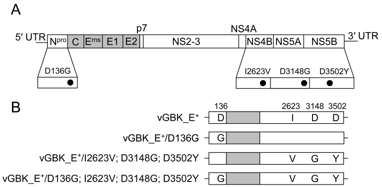
Three full-length cDNA clones with combinations of the four amino acid substitutions D136G, I2623V, D3148G and D3502Y were constructed in the pGBK_E+ backbone using the QuickChange Lightning Multi Mutagenesis Kit (Agilent Technologies, Santa Clara, CA, U.S.A.) and the In-Fusion HD Cloning Kit. The plasmid pGBK_E+/D136G has a single amino acid substitution at position 136 of GBK_E+, whereas the plasmids pGBK_E+/I2623V; D3148G; D3502Y and pGBK_E+/D136G; I2623V; D3148G; D3502Y have multiple amino acid substitutions at positions 2623, 3148 and 3502 of GBK_E+ and at positions 136, 2623, 3148 and 3502 of GBK_E+, respectively.
Full-length PCR amplification, in vitro RNA transcription, transfection and viral recovery: The cDNA-derived viruses were rescued as described previously [34] with some modifications. The full-length genome amplification strategy [22] was employed to obtain a full-length PCR amplicon for in vitro RNA transcription. The cDNA clones were amplified using primers 5GBKE+_T7 (5′-taa tac gac tca cta ta GTA TAC GAG ATT AGC TAA AGT ACT CG −3′, T7 promoter sequence underlined) and 3GBKE+ (5′- GGG GCT GTT AGA GGC ATC CTC TAG TC −3′) with AccuPrime Taq DNA polymerase High Fidelity (Life Technologies). Viral RNA was transcribed in vitro from the purified full-length PCR amplicon using the MEGAscript T7 Kit (Life Technologies), the remaining PCR amplicon was digested using DNase (Life Technologies), and the viral RNA was then purified with a MicroSpin S-400 column (GE Healthcare, Buckinghamshire, U.K.). MDBK-HS cells (3 × 10⁶ cells) were transfected with 10 µg of viral RNA in a 0.4 cm cuvette using the Gene Pulser Xcell Electroporation system (Bio-Rad, Hercules, CA, U.S.A.) set at 180 V and 950 µF. The cells were incubated at 37°C in 5% CO2 for 3 days, and the supernatants were then transferred onto naive MDBK-HS cells to obtain infectious viruses.
The viruses were named according to the plasmid from which they were rescued, replacing “p” with “v” in the nomenclature. The cDNA-derived viruses generated in the present study are shown in Fig. 1.
Fig. 1.
Schematic representation of the genomic differences between GBK_E+ virus and GBK_E− virus, and mutant viruses derived from cDNA clones generated in the present study. (A) Four amino acid substitutions, D136G, I2623V, D3148G and D3502Y, were found in the GBK_E− virus in comparison with the GBK_E+ virus. (B) Three recombinant viruses with combinations of amino acid substitutions were generated in the GBK_E+ backbone by site-directed mutagenesis and In-Fusion techniques. The white and gray boxes indicate the nonstructural and structural proteins, respectively.
END assay and measurements of type I interferon production: The END assay was conducted using BFM cells as described previously [10]. In brief, BFM cells grown in 96-well plates were infected with BVDV and incubated for 5 days at 37°C in 5% CO2. After removal of the supernatants, the cells were superinfected with 1 HA/ml of the NDV Miyadera strain. The END phenomenon was regarded as positive, if strong CPE was observed in NDV-infected cells.
Measurements of type I interferon production in BVDV-infected cells were performed using a previously described plaque reduction method with VSV as a challenge virus [20], with some modifications. In brief, the supernatants from BVDV-infected cells were inactivated by exposure to UV light (254 nm) in the UV Crosslinker (ATTO Corporation, Tokyo, Japan) under the condition of 500 mJ/cm2. Viral inactivation of samples was confirmed using indirect FA techniques with anti-BVDV NS3 monoclonal antibody #46/1 [13] after inoculation of samples onto MDBK-HS cells and incubation for 2 days at 37°C in 5% CO2. Then, monolayers of SK-L cells in 12-well plates were inoculated with 1 ml of a four-fold dilution of UV-inactivated supernatant and incubated for 24 hr at 37°C in 5% CO2. The supernatants were removed, and the cells were inoculated with VSV. Interferon titers were expressed as reciprocals of dilutions that reduced the number of challenge viral plaques by 50%.
Detection of IRF-3 in BVDV-infected cells by sodium dodecyl sulfate-polyacrylamide gel electrophoresis (SDS-PAGE) and western blotting: SDS-PAGE and western blotting were performed as described previously [12]. BFM cells were infected with BVDV (m.o.i. of 1) in six-well plates and incubated for 5 days at 37°C in 5% CO2. Lysates of cells were separated with SDS-PAGE. After transferring the proteins from the gels to the Immunobilion-P Transfer Membrane (Millipore, Billerica, MA, U.S.A.), the membranes were treated with anti-human IRF-3 rabbit polyclonal antibodies (GeneTex, Hsinchu City, Taiwan), goat anti-rabbit IgG-HRP conjugate (Bio-Rad) and Immunobilion Western Detection Reagents (Millipore), in that order. The membranes were read using Lumi Vision PRO (Aishin Seiki, Kariya, Japan), and specific bands for IRF-3 were detected.
RESULTS
Cloning of a pair of END+/ END− viruses from the BVDV GBK strain and determining their whole genome sequences: A pair of END+ and END− viruses (GBK_E+ and GBK_E−, respectively) was cloned from the BVDV GBK strain by means of reverse plaque formation techniques and limited dilution. The genomes of GBK_E+ and GBK_E− viruses were both 12,284 nucleotides in length and encoded 3,897 deduced amino acids. Comparison of the complete genome sequences of both viruses revealed only six nucleotide and four amino acid differences (D136G, I2623V, D3148G and D3502Y, the numbers refer to the amino acid position in GBK_E+) between the two viruses (Table 1). The complete genome sequences of the GBK_E+ and GBK_E− strains were deposited in the DDBJ/EMBL/GenBank databases under accession numbers AB894423 and AB894424, respectively.
Table 1. Differences of amino acid sequences in the genome of GBK_E+ and GBK_E−.
| Viral protein | Npro | NS4B | NS5A | NS5B |
|---|---|---|---|---|
| Amino acid position | 136 | 2,623 | 3,148 | 3,502 |
| GBK_E+ | Da) | I | D | D |
| GBK_E− | G | V | G | Y |
a) D: Aspartic acid, I: Isoleucine, G: Glycine, V: Valine, Y: Tyrosine.
Generation and characterization of in vitro-rescued viruses: The infectious BVDV vGBK_E+ was successfully rescued by electroporation of MDBK-HS cells with viral RNA transcribed in vitro from a full-length PCR amplicon obtained from a cDNA clone pGBK_E+. The complete sequence of vGBK_E+ was entirely identical to that of GBK_E+. The mutant viruses vGBK_E+/D136G, vGBK_E+/I2623V; D3148G; D3502Y and vGBK_E+/D136G; I2623V; D3148G; D3502Y were also recovered from full-length cDNA clones. Sequencing of the complete genomes of these three viruses confirmed the mutations at the desired amino acid positions and demonstrated the lack of any other mutations in comparison with the parental GBK_E+ virus.
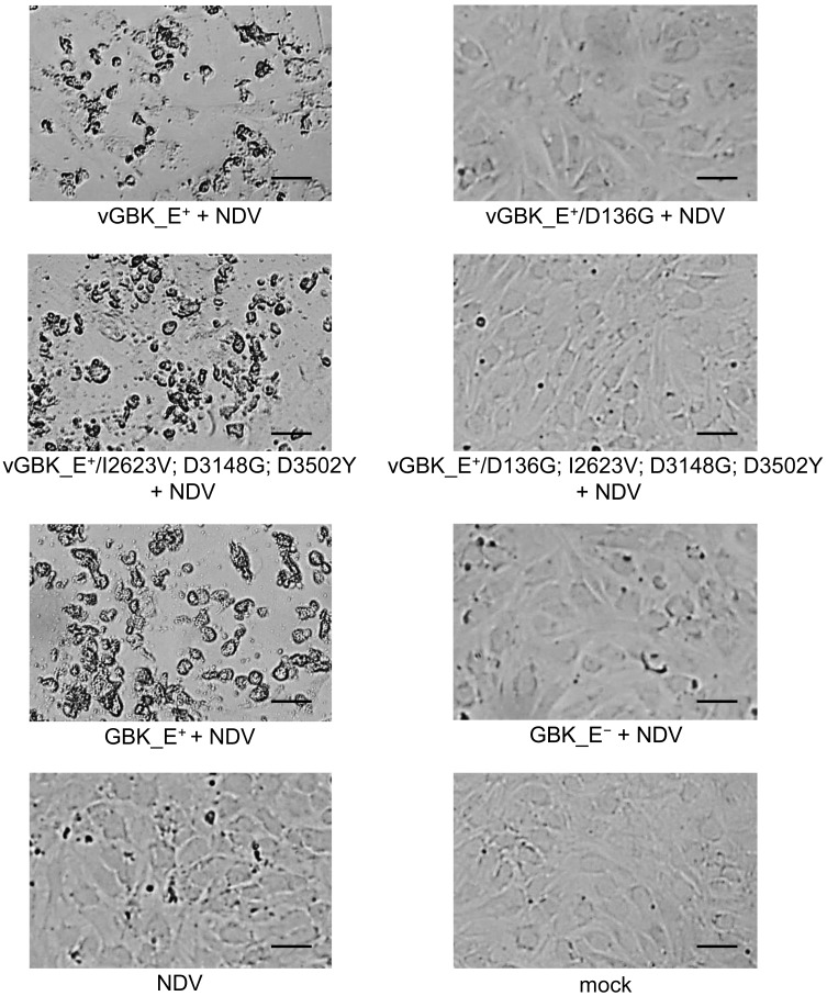
To investigate the biological properties of the mutant viruses, BFM cells were inoculated with both the parental GBK_E+ and the in vitro-rescued vGBK_E+ viruses. The results revealed that both GBK_E+ and vGBK_E+ were ncp (data not shown) and exhibited the END phenomenon (END+) (Fig. 2). In addition, titration of both viruses in MDBK-HS cells revealed that vGBK_E+ exhibited the same growth characteristics as wild-type GBK_E+ (data not shown). Moreover, compared with the wild-type GBK_E+ strain, other in vitro-rescued viruses grew equally in MDBK-HS cells (data not shown).
Fig. 2.
Results of END assays. BFM cells infected with GBK_E+, GBK_E− or the in vitro-rescued viruses were superinfected with Newcastle disease virus (NDV). Cells infected with GBK_E+, vGBK_E+ or vGBK_E+/I2623V; D3148G; D3502Y exhibited distinguishable CPE after superinfection with NDV (END+), whereas those infected with GBK_E−, vGBK_E+/D136G or vGBK_E+/D136G; I2623V; D3148G; D3502Y did not exhibit CPE (END−). Scale bar, 5 µm.
Identification of the amino acid determinants responsible for inhibition of type I interferon production: As expected, the mutant viruses vGBK_E+/D136G, vGBK_E+/I2623V; D3148G; D3502Y and vGBK_E+/D136G; I2623V; D3148G; D3502Y remained ncp (data not shown). An END assay revealed that the vGBK_E+/D136G and vGBK_E+/D136G; I2623V; D3148G; D3502Y viruses were changed to END−; BFM cells infected with these viruses did not exhibit CPE after NDV superinfection. In contrast, the vGBK_E+/I2623V; D3148G; D3502Y virus remained END+; BFM cells infected with this virus exhibited clear CPE after superinfection with NDV, as observed in BFM cells infected with GBK_E+ (Fig. 2).
The amount of type I interferon in supernatants from BFM cells infected with the parent or one of the mutant viruses GBK_E+, GBK_E−, vGBK_E+, vGBK_E+/D136G, vGBK_E+/I2623V; D3148G; D3502Y or vGBK_E+/D136G; I2623V; D3148G; D3502Y was measured in SK-L cells. Secretion of type I interferon was inhibited in BFM cells infected with GBK_E+, vGBK_E+ or the virus carrying the mutations without Npro (vGBK_E+/I2623V; D3148G; D3502Y), which is in consistent with the results from the END assay. Cells infected with GBK_E− or viruses carrying mutations in Npro(vGBK_E+/D136G and vGBK_E+/D136G; I2623V; D3148G; D3502Y) did induce type I interferon (Table 2).
Table 2. Measurements of type I interferon in cells infected with wild-type and in-vitro rescued viruses.
| Viruses | type I interferon |
|---|---|
| GBK_E+ | <4 |
| vGBK_E+ | <4 |
| vGBK_E+/D136G | 4 |
| vGBK_E+/I2623V; D3148G; D3502Y | <4 |
| vGBK_E+/D136G; I2623V; D3148G; D3502Y | 4 |
| GBK_E− | 4 |
These results clearly indicate that the aspartic acid of Npro (at position 136 of the GBK_E+ strain) is the key amino acid residue that determines the capacity to inhibit the production of type I interferon and induce the END phenomenon.
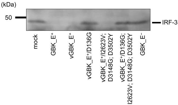
Detection of IRF-3 in cells infected with the parental or mutant viruses: To investigate the expression levels of IRF-3 in BVDV-infected cells, IRF-3 in the lysates of BFM cells infected with GBK_E+, GBK_E−, vGBK_E+ or one of the three mutant viruses was detected by western blotting. IRF-3 was not detected in the lysates of cells infected with GBK_E+, vGBK_E+ or vGBK_E+/I2623V; D3148G; D3502Y, whereas clear bands of IRF-3 appeared in the lysates of cells infected with the END− GBK_E− virus or one of the Npro mutant viruses (vGBK_E+/D136G and vGBK_E+/D136G; I2623V; D3148G; D3502Y) (Fig. 3).
Fig. 3.
Detection of IRF-3 in BFM cells infected with parent or mutant viruses. IRF-3 was not detected in western blots of the lysates of cells infected with GBK_E+, vGBK_E+ or vGBK_E+/I2623V; D3148G; D3502Y, whereas clear bands of IRF-3 appeared in western blots of the lysates of cells infected with GBK_E−, vGBK_E+/D136G or vGBK_E+/D136G; I2623V; D3148G; D3502Y.
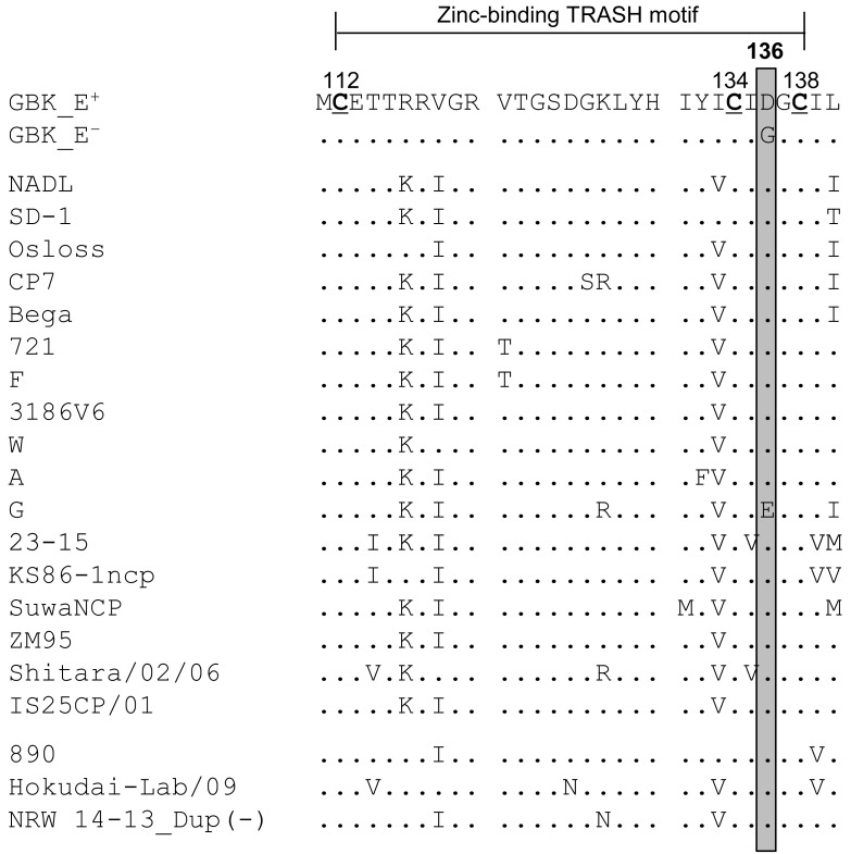
Comparison of amino acid sequences of zinc-binding TRASH motif in Npro: The amino acid sequences of the zinc-binding TRASH motifs in the Npro proteins from various BVDV strains deposited in the DDBJ/EMBL/GenBank databases were compared with those of the GBK_E+ and GBK_E− strains. At least one strain per subgenotype [1a−1o (except for 1l) and 2a−2c] [11, 18] was chosen in the present study. As a result, BVDV strains, except for the G strain, contain amino acid residues Cys112-Cys134-Asp136-Cys138 in the zinc-binding TRASH motif of Npro, as observed in GBK_E+ (Fig. 4).
Fig. 4.
Comparison of amino acid sequences of the zinc-binding TRASH motifs in Npro. The amino acid sequences of the zinc-binding TRASH motifs in the Npro proteins of various BVDV strains deposited in the DDBJ/EMBL/GenBank databases were compared with those of GBK_E+ and GBK_E−. At least one strain per subgenotype [1a−1o (except for 1l) and 2a−2c] was chosen. The cysteines (Cs) at positions 112, 134 and 138 of the zinc-binding TRASH motif are highlighted in bold and underlined. The amino acid residue at position 136 is boxed in gray. The accession numbers of strains from the DDBJ/EMBL/GenBank are as follows: NADL (M31182), SD-1 (M96751), Osloss (M96687), CP7 (U63479), Bega (AF049221), 721 (AF144463), F (AF287284), 3186V6 (AF287282), W (AF287290), A (AF287283), G (AF287285), 23-15 (AF287279), KS86-1ncp (AB078950), SuwaNCP (KC853440), ZM-95 (AF526381), Shitara/02/06 (AB359930), IS25CP/01 (AB359931), 890 (U18059), Hokudai-Lab/09 (AB567658) and NRW 14-13_Dup (−) (HG426485).
DISCUSSION
Both BVDV and CSFV inhibit the production of type I interferon in vitro through proteasomal degradation of cellular IRF-3 [1, 4, 8, 28]. Cells infected with these (END+) viruses exhibit strong CPE after superinfection with NDV or some orbiviruses [15, 20]. One of the two domains of pestivirus Npro, the C-terminal domain containing a zinc-binding TRASH motif consisting of Cys112-Cys134-Asp136-Cys138, is required for IRF-3 binding and prevention of type I interferon production [7, 32]. For CSFV, it has been reported that mutations at positions 112, 134, 136 or 138 in Npro of END+ viruses abolish the inhibition of type I interferon production, while mutations at positions 112 or 136 in Npro of END− viruses restore this function [24, 32, 33]. For BVDV, previous studies have revealed that amino acid substitutions L8P, E22V or H49L in Npro abolish its capacity to interfere with type I interferon production [2, 6]. However, to the best of our knowledge, there were no studies with BVDV that had approached the basic mechanism of the inhibition of type I interferon production using the amino acid differences between a pair of END+ and END− viruses as well as viruses genetically engineered on the basis of these differences. Pestiviruses are quasispecies; single strains consist of populations with various characteristics, such as END+ and END− [17]. We cloned a pair of END+ and END− viruses (GBK_E+ and GBK_E−) from the BVDV GBK strain using reverse plaque formation techniques [5, 19]. Determination of complete genome sequences of these viruses revealed that there were only four amino acid differences between them (Table 1). To clarify the molecular mechanism of the inhibition of type I interferon production and the END phenomenon, we generated a full-length cDNA clone of GBK_E+ (pGBK_E+) as well as three other full-length cDNA clones with single (D136G) or multiple (I2623V; D3148G; D3502Y and D136G; I2623V; D3148G; D3502Y) mutations. Four viruses were rescued from these full-length cDNA clones (Fig. 1), and their properties were investigated.
The END assay and the IFN bioassay of vGBK_E+ and three mutant viruses revealed that vGBK_E+ and vGBK_E+/I2623V; D3148G; D3502Y exhibited the END phenomenon and inhibited the production of type I interferon, whereas the Npro mutant viruses vGBK_E+/D136G and vGBK_E+/D136G; I2623V; D3148G; D3502Y were END− and did not inhibit the production of type I interferon (Fig. 2, Table 2). This result demonstrates that the single aspartic acid residue in the zinc-binding TRASH motif of Npro, which occurs at position 136 of the genome of the GBK_E+ virus, is the key to determine viral capacity to inhibit type I interferon production and display the END phenomenon. Because it has been reported that the Npro proteins of both BVDV and CSFV inhibit the production of type I interferon by proteasomal degradation of cellular IRF-3 [1, 4, 8, 28], we assessed whether Npro of wild-type GBK_E+, wild-type GBK_E−, vGBK_E+ and the three mutant viruses reduced the amount of cellular IRF-3 in infected cells by western blotting. An apparent reduction in cellular IRF-3 was observed in cells infected with wild-type GBK_E+, vGBK_E+ or vGBK_E+/I2623V; D3148G; D3502Y, whereas wild-type GBK_E− and mutant viruses with mutations in Npro showed no reduction in cellular IRF-3 (Fig. 3). Therefore, the results indicate that the inhibition of type I interferon production and the END phenomenon displayed by GBK_E+ occurs by the degradation of cellular IRF-3 caused by combining with the zinc-binding TRASH motif. Furthermore, this function was abolished by mutation of Npro at position 136. Comparison of vGBK_E+/D136G; I2623V; D3148G; D3502Y (the same amino acid sequence as GBK_E−) with vGBK_E+/I2623V; D3148G; D3502Y revealed that the function of Npro was restored by a single G136D mutation in Npro of the GBK_E− virus.
In the present study, a comparison of the amino acid sequences of GBK_E+, GBK_E− and viruses from DDBJ/EMBL/GenBank databases revealed that amino acid residues Cys112-Cys134-Asp136-Cys138 in the zinc-binding TRASH motif of Npro were well conserved among field BVDV isolates (Fig. 4). It is reported that these four residues are required for the inhibition of type I interferon production [7, 32]. Taken together, the above-mentioned results suggest that these field isolates inhibit the type I interferon production in infected cells, although there remains a possibility that amino acid residues other than those of TRASH motif contribute to this phenomenon. The G strain contains the amino acid residue Glu136 in the TRASH motif of Npro (Fig. 4), and this may affect the structure of Npro and result in the production of type I interferon in vitro. It was reported that 35 out of 45 (77.8%) field isolates of BVDV in Japan contained END+ virus as the predominant virus population compared with END− virus and seven isolates (15.6%) contained similar titers of END+ and END− viruses, whereas two isolates (4.4%) contained only END− virus [21]. Therefore, further studies are needed to investigate how quasispecies of BVDV (END+ and END−) contribute in vivo.
In conclusion, our results indicate that a single mutation in the TRASH motif at position 136 of BVDV Npro abolishes its interaction with bovine IRF-3 and halts the degradation of IRF-3. Moreover, the mutation of the amino acid residue at position 136 of GBK_E− restores its function as an interferon antagonist, as shown for CSFV. However, how Npro interacts with IRF-3 and the nature of the cascade after interaction with Npro and IRF-3 are hardly understood. In addition, it is unknown how the inhibition of type I interferon production contributes to the viral infection strategy when cells are infected with BVDV in vivo. Therefore, an additional study is also required to reveal the fundamental mechanism by which pestiviruses inhibit the production of type I interferon and how this mechanism functions in vivo.
Acknowledgments
We are grateful to Dr. Nicolas Ruggli (Institute of Virology and Immunology, Switzerland) for providing the full-length CSFV cDNA clone pA187-1.
REFERENCES
- 1.Bauhofer O., Summerfield A., Sakoda Y., Tratschin J. D., Hofmann M. A., Ruggli N.2007. Classical swine fever virus Npro interacts with interferon regulatory factor 3 and induces its proteasomal degradation. J. Virol. 81: 3087–3096. doi: 10.1128/JVI.02032-06 [DOI] [PMC free article] [PubMed] [Google Scholar]
- 2.Chen Z., Rijnbrand R., Jangra R. K., Devaraj S. G., Qu L., Ma Y., Lemon S. M., Li K.2007. Ubiquitination and proteasomal degradation of interferon regulatory factor-3 induced by Npro from a cytopathic bovine viral diarrhea virus. Virology 366: 277–292. doi: 10.1016/j.virol.2007.04.023 [DOI] [PMC free article] [PubMed] [Google Scholar]
- 3.Collett M. S., Larson R., Belzer S. K., Retzel E.1988. Proteins encoded by bovine viral diarrhea virus: the genomic organization of a pestivirus. Virology 165: 200–208. doi: 10.1016/0042-6822(88)90673-3 [DOI] [PubMed] [Google Scholar]
- 4.Fiebach A. R., Guzylack-Piriou L., Python S., Summerfield A., Ruggli N.2011. Classical swine fever virus Npro limits type I interferon induction in plasmacytoid dendritic cells by interacting with interferon regulatory factor 7. J. Virol. 85: 8002–8011. doi: 10.1128/JVI.00330-11 [DOI] [PMC free article] [PubMed] [Google Scholar]
- 5.Fukusho A., Ogawa N., Yamamoto H., Sawada M., Sazawa H.1976. Reverse plaque formation by hog cholera virus of the GPE− strain inducing heterologous interference. Infect. Immun. 14: 332–336. [DOI] [PMC free article] [PubMed] [Google Scholar]
- 6.Gil L. H., Ansari I. H., Vassilev V., Liang D., Lai V. C., Zhong W., Hong Z., Dubovi E. J., Donis R. O.2006. The amino-terminal domain of bovine viral diarrhea virus Npro protein is necessary for alpha/beta interferon antagonism. J. Virol. 80: 900–911. doi: 10.1128/JVI.80.2.900-911.2006 [DOI] [PMC free article] [PubMed] [Google Scholar]
- 7.Gottipati K., Ruggli N., Gerber M., Tratschin J. D., Benning M., Bellamy H., Choi K. H.2013. The structure of classical swine fever virus Npro: a novel cysteine autoprotease and zinc-binding protein involved in subversion of type I interferon induction. PLoS Pathog. 9: e1003704. doi: 10.1371/journal.ppat.1003704 [DOI] [PMC free article] [PubMed] [Google Scholar]
- 8.Hilton L., Moganeradj K., Zhang G., Chen Y. H., Randall R. E., McCauley J. W., Goodbourn S.2006. The Npro product of bovine viral diarrhea virus inhibits DNA binding by interferon regulatory factor 3 and targets it for proteasomal degradation. J. Virol. 80: 11723–11732. doi: 10.1128/JVI.01145-06 [DOI] [PMC free article] [PubMed] [Google Scholar]
- 9.Hirano N., Sato F., Ono K., Murakami T., Matumoto M.1987. A sensitive plaque assay for bovine rotavirus in cultures of the bovine cell line GBK. Vet. Microbiol. 13: 383–387. doi: 10.1016/0378-1135(87)90069-1 [DOI] [PubMed] [Google Scholar]
- 10.Inaba Y., Tanaka Y., Kumagai T., Omori T., Ito H.1968. Bovine diarrhea virus. II. END phenomenon: exaltation of Newcastle disease virus in bovine cells infected with bovine diarrhea virus. Jpn. J. Microbiol. 12: 35–49. doi: 10.1111/j.1348-0421.1968.tb00367.x [DOI] [PubMed] [Google Scholar]
- 11.Jenckel M., Hoper D., Schirrmeier H., Reimann I., Goller K. V., Hoffmann B., Beer M.2014. Mixed triple: allied viruses in unique recent isolates of highly virulent type 2 bovine viral diarrhea virus detected by deep sequencing. J. Virol. 88: 6983–6992. doi: 10.1128/JVI.00620-14 [DOI] [PMC free article] [PubMed] [Google Scholar]
- 12.Kameyama K., Sakoda Y., Tamai K., Nagai M., Akashi H., Kida H.2006. Genetic recombination at different points in the Npro-coding region of bovine viral diarrhea viruses and the potentials to change their antigenicities and pathogenicities. Virus Res. 116: 78–84. doi: 10.1016/j.virusres.2005.08.016 [DOI] [PubMed] [Google Scholar]
- 13.Kameyama K., Sakoda Y., Tamai K., Igarashi H., Tajima M., Mochizuki T., Namba Y., Kida H.2006. Development of an immunochromatographic test kit for rapid detection of bovine viral diarrhea virus antigen. J. Virol. Methods 138: 140–146. doi: 10.1016/j.jviromet.2006.08.005 [DOI] [PubMed] [Google Scholar]
- 14.Kozasa T., Aoki H., Nakajima N., Fukusho A., Ishimaru M., Nakamura S.2011. Methods to select suitable fetal bovine serum for use in quality control assays for the detection of adventitious viruses from biological products. Biologicals 39: 242–248. doi: 10.1016/j.biologicals.2011.06.001 [DOI] [PubMed] [Google Scholar]
- 15.Kumagai T., Shimizu T., Matumoto M.1958. Detection of hog cholera virus by its effect on Newcastle disease virus in swine tissue culture. Science 128: 366. doi: 10.1126/science.128.3320.366 [DOI] [PubMed] [Google Scholar]
- 16.Meyers G., Thiel H.1996. Molecular characterization of pestiviruses. Adv. Virus Res. 47: 53–118. doi: 10.1016/S0065-3527(08)60734-4 [DOI] [PubMed] [Google Scholar]
- 17.Muhsen M., Aoki H., Ikeda H., Fukusho A.2013. Biological properties of bovine viral diarrhea virus quasispecies detected in the RK13 cell line. Arch. Virol. 158: 753–763. doi: 10.1007/s00705-012-1538-x [DOI] [PubMed] [Google Scholar]
- 18.Nagai M., Hayashi M., Itou M., Fukutomi T., Akashi H., Kida H., Sakoda Y.2008. Identification of new genetic subtypes of bovine viral diarrhea virus genotype 1 isolated in Japan. Virus Genes 36: 135–139. doi: 10.1007/s11262-007-0190-0 [DOI] [PubMed] [Google Scholar]
- 19.Nakamura S., Fukusho A., Inoue Y., Sasaki H., Ogawa N.1993. Isolation of different non-cytopathogenic bovine viral diarrhoea (BVD) viruses from cytopathogenic BVD virus stocks using reverse plaque formation method. Vet. Microbiol. 38: 173–179. doi: 10.1016/0378-1135(93)90084-K [DOI] [PubMed] [Google Scholar]
- 20.Nakamura S., Shimazaki T., Sakamoto K., Fukusho A., Inoue Y., Ogawa N.1995. Enhanced replication of orbiviruses in bovine testicle cells infected with bovine viral diarrhoea virus. J. Vet. Med. Sci. 57: 677–681. doi: 10.1292/jvms.57.677 [DOI] [PubMed] [Google Scholar]
- 21.Nishine K., Aoki H., Sakoda Y., Fukusho A.2014. Field distribution of END phenomenon-negative bovine viral diarrhea virus. J. Vet. Med. Sci. 76: 1635–1639. doi: 10.1292/jvms.14-0220 [DOI] [PMC free article] [PubMed] [Google Scholar]
- 22.Rasmussen T. B., Reimann I., Uttenthal A., Leifer I., Depner K., Schirrmeier H., Beer M.2010. Generation of recombinant pestiviruses using a full-genome amplification strategy. Vet. Microbiol. 142: 13–17. doi: 10.1016/j.vetmic.2009.09.037 [DOI] [PubMed] [Google Scholar]
- 23.Ruggli N., Tratschin J. D., Mittelholzer C., Hofmann M. A.1996. Nucleotide sequence of classical swine fever virus strain Alfort/187 and transcription of infectious RNA from stably cloned full-length cDNA. J. Virol. 70: 3478–3487. [DOI] [PMC free article] [PubMed] [Google Scholar]
- 24.Ruggli N., Summerfield A., Fiebach A. R., Guzylack-Piriou L., Bauhofer O., Lamm C. G., Waltersperger S., Matsuno K., Liu L., Gerber M., Choi K. H., Hofmann M. A., Sakoda Y., Tratschin J. D.2009. Classical swine fever virus can remain virulent after specific elimination of the interferon regulatory factor 3-degrading function of Npro. J. Virol. 83: 817–829. doi: 10.1128/JVI.01509-08 [DOI] [PMC free article] [PubMed] [Google Scholar]
- 25.Rümenapf T., Unger G., Strauss J. H., Thiel H. J.1993. Processing of the envelope glycoproteins of pestiviruses. J. Virol. 67: 3288–3294. [DOI] [PMC free article] [PubMed] [Google Scholar]
- 26.Rümenapf T., Stark R., Heimann M., Thiel H. J.1998. N-terminal protease of pestiviruses: identification of putative catalytic residues by site-directed mutagenesis. J. Virol. 72: 2544–2547. [DOI] [PMC free article] [PubMed] [Google Scholar]
- 27.Sakoda Y., Fukusho A.1998. Establishment and characterization of a porcine kidney cell line, FS-L3, which forms unique multicellular domes in serum-free culture. In Vitro Cell. Dev. Biol. Anim. 34: 53–57. doi: 10.1007/s11626-998-0053-6 [DOI] [PubMed] [Google Scholar]
- 28.Seago J., Hilton L., Reid E., Doceul V., Jeyatheesan J., Moganeradj K., McCauley J., Charleston B., Goodbourn S.2007. The Npro product of classical swine fever virus and bovine viral diarrhea virus uses a conserved mechanism to target interferon regulatory factor-3. J. Gen. Virol. 88: 3002–3006. doi: 10.1099/vir.0.82934-0 [DOI] [PubMed] [Google Scholar]
- 29.Shimizu Y., Furuuchi S., Kumagai T., Sasahara J.1970. A mutant of hog cholera virus inducing interference in swine testicle cell cultures. Am. J. Vet. Res. 31: 1787–1794. [PubMed] [Google Scholar]
- 30.Simmonds P., Becher P., Collett M. S., Gould E. A., Heinz F. X., Meyers G., Monath T., Pletnev A., Rice C. M., Stiasny K., Thiel H.-J., Weiner A., Bukh J.2012. Family Flaviviridae. pp. 1003–1020. In: Virus Taxonomy. Ninth Report of the International Commitee on Taxonomy of Viruses (King, A. M. Q., Adams, M. J., Carstens, E. B. and Lefkowitz, E. J. eds.), Elsevier Academic Press, San Diego. [Google Scholar]
- 31.Stark R., Meyers G., Rümenapf T., Thiel H. J.1993. Processing of pestivirus polyprotein: cleavage site between autoprotease and nucleocapsid protein of classical swine fever virus. J. Virol. 67: 7088–7095. [DOI] [PMC free article] [PubMed] [Google Scholar]
- 32.Szymanski M. R., Fiebach A. R., Tratschin J. D., Gut M., Ramanujam V. M., Gottipati K., Patel P., Ye M., Ruggli N., Choi K. H.2009. Zinc binding in pestivirus Npro is required for interferon regulatory factor 3 interaction and degradation. J. Mol. Biol. 391: 438–449. doi: 10.1016/j.jmb.2009.06.040 [DOI] [PubMed] [Google Scholar]
- 33.Tamura T., Nagashima N., Ruggli N., Summerfield A., Kida H., Sakoda Y.2014. Npro of classical swine fever virus contributes to pathogenicity in pigs by preventing type I interferon induction at local replication sites. Vet. Res. 45: 47. doi: 10.1186/1297-9716-45-47 [DOI] [PMC free article] [PubMed] [Google Scholar]
- 34.Tamura T., Sakoda Y., Yoshino F., Nomura T., Yamamoto N., Sato Y., Okamatsu M., Ruggli N., Kida H.2012. Selection of classical swine fever virus with enhanced pathogenicity reveals synergistic virulence determinants in E2 and NS4B. J. Virol. 86: 8602–8613. doi: 10.1128/JVI.00551-12 [DOI] [PMC free article] [PubMed] [Google Scholar]
- 35.Tautz N., Elbers K., Stoll D., Meyers G., Thiel H. J.1997. Serine protease of pestiviruses: determination of cleavage sites. J. Virol. 71: 5415–5422. [DOI] [PMC free article] [PubMed] [Google Scholar]


