Abstract
Background: In the early 1990s, the biological significance of light-emitting diodes was realized. Since this discovery, various light sources have been investigated for their cutaneous effects. Study design: A Medline search was performed on light-emitting diode lights and their therapeutic effects between 1996 and 2010. Additionally, an open-label, investigator-blinded study was performed using a yellow light-emitting diode device to treat acne, rosacea, photoaging, alopecia areata, and androgenetic alopecia. Results: The authors identified several case-based reports, small case series, and a few randomized controlled trials evaluating the use of four different wavelengths of light-emitting diodes. These devices were classified as red, blue, yellow, or infrared, and covered a wide range of clinical applications. The 21 patients the authors treated had mixed results regarding patient satisfaction and pre- and post-treatment evaluation of improvement in clinical appearance. Conclusion: Review of the literature revealed that differing wavelengths of light-emitting diode devices have many beneficial effects, including wound healing, acne treatment, sunburn prevention, phototherapy for facial rhytides, and skin rejuvenation. The authors’ clinical experience with a specific yellow light-emitting diode device was mixed, depending on the condition being treated, and was likely influenced by the device parameters.
Light-emitting diode (LED) sources are unique in that they emit a narrow spectrum of light in a noncoherent manner. The LED was invented in 1962, but early LEDs lacked the capability to produce biologically relevant energies. In addition, wavelengths emitted were broad and varied by as much as 100nm. In the 1990s, the National Aeronautics and Space Administration (NASA) developed LEDs that produced a very narrow spectrum of light that in turn allowed for their first clinical applications.1 Within the past 15 years, a better understanding of photobiology and an increased demand for minimally invasive yet effective dermatologic treatments has led to a growing interest in LED devices.
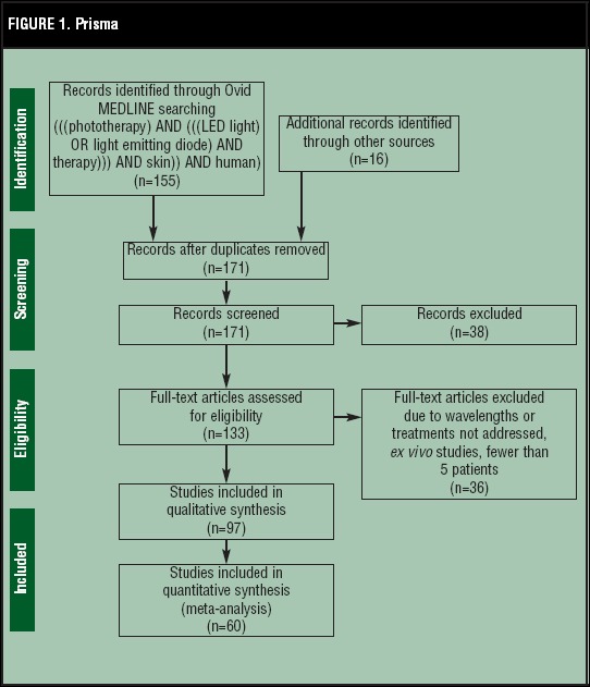
To the authors’ knowledge, there has never been a comprehensive overview of LED-based therapies. The authors searched Ovid MEDLINE® from 1996 through December 2013 for articles in which “LED light” or “light emitting diodes” were stringed with “therapy” and “skin.” There were 155 total results, and Figure 1 details how the articles were selected in a PRISMA flow diagram. Here the authors review the science behind LEDs and then expand upon the clinical application of red, yellow, blue, and near infrared (IR) LEDs. Finally, they discuss their own experiences using a yellow LED device.
Figure 1.

PRISMA flow diagram showing how the articles were selected
LED TECHNOLOGY
Light-emitting diodes comprise a semiconductor chip situated upon a reflective surface. Light is produced when electricity is run through the semiconductor. The wavelength of light produced is dependent on the composition of the semiconductor chip. Depth of tissue penetration, and therefore the light’s target, is primarily dependent upon the wavelength of the light. A summary of the parameters of different wavelengths of LED light, along with their clinical applications, is presented in Table 1.
TABLE 1.
Parameters of different wavelengths of LED light and their clinical applications
| BLUE | YELLOW | RED | IR | COMBINED | |
|---|---|---|---|---|---|
| Wavelength (nm) | 400-170 | 570-590 | 630-700 | 800-1200 | Variable |
| Depth of LED light penetration | <1mm | 0.5-2mm | 2-3mm | 5-10mm | Variable |
| Deepest target | Epidermis | Papillary dermis | Adnexa | Adnexa and reticular dermis | Variable |
| Studied therapeutic uses |
|
|
|
|
|
Light delivery by LED devices is either continuous or photomodulated. Photomodulated light is delivered in a pulsed mode with specific pulse sequences and durations. There is evidence that photo-modulated light affects cells differently than continuous light.1 Commercially available LED units include wavelengths in the red, yellow, blue, and near infrared portions of the spectrum.
MECHANISM
Research on LED mechanisms has yielded multiple pathways by which clinical benefit is achieved. LEDs appear to affect cellular metabolism by triggering intracellular photobiochemical reactions. Observed effects include increased ATP, modulation of reactive oxygen species, the induction of transcription factors, alteration of collagen synthesis, stimulation of angiogenesis, and increased blood flow.2
Red LEDs specifically have been shown to activate fibroblast growth factor, increase type 1 pro-collagen, increase matrix metallo-proteinase-9 (MMP-9), and decrease MMP-1. An increase in fibroblast number and a mild inflammatory infiltrate following exposure has been demonstrated histologically.3,4
Photomodulated yellow light alters ATP production, gene expression, and fibroblast activity.5-7 Increased ATP production is thought to be mediated via the absorption of photons by mitochondrial protoporphyrin IX. Interestingly, only photomodulated yellow LED has been shown to produce a tissue response implying that the light’s ability to affect cells is dependent on the number and pattern of photon delivery.8
Blue light appears to exert its effect on acne via its influence on Propionibacterium acnes and its antiinflammatory properties. P. acnes contains naturally occurring porphyrins, mainly coproporphyrin and protoporphyrin IX. Absorption of blue light by these molecules is believed to induce a natural photodynamic therapy (PDT) effect with destruction of the bacteria via the formation of oxygen free radicals. Blue light’s anti-inflammatory effect appears to be the result of a shift in cytokine production.9
Near infrared light, also known as monochromatic infrared energy (MIRE), is believed to stimulate circulation by inducing the release of guanylate cyclase and nitrous oxide, which, in turn, promotes vasodilation and growth factor production as well as angiogenesis, leading to subsequent wound healing.10
FDA APPROVED DEVICES AND INDICATIONS
There are numerous manufacturers of LED lights and some produce systems of different wavelengths. Photo Therapeutics, Inc. of Carlsbad, California, markets several LED systems under the brand name Omnilux©. Omnilux PDT™ (633nm) is indicated for PDT of nonmelanoma skin cancer (NMSC). Omnilux Revive™ (633nm) is a red light device marketed for skin rejuvenation. It produces about 30 percent more output energy than the PDT device. Omnilux Blue™ (415nm) is approved for the treatment of acne and actinic keratoses (AKs). Omnilux Plus™ (830nm) is an infrared device indicated for skin rejuvenation and wound healing.11
More recently, Ambicare Health of Scotland has created a portable adhesive PDT device called Ambulight PDT™. Ibbotson and Ferguson12 showed it to be as effective and less painful in the treatment of nonmelanoma skin cancers as conventional PDT due to its lower irradiance. It may be considered as an alternative treatment for isolated lesions.
Light Bioscience of Virginia Beach, Virginia, manufactures a yellow light device, GentleWaves® LED Photomodulation® (590nm). It delivers a 35-second treatment in a patented 102ms pulsed cycle. While pulsed light has been shown to be efficacious during testing, continuous delivery of light was not shown to be efficacious during initial testing of the device.6
Anodyne Therapy, LLC, of Tampa, Florida, markets the MIRE therapy system (890nm). The device is marketed for improving circulation and decreasing pain, stiffness, and muscle spasm.
RED LIGHT-EMITTING DIODES
Red LEDs have the deepest tissue penetration of the visible wavelengths and are therefore used to target dermal structures, such as adnexa and fibroblasts.13 Red LEDs have been studied for a wide variety of uses, including wound healing, photodamage, the treatment of NMSCs, precancers, warts, and the prevention of oral mucositis in cancer patients.
A split-face study of red LED (633nm) in patients who had undergone blepharoplasty and periocular resurfacing demonstrated a statistically significant improvement of edema, erythema, bruising, and pain on the treated side of the face.14 Red LED (633nm) following erbium-doped yttrium aluminum garnet (Er:YAG) ablation of palmoplantar verrucae has been shown to speed recovery.15 A retrospective blinded study by Sakamoto et al16 found aminolevulinic acid (ALA) or methylester aminolevulinic acid (MAL) combined with red LED to statistically improve scar appearance after two or more treatments. In 2011, a prospective, split-face, double-blind, randomized, controlled trial by Sanclemente et al17 found that MAL combined with red LED demonstrated superior efficacy in treatment of global facial photodamage compared with placebo and red LED based on Dover’s modified global photodamage score. The treatment was well-tolerated and resulted in high patient satisfaction in 80.4 percent of subjects.17 A similar prospective randomized trial of MAL-PDT with red light also found global clinical improvement in 10 of 14 patients and histologically found increased collagen fibers and decreased elastic fibers.18
Red LED light appears to be a promising treatment option for premalignant and malignant lesions. Successful treatment of NMSC with red LED was demonstrated by Calzavara-Pinton et al19 who used two MAL-PDT sessions to treat 112 biopsy-proven Bowen’s disease (BD) lesions. The complete response rates were 73.2 percent at three months and 53.6 percent at 24 months post-treatment. They found that the best clinical response was in well-differentiated (Broders’ scores I and II) BD lesions and worst in nodular, invasive, and/or poorly differentiated (Broders’ scores III and IV) BD lesions.19 NMSC with red LED PDT was demonstrated by Wong et al20 who used a red (630nm) custom-made LED array in conjunction with 2% ALA to treat Bowen’s disease of the digit. The treatment was delivered at 240J/cm2 in two 50-minute sessions. Complete clinical clearance occurred in 3 out of 4 patients, all of which healed without scarring. Histologic clearance was confirmed in one of these patients.20 Lopez et al21 demonstrated effective treatment of extensive Bowen’s disease using red LED PDT preceded by application of MAL cream. Eighteen patients were treated and 90 percent of their lesions showed a complete clinical response after 12 weeks with good or excellent cosmetic outcomes in 94 percent of patients at a 12-month follow-up.21 Indeed, a 2011 review article of three databases found that MAL combined with LED had the highest response rates of 95 percent compared with 82 percent with ALA-PDT.22 In the United Kingdom, Baas et al23 have shown promising results in basal cell carcinoma (BCC) treatment after use of a second-generation intravenous photosensitizer, meta-tetrahydroxyphenylchlorin (mTHPC), in conjunction with a red LED (652 nm).23
Additional studies have evaluated the effectiveness of red LED PDT in the treatment of AKs. Wiegell et al24 have shown red LED to be more effective than continuous, ultra-low-intensity artificial daylight in AK therapy.24 In two other studies, patients underwent two MAL-PDT treatments one week apart. The first study found a complete response rate of 59.2 percent in the treatment group versus 14.9 percent in the placebo group.25 The second study found 68.4 percent a complete response rate for the treatment group versus 6.9 percent in the placebo group.26 A recent study of 50 patients, however, showed no difference between the effectiveness of MAL-PDT and pulsed dye laser (PDL) on AKs, although PDL appears to be easier to use and less painful.27
A retrospective analysis of off-label PDT with MAL in Italy suggested a therapeutic role for treatment of granulomatous dermal disorders and follicular inflammatory diseases, such as acne vulgaris, granuloma annulare, and necrobiosis lipoidica.28 Despite these suggestions, a 2011 multicenter study by Berking et al29 did not recommend MAL-PDT as first-line therapy of necrobiois lipoidica due to its response rate of 39 percent.
Red LED (660nm) has been shown to prevent ultraviolet (UV)-induced erythema. In a study by Barolet et al,30 subjects experienced an increase in minimal erythema dose (MED) corresponding to approximately a sun protection factor (SPF) of 15 after a series of 5 to 10 red LED treatments.30 Whether LED exposure provides a true reduction in UV damage or just a reduction in erythema was not assessed.
Lastly, Whelen et al31 found a beneficial effect of daily red LED (670nm) treatments on the incidence and severity of oral mucositis (OM) in pediatric patients undergoing myeloablative therapy.31 Similarly, Corti et al32 reported that red LED treatment is safe and capable of reducing the duration of chemotherapy-induced OM in adults.
YELLOW LIGHT-EMITTING DIODES
Yellow LEDs penetrate the skin between 0.5 and 2mm. Much of yellow LED application has been focused on photoaging and as an adjuvant therapy to laser treatment. Recently, it has also been shown to decrease the intensity and duration of erythema after fractional laser skin resurfacing.33
In a large study, Weiss et al6 reported their clinical experience with photomodulated yellow LED (590nm) in a total of 900 patients with photoaged skin. Patients received LED treatment alone or in combination with intense pulsed light (IPL), PDL, potassium-titanyl-phosphate (KTP) laser, or infrared lasers. Patients who received LED alone self-reported a softening of skin and a reduction in fine lines. Post-thermal/nonablative-treated patients self-reported a reduction in post-treatment erythema of the primary treatment. In two studies by Weiss et al, a yellow LED (590nm) was used on 93 and 90 patients, respectively, with mild-to-moderate photoaging. In the first study, an independent observer determined that photoaging was decreased by one Fitzpatrick wrinkle class in 90 percent of subjects.8 In the second study, optical profilometry showed a 10-percent improvement by surface topographical measurements, and histology showed increased collagen in 100 percent of post-treatment subjects.34
Despite these promising results, a study by Boulos et al35 suggests that these results are complicated by placebo effect or observer bias. They conducted a study designed to replicate the results of Weiss. They found similar patient perceptions, but were unable to replicate the objective data using a panel of 30 blinded experts, including ophthalmologists and oculoplastic surgeons.35 The authors’ experience, which will be discussed later, is consistent with Boulos’ results.
Khoury and Goldman36 performed a split-face study in which subjects received two photomodulated yellow LED treatments after IPL. A blinded observer determined an approximate 10-percent reduction in erythema on the treated side. Four patients also reported decreased pain.36 Similarly, photomodulated yellow LED has also been shown to speed healing and reduce erythema after fractionated laser therapy.33
DeLand et al37 investigated the value of yellow LED photomodulation therapy (590nm) in regards to preventing or improving the skin’s tolerance to radiation dermatitis. Patients were treated with yellow LED after a series of intensity-modulated radiation treatments. The majority of patients experienced a minimal skin reaction to radiation (grade 0 or 1 radiation dermatitis) and only 5.3 percent of patients had to interrupt radiation therapy due to a skin reaction as compared to 68 percent of controls. This suggests that LED treatments reduced the incidence and degree of radiation-induced skin reactions as well as the incidence of treatment interruption because of skin reaction. However, in a similarly sized study that evaluated yellow LED photomodulation in patients with radiation dermatitis, the authors found statistically insignificant differences between the treatment groups’ and the control groups’ graded reactions post-radiation therapy. The percentage of LED-treated patients with grade 0, grade 1, grade 2, and grade 3 reactions were 0, 33, 67, and 0 percent, respectively; the non-treated groups were 7, 27, 60, and 7 percent, respectively. The authors concluded that it did not reduce any incidence of radiation-induced skin reactions.38
BLUE LIGHT-EMITTING DIODES
Blue LED light (400-470nm) has a maximal penetration of up to 1mm.2 It is best suited for the treatment of more superficial conditions, such as AKs, or to target P. acnes in acne vulgaris. Morton et al39 treated 30 patients with mild-to-moderate acne with 8-, 10-, or 20-minute blue LED (415nm) treatments over a period of four weeks. Mean inflammatory lesion counts decreased at Weeks 5, 8, and 12 by 25, 53, and 60 percent, respectively, with minimal effect on noninflammatory lesions.39 Tremblay et al40 gave patients with mild-to-moderate inflammatory acne two 20-minute treatments of blue LED (415nm) per week for 4 to 8 weeks. Ninety percent of patients were satisfied with the result.40 Objectively, patients had a 50-percent reduction in lesion counts and nine patients were completely clear. Two similar clinical studies showed reductions in lesion size, number, and erythema in patients as evaluated by physician and patients after treatment with blue LED.41,42 Although blue light has been tried in conjunction with ALA in the treatment of acne of 20 patients, patients experienced greater side effects and the results were not clinically significant when compared with blue LED alone.43
Recently, blue LED has also shown promise in the treatment of thicker lesions such as psoriasis. A 2011 prospective, randomized study of 37 patients showed statistically significant improvement of irradiated plaques after four weeks of treatment with an at-home LED-based on the Local Psoriasis Severity Index (LPSI).44
INFRARED
IR LED treatment can penetrate the skin between 5 and 10mm and has been used to treat wounds, ulcers, recalcitrant lesions, cutaneous scleroderma, and has even been shown to treat cellulite.45-47 They are frequently used in combination therapy with other light devices.
Data on monotherapy with IR LEDs is limited. In 2007, Hunter et al48 reviewed the use of an infrared LED device on several patients: a diabetic with non-healing wounds, a bed-bound patient with methicillin-resistant Staphylococcus aureus furuncles, and a patient with painful full-thickness pressure wounds. Patients received 30-minute IR treatments 2 to 5 times a week coupled with a topical treatment of the clinician’s choice. In each case, there was noted to be increased wound contraction and granulation and decreased pain, edema, and infection.48 In 2013, Lev-Tov et al49 concluded that certain low-level IR fluences resulted in a statistically significant reduction in fibroblast proliferation than in controls without a reduction in cellular viability. With more study, this could prove to be beneficial for scar treatment and wound healing.
Combination therapy using IR A plus visible light treatment has been shown to be effective in the treatment of patients with cutaneous scleroderma. Durometry measurements in 7 out of 10 patients showed a persistent marked improvement in hardness of the skin after therapy.46 This could prove to be beneficial in the future treatment of dysmorphism, contractures, and restricted movement.
COMBINATION TREATMENT
A number of studies have indicated that exposing patients to a combination of LED wavelengths is more effective than monotherapy.50-56 This synergistic effect has been investigated on a variety of skin disorders, most notably photoaging and acne.
A prospective, placebo-controlled, double-blind, split-face trial by Lee et al50 randomized patients with facial rhytides to receive red LED (640nm), IR (830nm), both, or sham treatments. Patients demonstrated a statistically significant reduction in wrinkle severity across all treatment groups; 26, 33, and 36 percent, respectively. Skin elasticity also improved. Tissue assays were notable for an increase in collagen and elastic fibers adjacent to highly active fibroblasts. The pro-inflammatory cytokines interleukin 1β (IL-1β) and tumor necrosis factor-α (TNF-α) were increased while interleukin 6 (IL-6) was decreased. In a separate study, Goldberg et al51 investigated the combination of red (633nm) and IR (830nm) LED treatment on photodamaged skin and reported softening of periorbital wrinkles in 80 percent of subjects. There was subjective improvement of softness, smoothness, and firmness. Histologic examination demonstrated increased number and thickness of collagen fibrils.51 A similar study in 2012 showed increases type I collagen expression and the number of viable fibroblasts when treated with different combinations of 630nm, 830nm, and varying wavelengths of red and IR light.57 Three additional studies investigating the effects of combination red and IR LED on photodamaged skin were also reviewed and showed similar results. In all three of the studies, patients reported subjective improvement and a slight-to-moderate objective improvement was observed.52-54
Combination LED for the treatment of acne is also promising. Lee et al55 treated patients with moderate acne with a combination of blue (415nm) and red (640nm) LED devices. A 34-percent improvement in comedone count and a 78-percent improvement in the number of inflammatory lesions were observed. Reduced melanin levels were measured with a Mexameter™ (Courage+Khazaka electronic GmbH, Cologne, Germany), corresponding to an overall perception of improved complexion. A similar study by Sadick et al58 using combination blue and near infrared (830nm) LED therapy showed improvement in 11 individuals’ lesions an average of 48.8 percent. Goldberg et al56 treated patients with mild-to-severe acne with dermabrasion followed by alternating red (633nm) and blue (415nm) LEDs. There was a 46 percent and 81 percent decrease in lesion count at four and 12 weeks, respectively. More recently, Kwon et al59 demonstrated a decrease of both inflammatory and noninflammatory acne lesions by 77 percent and 54 percent, respectively, following home-use combined blue and red LED.
Preliminary research on nine patients using combination 830nm and 633nm light for the treatment of recalcitrant psoriasis is promising. Clearance rates by the end of the follow-up period ranged from 60 to 100 percent with universally high satisfaction rates.60
THE AUTHORS’ EXPERIENCE
At the authors’ institution, they have used the GentleWaves® LED Photomodulation® yellow LED device, which is FDA approved for the treatment of photoaging, and delivers 250ms pulses of 0.1J/cm2 of energy in a patented 35-second treatment. However, FDA approval for these devices did not necessitate demonstration of efficacy. The authors undertook an open-label, Institutional Review Board (IRB)-approved trial, with the goal of examining the effectiveness of photomodulated yellow LED in the treatment of several common skin conditions. Signed informed consent was obtained. They enrolled patients with acne (N=3), rosacea (N=6), photoaging (N=10), alopecia areata, and androgenetic alopecia (N=2). Study patients received weekly treatments for eight weeks. A blinded observer evaluated pre- and post-treatment photographic images for clinical efficacy and participants were asked to subjectively rate changes in their skin. Overall treatments were well-tolerated and most patients completed the eight-week protocol. Those who discontinued early cited minimal perceived benefit and the inconvenience of office visits.
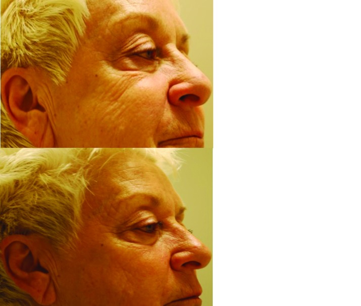
Ten patients were enrolled for photoaging. A modest improvement in the appearance of fine periocular rhytides was observed in 8 out of 10 patients, which is similar to results published elsewhere.6,8 Most patients reported an overall perceived benefit, although photographic evidence is subtle (Figure 2). Unlike other studies, none of the authors’ patients improved their Glogau photoaging score. One patient, however, perceived such satisfactory improvement in her photoaging that after the study she has continued twice-monthly treatments.
Figure 2.
Evaluation of photoaging in a 70-year-old patient with rhytides for 30 years who received eight total treatments on the forehead and around the eyes
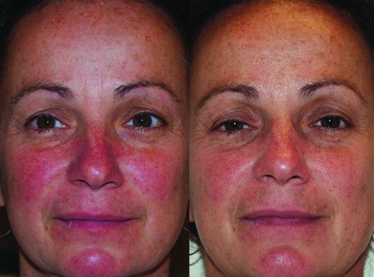
Two of four rosacea patients noted a reduction of erythema that was confirmed with photography (Figure 3). There was no change in the papulopustular component of their disease. The effect of yellow LEDs on vasculature has been demonstrated previously,33,35-38 so it is not surprising that the erythematotelangiectatic component of rosacea was more responsive to treatment than the inflammatory component.
Figure 3.
Evaluation of rosacea in a 44-year-old patient with rosacea for 15 years who received nine total treatments on the nose and left and right malar cheeks
In a patient with a nine-month history of erosive pustular dermatosis of the scalp, the authors saw a dramatic improvement after one treatment. However, the condition recurred about three months later and has not yet been retreated pending a scheduled visit. Another patient was given a yellow LED treatment immediately following a 30-percent glycolic acid peel. She reported less post-procedure erythema than she had experienced after previous glycolic peels.
No clinical benefit was seen in a patient with alopecia totalis. One woman with androgenetic alopecia was treated; she reported a slight re-growth over the anterior hairline, but this was not collaborated with clinical photos. Several patients were treated for acne and had no clinical improvement. One patient quit the study after three treatments because she noticed a flare of her acne.
SIDE EFFECTS
Generally, minimal to no side effects were experienced. One patient reported post-treatment erythema that lasted 24 hours. The authors were unable to examine this patient while symptoms were present and she received additional treatments without complication. In the other literature reviewed, side effects were either mild or not reported. It remains prudent to screen individuals with photosensitive dermatoses, or those taking photosensitizing medications, as these are contraindications to treatment.13-15,20,36-40,45
DISCUSSION
While much of the data on LED use in dermatologic therapy is promising, it is important to highlight the value of randomized, controlled trials and randomized, blinded trials for their increased objectivity. From the authors’ review, it appears as though combination blue-red PDT, ALA/MAL-PDT, and IR therapies have been shown to have the greatest success as dermatologic therapies for acne, photodamage, wrinkles, and scar appearance. The double-blind, randomized, control trial by Kwon et al59 has shown effective treatment of mild-to-moderate acne using combination blue-red LED phototherapy. In treating global photodamage, fine lines, mottled pigmentation, tactile roughness, sallowness, erythema, and telangiectasia, a prospective, split-face, double-blind, randomized, placebo-controlled trial by Sanclemente et al17 on 48 patients found that MAL-red PDT had superior efficacy compared to placebo. A similar prospective, randomized trial also found global clinical improvement of photodamage treated with MAL-red PDT.18 Wrinkle severity was statistically significantly reduced by red LED, IR, and a combination of both in a prospective, placebo-controlled, double-blind, split-face trial by Lee et al.50
Scar appearance has been shown to have statistically significant improvement as determined by three board-certified dermatologists after ALA/MAL-PDT treatments based on the retrospective study of 21 patients by Sakamoto et al.16
Although these studies have demonstrated some of the beneficial uses of LED therapy, there are other randomized, controlled trials that have shown the ineffectiveness of LED therapies. For example, yellow LED alone has been shown in a randomized, controlled, double-blind study to not prevent radiation dermatitis in patients with breast cancer.38 A retrospective study by Berking et al29 on the treatment of necrobiosis lipoidica with MAL/ALA-PDT showed a low response rate suggesting that it not be used as first-line therapy.
The authors’ own experience with photomodulated yellow LED studied a small number of patients and does not allow for any definitive statements to be made about the yellow LED device. Nevertheless, the mixed results, they believe, are more a function of the energy settings used than a reflection on the technology as a whole.
Indeed, the beneficial use of LED for phototherapy exists, but improved data in fluence parameter use must be explored. The authors also suggest that additional randomized, controlled, blinded trials be completed with red, yellow, blue, and IR LED in order for definitive practice guidelines to be made. As LED device use expands and their indications become better defined, more efficacious treatments will be available. This is an exciting field that has yet to reach its full potential.
Footnotes
DISCLOSURE:The authors report no relevant conflicts of interest.
References
- 1.Calderhead RG. The photobiological basics behind light-emitting diode (LED) phototherapy. Laser Therapy. 2007;16:97–108. [Google Scholar]
- 2.Barolet DB. Light-emitting diodes (LEDs) in dermatology. Semin Cutan Med Surg. 2008;27:227–238. doi: 10.1016/j.sder.2008.08.003. [DOI] [PubMed] [Google Scholar]
- 3.Barolet D, Roberge C, Auger F, et al. Regulation of skin collagen metabolism in vitro using a pulsed 660 nm LED light source: clinical correlation with a single-blinded study. J Invest Dermatol. 2009;129:2751–2759. doi: 10.1038/jid.2009.186. [DOI] [PubMed] [Google Scholar]
- 4.Almeida Issa MC, Piñeiro-Maceira J, et al. Immunohistochemical expression of matrix metalloproteinases in photodamaged skin by photodynamic therapy. Br J Dermatol. 2009;161:647–653. doi: 10.1111/j.1365-2133.2009.09326.x. [DOI] [PubMed] [Google Scholar]
- 5.McDaniel DH, Weiss RA, Geronemus R. Light-tissue interactions I: photothermolysis vs photomodulation laboratory findings. Lasers Surg Med. 2002;14:25. [Google Scholar]
- 6.Weiss RA, McDaniel DH, Geronemus RG, et al. Clinical experience with light-emitting diode (LED) photomodulation. Dermatol Surg. 2005;31(9 Pt 2):1199–1205. doi: 10.1111/j.1524-4725.2005.31926. [DOI] [PubMed] [Google Scholar]
- 7.McDaniel DH, Weiss RA, Geronemus RG, Mazur C, Wilson S, Weiss MA. Varying ratios of wavelengths in dual wavelength LED photomodulation alters gene expression profiles in human skin fibroblasts. Lasers Surg Med. 2010;42:540–545. doi: 10.1002/lsm.20947. [DOI] [PubMed] [Google Scholar]
- 8.Weiss RA, Weiss MA, Geronemus RG, McDaniel DH. A novel non-thermal non-ablative full panel LED photomodulation device for reversal of photoaging: digital microscopic and clinical results in various skin types. J Drugs Dermatol. 2004;3:605–610. [PubMed] [Google Scholar]
- 9.Shnitkind E, Yaping E, Geen S, et al. Anti-inflammatory properties of narrow-band blue light. J Drugs Dermatol. 2006;(7):605–610. [PubMed] [Google Scholar]
- 10.Burke T. [March 20, 2011]. http://www.photonicenergetics.com/Diabetic%20Issues%20Nitric%20Oxide.pdf Diabetes in control. Nitric oxide and its role in health and diabetes—part 9: how light (photo energy) may increase local NO and vasodilation. Available at:
- 11.Omnilux: World Leaders in Light Therapy. [March 25, 2011]. http://www.omnilux.co.uk/
- 12.Ibbotson SH, Ferguson J. Ambulatory photodynamic therapy using low irradiance inorganic light-emitting diodes for the treatment of non-melanoma skin cancer: an open study. Photodermatol Photoimmunol Photomed. 2012;28:235–239. doi: 10.1111/j.1600-0781.2012.00681.x. [DOI] [PubMed] [Google Scholar]
- 13.Simpson CR, Kohl M, Essenpreis M, et al. Near infrared optical properties of ex-vivo human skin and subcutaneous tissues measured using the Monte Carlo inversion technique. Phys Med Biol. 1998;43:2465–2478. doi: 10.1088/0031-9155/43/9/003. [DOI] [PubMed] [Google Scholar]
- 14.Trelles M, Allones I. Red light-emitting diode (LED) therapy accelerates wound healing post-blepharoplasty and periocular laser ablative resurfacing. J Cosmet Laser Ther. 2006;8:39–42. doi: 10.1080/14764170600607731. [DOI] [PubMed] [Google Scholar]
- 15.Trelles MA, Allones I, Mayo E. Er:YAG laser ablation of plantar verrucae with red LED therapy-assisted healing. Photomed Laser Surg. 2006;24:494–498. doi: 10.1089/pho.2006.24.494. [DOI] [PubMed] [Google Scholar]
- 16.Sakamoto FH, Izikson L, Tannous Z, et al. Surgical scar remodeling after photodynamic therapy using aminolaevulinic acid or its methylester: a retrospective, blinded study of patients with field cancerization. Br J Dermatol. 2012;166:413–416. doi: 10.1111/j.1365-2133.2011.10576.x. [DOI] [PubMed] [Google Scholar]
- 17.Sanclemente G, Medina L, Villa JF, Barrera LM, Garcia HI. A prospective split-face double-blind randomized placebo-controlled trial to assess the efficacy of methyl aminolev-ulinate + red-light in patients with facial photodamage. J Eur Acad Dermatol Venereol. 2011;25:49–58. doi: 10.1111/j.1468-3083.2010.03687.x. [DOI] [PubMed] [Google Scholar]
- 18.Issa MC, Pineiro-Maceira J, Vieira MT, et al. Photorejuvenation with topical methyl aminolevulinate and red light: a randomized, prospective, clinical, histopathologic, and morphometric study. Dermatol Surg. 2010;36:39–48. doi: 10.1111/j.1524-4725.2009.01385.x. [DOI] [PubMed] [Google Scholar]
- 19.Calzavara-Pinton PG, Venturini M, Sala R, et al. Methylaminolaevulinate-based photodynamic therapy of Bowen’s disease and squamous cell carcinoma. Br J Dermatol. 2008;159:137–144. doi: 10.1111/j.1365-2133.2008.08593.x. [DOI] [PubMed] [Google Scholar]
- 20.Wong TW, Sheu HM, Lee JY, Fletcher RJ. Photodynamic therapy for Bowen’s disease (squamous cell carcinoma in situ) of the digit. Dermatol Surg. 2001;27:452–456. doi: 10.1046/j.1524-4725.2001.00187.x. [DOI] [PubMed] [Google Scholar]
- 21.Lopez N, Meyer-Gonzalez T, Herrera-Acosta E, et al. Photodynamic therapy in the treatment of extensive Bowen’s disease. J Dermatolog Treat. 2012;23:428–430. doi: 10.3109/09546634.2011.590789. [DOI] [PubMed] [Google Scholar]
- 22.Calin MA, Diaconeasa A, Savastru D, Tautan M. Photosensitizers and light sources for photodynamic therapy of the Bowen’s disease. Arch Dermatol Res. 2011;303:145–151. doi: 10.1007/s00403-011-1122-3. [DOI] [PubMed] [Google Scholar]
- 23.Baas P, Saarnak AE, Oppelaar H, et al. Photodynamic therapy with meta-tetrahydroxyphenylchlorin for basal cell carcinoma: a phase I/II study. Br J Dermatol. 2001;145:75–78. doi: 10.1046/j.1365-2133.2001.04284.x. [DOI] [PubMed] [Google Scholar]
- 24.Weigell SR, Heydenreich J, Fabricius S, et al. Continuous ultra-low-intensity artificial daylight is not as effective as red LED light in photodynamic therapy of multiple actinic keratosis. Photodermatol Photoimmunol Photomed. 2011;27:280–285. doi: 10.1111/j.1600-0781.2011.00611.x. [DOI] [PubMed] [Google Scholar]
- 25.Pariser D, Loss R, Jarratt M, et al. Topical methyl-aminolevulinate photodynamic therapy using red light-emitting diode light for treatment of multiple actinic keratoses: a randomized, double-blind, placebo-controlled study. J Am Acad Dermatol. 2008;59:569–576. doi: 10.1016/j.jaad.2008.05.031. [DOI] [PubMed] [Google Scholar]
- 26.Szeimies R, Matheson R, Davis S, et al. Topical methyl aminolevulinate photodynamic therapy using red light-emitting diode light for multiple actinic keratoses: A randomized study. Dermatol Surg. 2009;35:589–592. doi: 10.1111/j.1524-4725.2009.01096.x. [DOI] [PubMed] [Google Scholar]
- 27.Kim BS, Kim JY, Son CH, et al. Light-emitting diode laser versus pulsed dye laser-assisted photodynamic therapy in the treatment of actinic keratosis and Bowen’s disease. Dermatol Surg. 2012;38:151–153. doi: 10.1111/j.1524-4725.2011.02240.x. [DOI] [PubMed] [Google Scholar]
- 28.Calzavara-Pinton PG, Rossi MT, Aronson E, et al. A retrospective analysis of real-life practice of off-label photodynamic therapy using methyl aminolevulinate (MAL-PDT) in 20 Italian dermatology departments. Part 1: inflammatory and aesthetic indications. Photochem Photobiol Sci. 2013;12:148–157. doi: 10.1039/c2pp25124h. [DOI] [PubMed] [Google Scholar]
- 29.Berking C, Hegyi J, Arenberger P, Ruzicka T, Jemec GB. Photodynamic therapy of necrobiosis lipoidica--a multicenter study of 18 patients. Dermatology. 2009;218:136–139. doi: 10.1159/000182259. [DOI] [PubMed] [Google Scholar]
- 30.Barolet D, Boucher A. LED photoprevention: reduced MED response following multiple LED exposures. Lasers Surg Med. 2008;40:106–112. doi: 10.1002/lsm.20615. [DOI] [PubMed] [Google Scholar]
- 31.Whelan HT, Connelly JF, Hodgson BD, et al. NASA light-emitting diodes for the prevention of oral mucositis in pediatric bone marrow transplant patients. J Clin Laser Med Surg. 2002;20:319–324. doi: 10.1089/104454702320901107. [DOI] [PubMed] [Google Scholar]
- 32.Corti L, Chiaron-Sileni V, Aversa S, et al. Treatment of chemotherapy-induced oral mucositis with light-emitting diode. Photomed Laser Surg. 2006;24:207–213. doi: 10.1089/pho.2006.24.207. [DOI] [PubMed] [Google Scholar]
- 33.Alster TS, Wanitphakdeedecha R. Improvement of postfractional laser erythema with light-emitting diode photomodulation. Dermatol Surg. 2009;25:813–315. doi: 10.1111/j.1524-4725.2009.01137.x. [DOI] [PubMed] [Google Scholar]
- 34.Weiss RA, McDaniel DH, Geronemus RG, Weiss MA. Clinical trial of a novel non-thermal LED array for reversal of photoaging: clinical, histologic, and surface profilometric results. Lasers Surg Med. 2005;36:85–91. doi: 10.1002/lsm.20107. [DOI] [PubMed] [Google Scholar]
- 35.Boulos P, Kelley JM, Falcao MF, et al. In the eye of the beholder - skin rejuvenation using a light-emitting diode photomodulation device. Dermatol Surg. 2009;35:229–239. doi: 10.1111/j.1524-4725.2008.34414.x. [DOI] [PubMed] [Google Scholar]
- 36.Khoury JG, Goldman MP. Use of light-emitting diode photomodulation to reduce erythema and discomfort after intense pulsed light treatment of photodamage. J Cosmet Dermatol. 2008;7:30–34. doi: 10.1111/j.1473-2165.2008.00358.x. [DOI] [PubMed] [Google Scholar]
- 37.DeLand MM, Weiss RA, McDaniel DH, Geronemus RG. Treatment of radiation-induced dermatitis with light-emitting diode (LED) photomodulation. Lasers Surg Med. 2007;39:164–168. doi: 10.1002/lsm.20455. [DOI] [PubMed] [Google Scholar]
- 38.Fife D, Rayhan DJ, Benham S, et al. A randomized, controlled, double-blind study of light emitting diode photomodulation for the prevention of radiation dermatitis in patients with breast cancer. Dermatol Surg. 2010;26:1921–1927. doi: 10.1111/j.1524-4725.2010.01801.x. [DOI] [PubMed] [Google Scholar]
- 39.Morton CA, Scholefield RD, Whitehurst C, Birch J. An open study to determine the efficacy of blue light in the treatment of mild to moderate acne. J Dermatol Treat. 2005;16:219–223. doi: 10.1080/09546630500283664. [DOI] [PubMed] [Google Scholar]
- 40.Tremblay JF, Sire DJ, Lowe NJ, Moy RL. Light-emitting diode 415 nm in the treatment of inflammatory acne: an open-label, multicentric, pilot investigation. J Gosmet Laser Ther. 2006;8:31–33. doi: 10.1080/14764170600607624. [DOI] [PubMed] [Google Scholar]
- 41.Gold MH, Sensing W, Biron JA. Clinical efficacy of home-use blue-light therapy for mild-to-moderate acne. J Gosmet Laser Ther. 2011;13:308–314. doi: 10.3109/14764172.2011.630081. [DOI] [PubMed] [Google Scholar]
- 42.Wheeland RG, Dhawan S. Evaluation of self-treatment of mild-to-moderate facial acne with a blue light treatment system. J Drugs Dermatol. 2011;10:596–602. [PubMed] [Google Scholar]
- 43.Akaraphanth R, Kanjanawanitchkul W, Gritiyarangsan P. Efficacy of ALA-PDT vs blue light in the treatment of acne. PhotodermatolPhotoimmunolPhotomed. 2007;23:186–190. doi: 10.1111/j.1600-0781.2007.00303.x. [DOI] [PubMed] [Google Scholar]
- 44.Weinstabl A, Hoff-Lesch S, Merk HF, von Felbert V. Prospective randomized study on the efficacy of blue light in the treatment of psoriasis vulgaris. Dermatology. 2011;223:251–259. doi: 10.1159/000333364. [DOI] [PubMed] [Google Scholar]
- 45.Horwitz LR, Burke TJ, Carnegie D. Augmentation of wound healing using monochromatic infrared energy: exploration of a new technology for wound management. Adv Wound Care. 1999;12:35–40. [PubMed] [Google Scholar]
- 46.von Felbert V, Kernland-Lang K, Hoffmann G, Wienert V, Simon D, Hunziker T. Irradiation with water-filtered infrared A plus visible light improves cutaneous scleroderma lesions in a series of cases. Dermatology. 2011;222:347–357. doi: 10.1159/000329024. [DOI] [PubMed] [Google Scholar]
- 47.Paolillo FR, Borghi-Silva A, Parizotto NA, et al. New treatment of cellulite with infrared-LED illumination applied during high-intensity treadmill training. J Gosmet Laser Ther. 2011;13:166–171. doi: 10.3109/14764172.2011.594065. [DOI] [PubMed] [Google Scholar]
- 48.Hunter S, Langemo D, Hanson D, et al. The use of monochromatic infrared energy in wound management. Adv Skin Wound Gore. 2007;20:265–266. doi: 10.1097/01.ASW.0000269312.45886.00. [DOI] [PubMed] [Google Scholar]
- 49.Lev-Tov H, Brody N, Siegel D, Jagdeo J. Inhibition of fibroblast proliferation in vitro using low-level infrared light- emitting diodes. Dermatol Surg. 2013;39(3 Pt l):422–425. doi: 10.1111/dsu.12087. [DOI] [PubMed] [Google Scholar]
- 50.Lee SY, Park KH, Choi JW, et al. A prospective, randomized, placebo-controlled, double-blinded, and split-face clinical study on LED phototherapy for skin rejuvenation: clinical, profilometric, histologic, ultrastructural, and biochemical evaluations and comparison of three different treatment settings. J Photochem PhotoMol B. 2007;88:51–67. doi: 10.1016/j.jphotobiol.2007.04.008. [DOI] [PubMed] [Google Scholar]
- 51.Goldberg DJ, Amin SA, Russell BA, et al. Combined 633 nm and 830 nm LED treatment of photoaging skin. J Drugs Dermatol. 2006;5:748–753. [PubMed] [Google Scholar]
- 52.Sadick N. A study to determine the efficacy of a novel handheld light-emitting diode device in the treatment of photoaged skin. J Gosmet Dermatol. 2008;7:263–267. doi: 10.1111/j.1473-2165.2008.00404.x. [DOI] [PubMed] [Google Scholar]
- 53.Baez F, Reilly LR. The use of light-emitting diode therapy in the treatment of photoaged skin. J Gosmet Dermatol. 2007;6:189–194. doi: 10.1111/j.1473-2165.2007.00329.x. [DOI] [PubMed] [Google Scholar]
- 54.Russell BA, Kellett N, Reilly LR. A study to determine the efficacy of combination LED light therapy (633nm and 830nm) in facial skin rejuvenation. J Gosmet Laser Ther. 2005;7(3-4):196–200. doi: 10.1080/14764170500370059. [DOI] [PubMed] [Google Scholar]
- 55.Lee SY, You CE, Park MY. Blue and red light combination LED phototherapy for acne vulgaris in patients with skin phototype IV. Lasers Surg Med. 2007;39:180–188. doi: 10.1002/lsm.20412. [DOI] [PubMed] [Google Scholar]
- 56.Goldberg DJ, Russell BA. Combination blue (415nm) and red (633nm) LED phototherapy in the treatment of mild to severe acne vulgaris. J Gosmet Laser Ther. 2006;8:71–75. doi: 10.1080/14764170600735912. [DOI] [PubMed] [Google Scholar]
- 57.Tian YS, Kim NH, Lee AY. Antiphotoaging effects of light-emitting diode irradiation on narrow-band ultraviolet B-exposed cultured human skin cells. Dermatol Surg. 2012;38:1695–1703. doi: 10.1111/j.1524-4725.2012.02501.x. [DOI] [PubMed] [Google Scholar]
- 58.Sadick N. A study to determine the effect of combination blue (415nm) and near-infrared (830nm) light-emitting diode (LED) therapy for moderate acne vulgaris. J Gosmet Laser Ther. 2009;11:125–128. doi: 10.1080/14764170902777349. [DOI] [PubMed] [Google Scholar]
- 59.Kwon HH, Lee JB, Yoon JY, et al. The clinical and histological effect of home-use, combination blue-red LED phototherapy for mild-to-moderate acne vulgaris in Korean patients: a double-blind, randomized controlled trial. Br J Dermatol. 2013;168:1088–1094. doi: 10.1111/bjd.12186. [DOI] [PubMed] [Google Scholar]
- 60.Ablon G. Combination 830-nm and 633-nm light-emitting diode phototherapy shows promise in the treatment of recalcitrant psoriasis: preliminary findings. Photomed Laser Surg. 2010;28:141–146. doi: 10.1089/pho.2009.2484. [DOI] [PubMed] [Google Scholar]


