Abstract
Purpose
To compare the outcomes of thoracic endovascular aortic repair (TEVAR) with those of open repair for descending thoracic aortic aneurysms (DTAA).
Materials and Methods
We compared the outcomes of 114 patients with DTAA and proximal landing zones 3 or 4 after TEVAR to those of 53 patients after conventional open repairs. Thirty-day and late mortality were the primary endpoints, and early morbidities, aneurysm-related death, and re-intervention were the secondary endpoints.
Results
The TEVAR group was older and had more incidences of dissecting aneurysm. The mean follow-up was 36±26 months (follow-up rate, 97.8%). The 30-day mortality in the TEVAR and open repair groups were 3.5% and 9.4% (p=0.11). Perioperative stroke and paraplegia incidences were similar between the groups [5.3% vs. 7.5% (p=0.56) and 7.5% vs. 3.5% (p=0.26), respectively]. Respiratory failure occurred more in the open repair group (1.8% vs. 26.4%, p<0.01). The incidence of acute kidney injury requiring dialysis was higher in the open repair group (1.8% vs. 9.4%, p<0.01). The cumulative survival rate was higher in the TEVAR group at 2 to 5 years (79.6% vs. 58.3%, p=0.03). The free from re-intervention was lower in the TEVAR group (65.3% vs. 100%, p=0.02), and the free from aneurysm-related death in the TEVAR and open repair groups were 88.5% and 86.1% (p=0.45).
Conclusion
TEVAR is safe and effective for treating DTAAs with improved perioperative and long-term outcomes compared with open repair.
Keywords: Aortic aneurysm, aorta, descending, endovascular procedures, cardiovascular surgical procedures, outcome assessment
INTRODUCTION
Since, the report on the first successful open repair of a thoracic aortic aneurysm with a prosthetic graft in 1953 by De Bakey and Cooley,1 an open surgical repair for treating thoracic aortic aneurysms has been the gold standard for 50 years. However, open aneurysm repair is associated with significantly high complications, including intraoperative and postoperative death, hemorrhage, stroke, and paraplegia. To reduce mortality and morbidity, this surgical procedure has been refined, and a multimodal approach has evolved to maximize organ protection. Nevertheless, it has still been associated with significant mortality and cumulative morbidity.
The first application of endovascular repair for thoracic aneurysms was in the mid-1990s, which caused a major paradigm shift with regard to thoracic aortic aneurysm treatment.2,3 Clinical outcomes, mortality, and major complications have been significantly improved with thoracic endovascular aortic repair (TEVAR).4,5 However, little is known about the follow-up outcomes of thoracic aortic aneurysms treated with TEVAR and the outcomes of TEVAR compared with conventional open surgery in patients with descending thoracic aortic aneurysm (DTAA). Recently, Desai, et al.6 reported that the overall survival rate at 8-10 years was similar between the open repair and TEVAR groups. They also reported that the use of TEVAR versus open repair did not influence late mortality in a risk adjusted Cox proportional hazard model.
The purpose of this study was to compare early and late mortality, early morbidity, and re-intervention between the open repair and TEVAR technique in patients with DTAA.
MATERIALS AND METHODS
Study population
From January 2006 to July 2013, 114 patients with isolated DTAA (proximal landing zones 3 or 4) underwent TEVAR. During the same period, comparators of 53 open repairs were identified from 96 thoracic/thoracoabdominal cases. The patients selected in the open repair group had a proximal anastomosis site below the left subclavian artery and had anatomy that was amenable to TEVAR. Exclusion criteria were >80 years old, because patients exceed 80 years old were not usually candidate for open repair in our institute during the study period. Disease extended to the abdominal aorta, and previous history of open repair or TEVAR for thoracic aortic aneurysm. The following preoperative variables were retrospectively reviewed and compared for each group: age, gender, body surface area, history of smoking, diabetes mellitus, cerebrovascular accident, coronary artery obstructive disease, renal insufficiency, previous coronary intervention, previous coronary artery bypass surgery, and pathology of aortic disease. The early postoperative outcomes, such as 30-day mortality, paraplegia, stroke, acute kidney injury (AKI), and prolonged mechanical ventilation as well as late outcomes, including the cause of mortality, readmission, and re-intervention were reviewed. This study was approved by the Institutional Review Board of Yonsei University College of Medicine (No. 4-2014-0033), and individual informed consent was waived.
Endpoints and definitions
Outcome criteria and definitions were based on recommended reporting standards for TEVAR.7 The primary endpoints of this study were 30-day mortality and late mortality. The secondary endpoints were early morbidities, including neurologic complications such as stroke and paraplegia, respiratory failure, AKI, need for dialysis, late aneurysm-related death, and re-intervention.
The proximal attachment zones were defined according to the proximal attachment site of the proximal edge of the covered endograft; zone 3 was defined as ≤2 cm of the left subclavian artery (without covering it), and zone 4 was defined when the proximal extent of the endograft was >2 cm distal to the left subclavian artery and ended within the proximal half of the DTAA (T6 level approximating the midpoint of the DTAA).7 Aneurysm-related death was defined as primary procedure-related mortality, secondary procedure-related mortality, and any death related to the aortic graft or aneurysm rupture at any time during the follow-up. Respiratory failure was defined as mechanical ventilation for longer than 24 h, need for re-intubation, tracheostomy, or adult respiratory distress syndrome. AKI was defined as any of the following: an increase in serum creatinine by ≥0.3 mg/dL within 48 h compared to that at baseline, an increase in serum creatinine to ≥1.5 times within 7 days compared to that at baseline, or a decrease in urine volume to <0.5 mL/kg/h for 6 h.8
Procedural techniques
The indications of TEVAR or open repair for isolated DTAA were as follows: 1) maximum aortic diameter ≥55 mm; 2) rapid aortic enlargement (≥10 mm per year); and 3) rupture or impending rupture. The treatment modality was decided collaboratively by the interventional cardiologist and surgeons that were involved in the patients' care, and it was based on the patients' co-morbidities, anatomical feature of the lesion, and the quality of vascular access.
TEVAR
All TEVAR procedures were performed with S & G SEAL endovascular stent-grafts (S & G Biotech, Seongnam, Korea), Gore TAG endoprostheses (W. L. Gore & Associates, Inc., Flagstaff, AZ, USA), or Cook Zenith TX2 stent grafts (Cook Endovascular, Bloomington, IN, USA). The procedures were performed in a hybrid operating room, which could accommodate a fixed high-quality floor-mounted image intensifier, transesophageal echocardiography, intravascular ultrasound, neuromonitoring equipment, a cardiopulmonary bypass pump, if necessary, and multiple movable viewing screens that display angiography findings usually after general anesthesia is administered.9 The TEVAR device was inserted through a 20 F, 22 F, or 24 F sheath, depending on the device size. Vascular access for angiograms was obtained percutaneously usually through the right femoral artery. Left carotid to subclavian artery bypass was performed before TEVAR procedure, following our institutional strategy, if there is significant stenosis in right vertebral artery, and thoracic aneurysm was involved just after the origin of left subclavian artery.
Cerebrospinal fluid (CSF) drainage was also performed in cases where the preoperative enhanced computed tomography (CT) angiogram showed a significant anterior spinal artery (the artery of Adamkiewicz) that was at risk of being occluded by the endovascular stent graft. Patients were injected with unfractionated heparin to maintain the activated clotting times above 250 s. The femoral vascular access was then isolated for device delivery. Angiographic landmarks guided the device positioning and deployment. After the procedure, a suture-mediated closure device (Perclose™; Abbott Lab., Menlo Park, CA, USA) was used for closure of the access site (Preclose technique).10
Open repair
All patients underwent DTAA repair via left thoracotomy. We used the left heart bypass technique (LHB) with a centrifugal pump in 20 patients (37.7%). In these patients, the blood was drained from the left atrial via the inferior pulmonary vein cannula and returned through a cannula in the femoral artery. The closed LHB circuit did not include a cardiotomy reservoir, an oxygenator, or a warming device.
In 33 patients (58.8%), the proximal aortic clamping was judged to be technically unsafe because of proximal calcification and thrombosis. Therefore, cardiopulmonary bypass was instituted, and the repair was completed in a bloodless surgical field with the patient under hypothermic circulatory arrest. Mean circulatory arrest time was 18.6 min (range, 14-33 min).
Data collection and clinical follow-up
Preoperative, intraoperative, and postoperative data were collected prospectively from the registry database, medical notes, and medical charts. Detailed data were collected on the index admission when surgery was performed. After discharge, the patients were regularly followed up at 1, 3, 6, and 12 months and yearly at outpatient clinics or by mail and telephone. Additionally, a CT angiogram was performed at least once a year for 5 years and every 2 or 3 years thereafter.
Statistical analysis
All data are expressed as mean±SD or frequency and percentage. Continuous variables were compared by use of the t-test and categorical variables were compared by use χ2 or Fisher's exact test. Early outcomes and late outcomes were compared using univariate statistics including the Student t-test for continuous data and Fisher's exact test for categorical data. Overall survival and free from aneurysm-related death and free from re-intervention were analyzed using the Kaplan-Meier survival technique, and the log-rank test (with adjustment for trend) was used for comparisons. A Cox proportional hazards regression model was used to calculate the odd ratios (ORs) and confidence intervals (CIs) of the various groups while adjusting for pre- and intraoperative factors including procedure techniques. The model selection was first done with a backward stepwise method, and variables with p-values of less than 0.05 were retained in the model as independent predictors. The model was then confirmed using a forward stepwise selection. Statistical calculations were performed using a computerized statistical program (SPSS 11.0, SPSS Inc., Chicago, IL, USA). p-values≥0.05 were considered as statically significant. All p-values are two-tailed.
RESULTS
Patient characteristics
Patients in the TEVAR group were significantly older (65.5±12.9 years in TEVAR group vs. 60.1±15.9 years in open repair group; p=0.02) and had a higher proportion of renal insufficiency than patients in the open repair group (33.0% in TEVAR group vs. 15.1% in open repair group; p=0.02). Sixty-nine patients (41.3%) were diagnosed with dissecting aneurysm. The incidence of dissection aneurysm was higher in the TEVAR group (47.4% in TEVAR group vs. 28.3% in open repair group, p=0.02), and the proportion of aortic rupture was higher in the open repair group (1.8% in TEVAR group vs. 9.4% in open repair group, p=0.02). The maximal diameters of the aneurysms were larger in the open repair group (56.2 mm in TEVAR group vs. 60.0 mm in open repair group, p=0.04). Table 1 summarizes the patients' baseline characteristics and aortic pathologies.
Table 1. Patient Characteristics.
| TEVAR (n=114) | Open repair (n=53) | p value | |
|---|---|---|---|
| Age, yrs | 65.5±12.9 | 60.1±15.9 | 0.02 |
| Female, n (%) | 27 (23.7) | 16 (30.2) | 0.37 |
| Smoker, n (%) | 46 (40.4) | 16 (30.2) | 0.3 |
| Body surface area, m2/kg | 1.7±0.2 | 1.7±0.2 | 0.77 |
| Diabetes mellitus, n (%) | 10 (8.8) | 7 (13.2) | 0.38 |
| Hypertension, n (%) | 84 (73.7) | 40 (75.5) | 0.81 |
| Previous CVA, n (%) | 4 (3.5) | 3 (5.7) | 0.52 |
| CAOD, n (%) | 13 (11.4) | 7 (13.2) | 0.74 |
| Renal insufficiency, n (%) | 37 (33.0) | 8 (15.1) | 0.02 |
| Previous PTCA, n (%) | 5 (4.4) | 4 (7.5) | 0.4 |
| Previous CABG, n (%) | 4 (3.5) | 1 (1.9) | 0.57 |
| Aortic pathology | |||
| Medial degeneration, n (%) | 50 (43.9) | 27 (50.1) | 0.08 |
| Dissecting aneurysm, n (%) | 54 (47.4) | 15 (28.3) | 0.02 |
| Traumatic dissection, n (%) | 7 (6.1) | 3 (5.7) | 0.9 |
| Marfan syndrome, n (%) | 1 (0.9) | 3 (5.7) | 0.06 |
| Aortic rupture, n (%) | 2 (1.8) | 5 (9.4) | 0.02 |
| Lesion | |||
| Zone 3, n (%) | 64 (56.1) | 37 (69.8) | 0.1 |
| Zone 4, n (%) | 50 (44.7) | 16 (30.2) | 0.07 |
| Aneurysm maximum size (mm) | 56.2±9.3 | 60.0±13.615 | 0.04 |
TEVAR, thoracic endovascular aortic repair; CVA, cerebrovascular accident; CAOD, coronary artery obstructive disease; PTCA, percutaneous transluminal coronary angioplasty; CABG, coronary artery bypass grafting.
Full cardiopulmonary bypass with circulatory arrest was predominantly performed during open repair (33/53, 58.5%), and the left atrial-femoral artery bypass was used for the remaining patients (20/53, 37.7%). A mean of 1.32±0.50 stents were used during TEVAR, and 14 of the patients (12.3%) underwent preoperative left carotid-subclavian bypass. Preoperative CSF drainage was performed in 67 patients (58.8%) of the TEVAR group and in 42 patients (79.2%) of the open repair group. Table 2 summarizes the operative data.
Table 2. Operative Characteristics.
| Operative data | |
|---|---|
| TEVAR | 114 |
| No. of device implanted, n | 1.32±0.50 |
| Previous left carotid-subclavian bypass, n (%) | 14 (12.3) |
| CSF drainage, n (%) | 67 (58.8) |
| Open repair | 53 |
| Left heart bypass, n (%) | 20 (37.7) |
| Cardiopulmonary bypass time (min) | 59.4±23.8 |
| Full bypass with circulatory arrest, n (%) | 33 (58.5) |
| Cardiopulmonary bypass time (min) | 153.2±51.6 |
| Circulatory arrest time (min) | 18.6±8.6 |
| Full bypass without circulatory arrest, n (%) | 2 (3.8) |
| Cardiopulmonary bypass time (min) | 115.5±54.4 |
| CSF drainage, n (%) | 42 (79.2) |
TEVAR, thoracic endovascular aortic repair; CSF, cerebrospinal fluid.
Endpoints
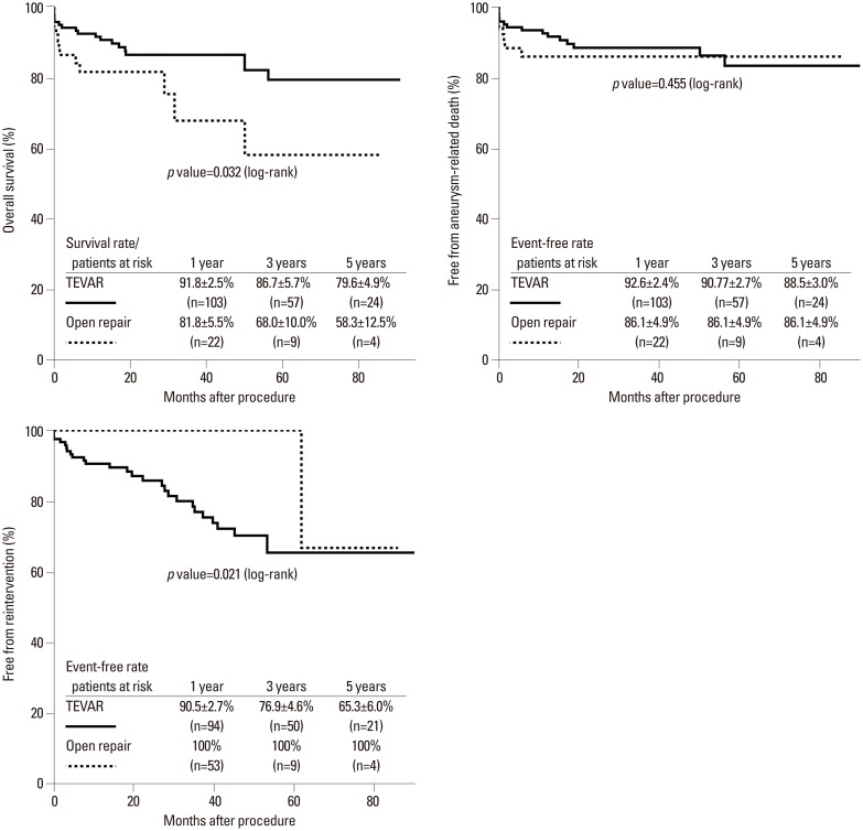
The completed follow-up rate was 97.8%, and the mean follow-up duration was 36±26 months. Overall, 30-day mortality was 3.5% and 9.4% in the TEVAR and open repair groups, respectively (p=0.14). Paraplegia occurred more in the open repair group (3.5% vs. 7.5%) but did not reach statistical difference (p=0.27). There was no difference in the incidence of stroke between the two groups (5.3% vs. 7.5%, p=0.73). The incidence of respiratory failure (1.8% vs. 26.4%, p<0.01), postoperative AKI (4.4% vs. 17.0%, p<0.01), and need for dialysis (1.8% vs. 9.4%, p=0.01) were significantly higher in the open repair group. Late mortality was 12.7% and 14.6% in the TEVAR and open repair groups, respectively (p=0.75). Late aneurysm related death occurred more in the TEVAR group (10.0% vs. 4.2%) but did not reach statistical difference (p=0.35). More patients who underwent TEVAR needed re-intervention significantly (21.8% vs. 2.1%, p<0.01). The Kaplan-Meier analysis revealed that free from re-intervention was significantly higher in the open repair group (log rank test, p=0.02) (Fig. 1). There were 24 patients (21.1%) with aortic re-intervention, including endoleak (17.4%) and retrograde dissection (3.5%), in the TEVAR group at the follow-up. One patient with endoleak and 4 patients with retrograde dissection were treated surgically. Table 3 summarizes the early and late outcomes (see discussion in the details of cases of late mortality).
Fig. 1. Rates of overall survival, free from aorta-related death, and free from re-intervention. TEVAR, thoracic endovascular aortic repair.
Table 3. Early and Late Outcomes.
| Variables | TEVAR (n=114) | Open repair (n=53) | p value |
|---|---|---|---|
| Early outcomes, n (%) | |||
| 30-day mortality | 4 (3.5) | 5 (9.4) | 0.14 |
| Paraplegia | 4 (3.5) | 4 (7.5) | 0.27 |
| Stroke | 6 (5.3) | 4 (7.5) | 0.73 |
| Respiratory failure | 2 (1.8) | 14 (26.4) | <0.01 |
| Acute kidney injury | 5 (4.4) | 9 (17) | 0.01 |
| Need for dialysis | 2 (1.8) | 5 (9.4) | 0.03 |
| Late outcomes* | |||
| Late death | 14 (12.7) | 7 (14.6) | 0.75 |
| Late aneurysm-related death | 11 (10.0) | 2 (4.2) | 0.35 |
| Re-intervention | 24 (21.8) | 1 (2.1) | <0.01 |
| Endoleak | 20 (17.4) | - | |
| Retrograde dissection | 4 (3.5) | - | |
| Leakage | - | 1 (2.1) |
TEVAR, thoracic endovascular aortic repair.
*Outcomes after 30 days postoperatively.
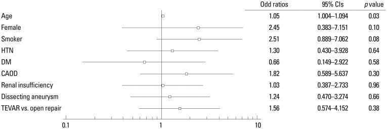
The cumulative survival rate at 3 to 5 years was significantly higher in the TEVAR group than in open repair patients (log rank test, p=0.03) (Fig. 1). However, free from aneurysm-related death was similar between the groups at 3 to 5 years (p=0.45). The Kaplan-Meier analysis revealed that free from re-intervention was significantly higher in the open repair group (log rank test, p=0.02) (Fig. 1). There were 24 patients (21.8%) with aortic re-intervention, including endoleak (18.2%) and retrograde dissection (3.6%), in the TEVAR group at the follow-up. One patient with endoleak and 4 patients with retrograde dissection were treated surgically. The variables for the Cox proportional hazard model that identified the predictors of aneurysm-related death included age, gender, history of smoking, hypertension, diabetes, coronary artery obstructive disease, renal insufficiency, aortic pathology, and the use of TEVAR versus open repair. The use of TEVAR and open repair did not influence aneurysm-related death (OR 1.557; 95% CIs 0.574-4.152; p=0.38). Old age was the only independent predictor for aneurysm-related death (OR 1.048; 95% CIs 1.004-1.094; p=0.03) (Fig. 2).
Fig. 2. Predictors of aneurysm-related death. HTN, hypertension; DM, diabetes mellitus; CAOD, coronary artery obstructive disease; TEVAR, thoracic endovascular aortic repair; CI, confidence interval.
DISCUSSION
This is a retrospective study assessed the efficacy and suitability of TEVAR in patients with isolated DTAA. The major finding of this study, based on 114 consecutive patients who underwent TEVAR, is that early mortality and neurologic complications did not differ between the TEVAR and open repair groups. However, the incidence of postoperative AKI and respiratory failure were significantly lower in the TEVAR group. In terms of late outcomes, the overall survival rate at 3 to 5 years was significantly higher in the TEVAR group, even though the patients of the TEVAR group had more risks. In our present study, the TEVAR patients were older than the open repair patients by almost 0.5 decade, and the TEVAR patients had a higher incidence of preoperative renal insufficiency. These differences occurred initially, because TEVAR was primarily used in high-risk patients who could not tolerate conventional open repair. The re-intervention rate and continued presence of complications, such as endoleaks, was higher in the TEVAR group, and free from aneurysm-related death at 3 to 5 years was not different between the groups, even though overall survival rate was better in the TEVAR group.
Treatment of DTAA is challenging. Although open repair for DTAA has become a refined surgical procedure, it has nevertheless been associated with significant perioperative mortality and neurologic complication. In recent series from high-volume centers of excellence, mortality and neurologic morbidity rates were shown to range from 5.4-7.2% for mortality, 2.1-6.2% for permanent stroke, and 0.8-2.3% for permanent paraplegia.11,12 The highly invasive nature of open repair necessitates a lesser invasive method for treating descending thoracic aortic disease. Since introduction of TEVAR, many reports have demonstrated that it is a safe and feasible alternative to conventional repair. The perioperative results for the three stent graft trials in the TEVAR arms showed rates of 1.9-2.1% for mortality, 2.4-4% for stroke, and 1.3-3% for permanent paraplegia.4,13,14
AKI is an another important complication and regarded as a marker of increased early or late morbidity and mortality after TEVAR or open repair for DTAA.15 The overall incidence of AKI after aortic surgery has been reported to be high compared with other cardiac surgeries.16 In addition to preoperative risk factors, thoracic aortic surgery itself is an independent risk factor for AKI because of the complexity of the procedure, which includes circulatory arrest.17 In our present study, 33 patients (58.5%) in the open repair group underwent total circulatory arrest with hypothermia, and AKI occurred in 9 patients (17%) of the group, including 5 patients (9.4%) who required dialysis. A wide range of incidence of renal dysfunction has been reported in patients undergoing TEVAR. Pisimisis, et al.18 reported that 11.9% of patients undergoing TEVAR had postoperative AKI. In our study, 5 TEVAR patients (4.4%) had an AKI with 2 requiring dialysis. Although there was a higher incidence of preoperative renal insufficiency in TEVAR patients, the incidence of postoperative AKI was lower in the TEVAR patients.
Respiratory failure was a major complication, especially after open repair. It affects postoperative morbidity and mortality, resulting in prolonged hospitalization. In addition to thoracotomy, cardiopulmonary bypass and hypothermia induced a systemic inflammatory reaction attributed to postoperative respiratory failure.19 Because of its less invasive procedure, respiratory failure was a relatively rare complication after TEVAR compared with open repair.5,6 In our study, 14 open repair patients (22.6%) developed respiratory failure compared with 2 patients (1.8%) in the TEVAR group.
Although the use of TEVAR has rapidly increased because of improved perioperative mortality and morbidity rates, there are no long-term data regarding the prevention of premature death using this technique. Especially, the durability and long-term complication rates of TEVAR have not yet been determined. Recent publications have cautioned that high re-intervention is associated with TEVAR.6 In our study, a significantly higher rate of re-intervention was associated with the TEVAR group (24 patients, 20.1%). The reasons for re-intervention were endoleak (20 patients, 17.4%) and retrograde dissection (4 patients, 3.5%). Both endoleaks and retrograde dissection after TEVAR potentially predisposes patients to catastrophic adverse events such as aneurysm progression and mortality.20 In the initially published studies on TEVAR, endoleaks after TEVAR seemed markedly less significant than that of aneurysms of the infra-renal aorta, however, it soon became obvious that endoleaks after TEVAR are a serious complication.21,22 Endoleak after TEVAR potentially predisposes patients to catastrophic adverse events such as aneurysm progression and mortality.20
The clinical importance of retrograde dissection of the ascending aorta after TEVAR has recently been developed as a clinical experience, and applications of TEVAR have been expanded. Previous studies have reported mortality rates ranging from 27% to 57%.23,24 In the present study, retrograde dissection was found to develop in 4 TEVAR patients, which mostly occurred in the early running period of TEVAR. All patients with retrograde dissection underwent surgical treatment, and 3 of these patients died 30 days postoperatively.
TEVAR also seems to be associated with the reduction of long-term mortality rates. The midterm follow-up of patients revealed a significant benefit in cumulative survival rates in the TEVAR group (77%) compared to that in the open repair group (58.4%), even though patients who underwent TEVAR were older than those who underwent open repair. There were 8 late deaths (3 in the TEVAR group and 5 in the open repair group) unrelated to aortic disease. In the TEVAR group, 2 patients died because of malignant tumor and 1 patient died with unknown reason. Among 5 deaths in the open repair group, 2 patients died because of pneumonia and sepsis, 1 patient malignant tumor, and 2 patients, who needed dialysis just after open repair, died with unknown reasons. A higher incidence of postoperative AKI and respiratory failure can affect long-term survival rates. Piffaretti, et al.25 reported that AKI was also associated with increased late mortality. We thought that higher rate of AKI and pneumonia after open repair could affect long-term survival and late death. Little is known about the mid-term or long-term outcomes after TEVAR in patients with DTAA. Recently, Desai, et al.6 reported outcomes similar to ours: the overall survival rate at 8-10 years was similar between the open repair and TEVAR groups, even though the TEVAR group was older and had more preoperative comorbidities than the open repair group. In our study, free from aneurysm-related death at 3 to 5 years was not different between the groups, even though overall survival rate was better in the TEVAR group.
The late aneurysm-related death rate was higher in TEVAR group (10.0% vs. 4.2%, p=0.35), although did not reach statistical difference. There were 11 late aneurysm-related deaths after TEVAR, which includes 3 deaths related to retrograde dissection, 4 related to surgical conversion due to endoleak, and 4 related to aneurysm rupture due to endoleak. There were 2 late aneurysm-related death after open repair, which includes 1 death related to rupture of anastomosis site, and 1 related to rupture of abdominal aortic aneurysm. As mentioned earlier, retrograde dissection and endoleak in the present study were significant complications, leading to catastrophic results. These significant complications may contribute to our relatively poor result of an aneurysm-related death rate after TEVAR. However, most of the late aneurysm-related deaths (8/11) occurred in patients who underwent TEVAR within the first 2 years of our institutional experience. The TEVAR device and skills of endovascular grafting have been improved since the first implantation of TEVAR at our institution; therefore, aneurysm-related death rates are expected to decrease.
Limitations
Limitations of this study include its small sample size, retrospective nature, and baseline differences between the treatment groups. These limitations affected the evaluation of long-term result efficacy of treatment. Difference of follow-up duration and follow-up frequency also could affect long term result. Usually, the patients who underwent TEVAR have more frequent follow-up to detect stent complication earlier. Indeed, in our study, there were 5 emergency cases (3 patients in TEVAR, 2 patients in open repair). We analyzed early and late outcomes only in the elective cases. There were no statistical differences in the 30-day mortality (2.7% in TEVAR vs. 7.8% in open repair, p=0.208), the incidences of paraplegia (2.7% in TEVAR vs. 5.9% in open repair, p=0.380), stroke (4.5% in TEVAR vs. 5.9% in open repair, p=0.708), late death (12% in TEVAR vs. 12.8% in open repair, p=0.90), and late aneurysm-related death (9.3% in TEVAR vs. 4.3% in open repair, p=0.35). However, rates of respiratory failure (0.9% in TEVAR vs. 13% in open repair, p<0.01), AKI (3.6% in TEVAR vs. 15.7% in open repair, p=0.01), and need for dialysis (0.9% in TEVAR vs. 7.8% in open repair, p=0.03) were higher in open repair group. We thought that sample size of emergency cases was so small that it did not influence early and late outcomes. Furthermore, the difference in aortic pathology between the groups was an another important limitation. The sample size of each pathologies were not large enough to compare early and late results between TEVAR and open repair. However, the number of patients with DTAA has been increasing in our institute, therefore, further study for each aortic pathologies could be possible in the near future. Additionally, this study is from a single institution, and the prospective multicenter data would be needed to confirm our findings. Even though the TEVAR patients were older than the open repair patients, compared with open repair, we deem that TEVAR is a safe and effective procedure for treating DTAAs with improved perioperative and long-term results. Further follow-up of these patients is planned, and larger multicenter studies will further confirm these findings.
Footnotes
The authors have no financial conflicts of interest.
References
- 1.De Bakey ME, Cooley DA. Successful resection of aneurysm of thoracic aorta and replacement by graft. J Am Med Assoc. 1953;152:673–676. doi: 10.1001/jama.1953.03690080017005. [DOI] [PubMed] [Google Scholar]
- 2.Dake MD, Miller DC, Semba CP, Mitchell RS, Walker PJ, Liddell RP. Transluminal placement of endovascular stent-grafts for the treatment of descending thoracic aortic aneurysms. N Engl J Med. 1994;331:1729–1734. doi: 10.1056/NEJM199412293312601. [DOI] [PubMed] [Google Scholar]
- 3.Mitchell RS, Dake MD, Sembra CP, Fogarty TJ, Zarins CK, Liddel RP, et al. Endovascular stent-graft repair of thoracic aortic aneurysms. J Thorac Cardiovasc Surg. 1996;111:1054–1062. doi: 10.1016/s0022-5223(96)70382-3. [DOI] [PubMed] [Google Scholar]
- 4.Andrassy J, Weidenhagen R, Meimarakis G, Rentsch M, Jauch KW, Kopp R. Endovascular versus open treatment of degenerative aneurysms of the descending thoracic aorta: a single center experience. Vascular. 2011;19:8–14. doi: 10.1258/vasc.2010.oa0256. [DOI] [PubMed] [Google Scholar]
- 5.Orandi BJ, Dimick JB, Deeb GM, Patel HJ, Upchurch GR., Jr A population-based analysis of endovascular versus open thoracic aortic aneurysm repair. J Vasc Surg. 2009;49:1112–1116. doi: 10.1016/j.jvs.2008.12.024. [DOI] [PubMed] [Google Scholar]
- 6.Desai ND, Burtch K, Moser W, Moeller P, Szeto WY, Pochettino A, et al. Long-term comparison of thoracic endovascular aortic repair (TEVAR) to open surgery for the treatment of thoracic aortic aneurysms. J Thorac Cardiovasc Surg. 2012;144:604–609. doi: 10.1016/j.jtcvs.2012.05.049. [DOI] [PubMed] [Google Scholar]
- 7.Fillinger MF, Greenberg RK, McKinsey JF, Chaikof EL Society for Vascular Surgery Ad Hoc Committee on TEVAR Reporting Standards. Reporting standards for thoracic endovascular aortic repair (TEVAR) J Vasc Surg. 2010;52:1022–1033. doi: 10.1016/j.jvs.2010.07.008. [DOI] [PubMed] [Google Scholar]
- 8.Levey AS, Levin A, Kellum JA. Definition and classification of kidney diseases. Am J Kidney Dis. 2013;61:686–688. doi: 10.1053/j.ajkd.2013.03.003. [DOI] [PubMed] [Google Scholar]
- 9.Brinkman WT, Szeto WY, Bavaria JE. Stent graft treatment for transverse arch and descending thoracic aorta aneurysms. Curr Opin Cardiol. 2007;22:510–516. doi: 10.1097/HCO.0b013e3282f04273. [DOI] [PubMed] [Google Scholar]
- 10.Lee WA, Brown MP, Nelson PR, Huber TS, Seeger JM. Midterm outcomes of femoral arteries after percutaneous endovascular aortic repair using the Preclose technique. J Vasc Surg. 2008;47:919–923. doi: 10.1016/j.jvs.2007.12.029. [DOI] [PubMed] [Google Scholar]
- 11.Wong DR, Parenti JL, Green SY, Chowdhary V, Liao JM, Zarda S, et al. Open repair of thoracoabdominal aortic aneurysm in the modern surgical era: contemporary outcomes in 509 patients. J Am Coll Surg. 2011;212:569–579. doi: 10.1016/j.jamcollsurg.2010.12.041. [DOI] [PubMed] [Google Scholar]
- 12.Estrera AL, Miller CC, 3rd, Chen EP, Meada R, Torres RH, Porat EE, et al. Descending thoracic aortic aneurysm repair: 12-year experience using distal aortic perfusion and cerebrospinal fluid drainage. Ann Thorac Surg. 2005;80:1290–1296. doi: 10.1016/j.athoracsur.2005.02.021. [DOI] [PubMed] [Google Scholar]
- 13.Fairman RM, Criado F, Farber M, Kwolek C, Mehta M, White R, et al. Pivotal results of the Medtronic Vascular Talent Thoracic Stent Graft System: the VALOR trial. J Vasc Surg. 2008;48:546–554. doi: 10.1016/j.jvs.2008.03.061. [DOI] [PubMed] [Google Scholar]
- 14.Matsumura JS, Cambria RP, Dake MD, Moore RD, Svensson LG, Snyder S, et al. International controlled clinical trial of thoracic endovascular aneurysm repair with the Zenith TX2 endovascular graft: 1-year results. J Vasc Surg. 2008;47:247–257. doi: 10.1016/j.jvs.2007.10.032. [DOI] [PubMed] [Google Scholar]
- 15.Huynh TT, van Eps RG, Miller CC, 3rd, Villa MA, Estrera AL, Azizzadeh A, et al. Glomerular filtration rate is superior to serum creatinine for prediction of mortality after thoracoabdominal aortic surgery. J Vasc Surg. 2005;42:206–212. doi: 10.1016/j.jvs.2005.03.062. [DOI] [PubMed] [Google Scholar]
- 16.Kuitunen A, Vento A, Suojaranta-Ylinen R, Pettilä V. Acute renal failure after cardiac surgery: evaluation of the RIFLE classification. Ann Thorac Surg. 2006;81:542–546. doi: 10.1016/j.athoracsur.2005.07.047. [DOI] [PubMed] [Google Scholar]
- 17.Arnaoutakis GJ, Bihorac A, Martin TD, Hess PJ, Jr, Klodell CT, Ejaz AA, et al. RIFLE criteria for acute kidney injury in aortic arch surgery. J Thorac Cardiovasc Surg. 2007;134:1554–1560. doi: 10.1016/j.jtcvs.2007.08.039. [DOI] [PubMed] [Google Scholar]
- 18.Pisimisis GT, Khoynezhad A, Bashir K, Kruse MJ, Donayre CE, White RA. Incidence and risk factors of renal dysfunction after thoracic endovascular aortic repair. J Thorac Cardiovasc Surg. 2010;140(6 Suppl):S161–S167. doi: 10.1016/j.jtcvs.2010.10.014. [DOI] [PubMed] [Google Scholar]
- 19.Morimoto K, Nishimura K, Miyasaka S, Maeta H, Taniguchi I. The effect of sivelestat sodium hydrate on severe respiratory failure after thoracic aortic surgery with deep hypothermia. Ann Thorac Cardiovasc Surg. 2011;17:369–375. doi: 10.5761/atcs.oa.10.01555. [DOI] [PubMed] [Google Scholar]
- 20.Hansen CJ, Bui H, Donayre CE, Aziz I, Kim B, Kopchok G, et al. Complications of endovascular repair of high-risk and emergent descending thoracic aortic aneurysms and dissections. J Vasc Surg. 2004;40:228–234. doi: 10.1016/j.jvs.2004.03.051. [DOI] [PubMed] [Google Scholar]
- 21.Neuhauser B, Perkmann R, Greiner A, Steingruber I, Tauscher T, Jaschke W, et al. Mid-term results after endovascular repair of the atherosclerotic descending thoracic aortic aneurysm. Eur J Vasc Endovasc Surg. 2004;28:146–153. doi: 10.1016/j.ejvs.2004.04.006. [DOI] [PubMed] [Google Scholar]
- 22.Scharrer-Pamler R, Kotsis T, Kapfer X, Görich J, Orend KH, Sunder-Plassmann L. Complications after endovascular treatment of thoracic aortic aneurysms. J Endovasc Ther. 2003;10:711–718. doi: 10.1177/152660280301000405. [DOI] [PubMed] [Google Scholar]
- 23.Eggebrecht H, Thompson M, Rousseau H, Czerny M, Lönn L, Mehta RH, et al. Retrograde ascending aortic dissection during or after thoracic aortic stent graft placement: insight from the European registry on endovascular aortic repair complications. Circulation. 2009;120(11 Suppl):S276–S281. doi: 10.1161/CIRCULATIONAHA.108.835926. [DOI] [PubMed] [Google Scholar]
- 24.Dong ZH, Fu WG, Wang YQ, Guo da Q, Xu X, Ji Y, et al. Retrograde type A aortic dissection after endovascular stent graft placement for treatment of type B dissection. Circulation. 2009;119:735–741. doi: 10.1161/CIRCULATIONAHA.107.759076. [DOI] [PubMed] [Google Scholar]
- 25.Piffaretti G, Mariscalco G, Bonardelli S, Sarcina A, Gelpi G, Bellosta R, et al. Predictors and outcomes of acute kidney injury after thoracic aortic endograft repair. J Vasc Surg. 2012;56:1527–1534. doi: 10.1016/j.jvs.2012.05.106. [DOI] [PubMed] [Google Scholar]


