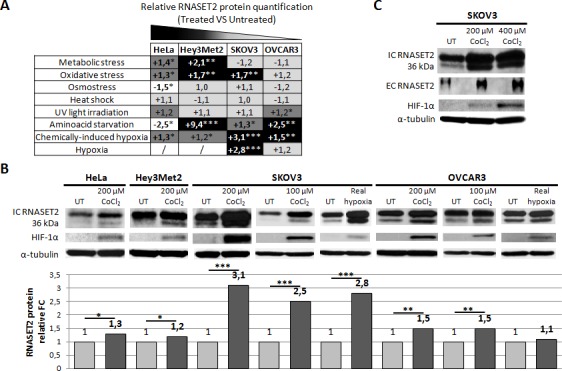
Figure 2. Human RNASET2 protein levels change following stress induction.

A) Eight different stress-inducing conditions were applied to four different cell lines and Western blot analysis for RNASET2 protein was performed. Relative fold-changes in RNASET2 expression are reported in treated vs. untreated cells, with dark and light shadows representing higher or lower levels with respect to untreated cells, respectively. B, C) Western Blot analysis data on total protein extracts (B) and supernatants (C). A significant increase in the levels of the intracellular 36 kDa RNASET2 protein was observed in response to both chemically-induced (CoCl2) and real (1% O2) hypoxic conditions. HIF-1α protein levels were assessed as an hypoxia stress marker. A concomitant increase of the secreted 36 kDa RNASET2 protein (EC RNASET2) was also observed in SKOV3 cells in response to chemically-induced hypoxia. Triplicate experiments were performed for each experimental condition assessed. Statistical analysis was performed using two-tailed Student's t-test. *p<0,05; **p<0,01; ***p<0,001. UT: untreated. IC: intracellular. EC: extracellular. FC: fold-change.
