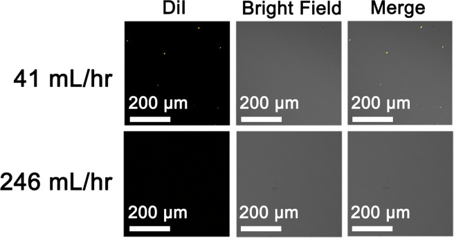
FIG. 5.
Confocal images of the aggregated lipid-PLGA NPs in complete medium containing serum. The DiI-labeled NPs (PL-41 and PL-246) are incubated with complete medium (DMEM with 10% FBS) for 3 h. The final concentration of DiI in medium is 25 ng/ml. After incubation, the NPs in medium are visualized with confocal laser scanner microscope and the aggregated NPs can be detected as fluorescent dots (orange dots).

