Abstract
Background:
Development of new drug carriers would be an interesting approach if it allowed increased efficacy of antibiotics and a reduction in doses, thus reducing the risk of developing resistance. As with most drug carriers, niosomes have been used to improve the selective delivery and the therapeutic index of antimicrobial agents.
Methods:
In this study, different formulation of niosomes containing ciprofloxacin (CPFX), Span (20, 60 or 80), Tween (20, 60 or 80) and cholesterol were prepared by film hydration method. The release of the drug from different formulations was studied by using Franz diffusion cell. The niosomes were further characterized by optical microscopy and particle size analysis, and their antimicrobial activity was evaluated.
Results:
Size of the niosomes was significantly dependent on the amount of cholesterol and surfactant type and varied from 8.56 to 61.3 μm. The entrapment efficiency of CPFX niosomes prepared by remote loading was more than 74%. Niosomes composed of Span/Tween 60 provided a higher CPFX release rate than other formulations. The obtained results indicated a diffusion-based mechanism for drug leakage through bilayers. All formulations presented more antibacterial activity as compared to free CPFX solution.
Conclusion:
Niosomal CPFX appears to be a promising approach in the management of bacterial infections, especially ophthalmic ones, and should be further evaluated by in vivo experiments.
Keywords: Ciprofloxacin, Niosomes, Release
Introduction
Ciprofloxacin (CPFX) has a wide spectrum, high bactericidal activity, and good ability to penetrate most tissues and accumulate in cells 1. However, as a water-soluble drug, CPFX efficacy is limited by poor ocular bioavailability. Marketed ophthalmic CPFX drops need to be administered 6 times daily reducing the patient compliance. The encapsulation of CPFX in carrier systems can improve ophthalmologic bioavailability and prolong the therapeutic action. Many different drug delivery systems were evaluated to increase its efficacy for treatment of different kinds of infectious diseases. The hydroxyapatite microspheres containing CPFX were used as an implantable drug delivery system for the treatment of bone infections 2. In another study disposition of CPFX-loaded polyisobutylcyanoacrylate nanoparticles was studied after intravenous infusion to rabbits and its efficacy against Mycobacterium avium (M. avium)3 complex in human macrophages was evaluated 4.
An important physical characteristic of drug delivery systems is drug releases pattern. Ke et al reported ocular sustained release formulation of CPFX which delivered 10-fold more drug into the aqueous humor than the standard solution formulation 5. In another study, CPFX-releasing osteoconductive bone defect filler based on controlled, long-acting CPFX release from a bioabsorbable matrix was efficient to provide targeted local bactericidal concentrations which were limited only to the tissue areas near the implantation site 6.
Vesicular systems were widely used to decrease the adverse effects and reduce the total amount of antimicrobial agents required 7. These systems are lipid based vesicles that self assemble into bilayers to entrap both hydrophilic and lipophilic compounds. Liposomes are composed of natural amphiphilc lipids, usually containing cholesterol 8,9. Niosomes are non-ionic surfactant vesicles that are made up of a synthetic amphiphilic bilayer and cholesterol. Niosomes exhibit preferred characteristics, such as being more chemically stable, easier to store, safer to handle, and less expensive than liposomes 10.
Niosomes were previously used for continuous and controlled release of drugs 10,11. Abdelkader et al prepared Span 60-based niosomes of naltrexone to evaluate its in vitro release parameters 12. They reported that niosomal encapsulating naltrexone could significantly control drug release rate and extent. In another study, Span 40-based niosomes of metformin showed a significant extended-release and better hypoglycemic efficiency compared to free metformin solution 13. Factors affecting release characteristics of niosomes are type of drug, amount and type of surfactant, cholesterol content, and methods of preparation.
Among the different factors that influence the drug release from vesicles, the bilayer composition is a critical factor. For example, Mokhtar et al reported that release profiles of niosomal flurbiprofen were affected by cholesterol amount 14. Another study reported that the alkyl chain length surfactant and the method of preparation influence acetazolamide release rate from niosomes as ophthalmic carriers 15. They observed that Multilamellar Vesicles (MLV) had higher entrapment efficiency and lower drug release after 8 hr and concluded acetazolamide was released form niosome by a diffusion controlled mechanism.
The purpose of the current study was to prepare and characterize niosomal formulation containing CPFX in order to be used as ocular prolonged-release carriers. In this study, the preparation of different niosomal formulations were reported using different molar ratios of Span 20 and Tween 20, Span 60 and Tween 60 or Span 80 and Tween 80, in combination with cholesterol. Also, the effect of formulation components on size and encapsulation efficiency of vesicles, in vitro release profile of CPFX from niosomes and in vitro antibacterial effect of CPFX loaded niosomes and free drug against Staphylococcus aureus (S. aureus), Pseudomonas aeruginosa (P. aeruginosa), Klebsiella pneumonia (K. pneumonia) and Escherichia coli (E. coli) were evaluated.
Materials and Methods
Chemical
CPFX was obtained from Bayer (Newbury, UK). Polysorbate 20, 60 and 80 (Tween 20, 60 and 80), and Sorbitan monopalmitate 20, 60 and 80 (Span 20, 60 and 80), and cholesterol were purchased from Fluka (Switzerland). All other chemicals and analytical grade solvents were obtained from Merck (Germany).
Bacteria
S. aureus (PTCC1112), P. aeruginosa (PTCC1074), E. coli (PTCC1330), K. pnuemonia (PTCC1053) were obtained from the Persian type culture collection. They were subcultured on Muller-Hinton agar plates and incubated at 37°C.
Niosome preparation
The compositions of different niosome formulations are presented in table 1. Nisomal formulations were prepared by film hydration (hand shaking method), as previously reported 16. Briefly, 400 μmol of surfactant (Span 20 and Tween 20, Span 60 and Tween 60 or Span 80 and Tween 80) and cholesterol were dissolved in chloroform in a round-bottomed flask. The organic solvent was evaporated at 55°C under reduced pressure, using a rotary evaporator at 120 rpm. The resultant thin lipid film produced on the inner wall of the flask was then hydrated using 5 ml of ammonium sulfate (250 mM, pH=2.5) for 1 hr at 55°C. The non-entrapped ammonium sulfate was removed from the niosomal suspension by dialysis against 10% (w/v) sucrose (pH=2.5). Five ml of CPFX solution (5 mg/ml) was added to this niosomal suspension at 55°C for 60 min. Residual CPFX was removed from formulations by dialysis against 10% (w/v) sucrose (pH=5). The final formulations were stored in refrigerator (4–8°C) for further studies.
Table 1.
The composition of niosomal prepared formulations (molar ratio)
| Formulation number | Span 20 | Span 60 | Span 80 | Tween 20 | Tween 60 | Tween 80 | Cholesterol |
|---|---|---|---|---|---|---|---|
| F1 | 3.5 | -- | -- | 3.5 | -- | -- | 3 |
| F2 | 3 | -- | -- | 3 | -- | -- | 4 |
| F3 | 2.5 | -- | -- | 2.5 | -- | -- | 5 |
| F4 | -- | 3.5 | -- | -- | 3.5 | -- | 3 |
| F5 | -- | 3 | -- | -- | 3 | -- | 4 |
| F6 | -- | 2.5 | -- | -- | 2.5 | -- | 5 |
| F7 | -- | -- | 3.5 | -- | -- | 3.5 | 3 |
| F8 | -- | -- | 3 | -- | -- | 3 | 4 |
| F9 | -- | -- | 2.5 | -- | -- | 2.5 | 5 |
Size and morphology of niosomes
The particle size and particle size distribution of niosomes were determined by laser-light scattering (Malvern Mastersizer 2000E, UK). Some micrographs were prepared by a camera attached to the optical microscope (Nikon HFX-DX, Japan) in 10×40 and 10×100 magnifications.
Drug loading
The amount of loaded CPFX was analyzed after disrupting the niosomes by isopropyl alcohol. The concentration of CPFX in niosomes was determined using a UV/visible spectrophotometer (Shimadzu, 2100, Japan) at 277 nm. The concentration of the samples was obtained from a linear equation of CPFX standard curve constructed previously.
To determine the encapsulation efficiency of niosomes, unentrapped CPFX was removed from the formulations by dialysis against 10% (w/v) sucrose (pH= 5). The concentration of CPFX niosmal formulation was determined before and after dialysis. The entrapment efficiency was defined as follows:
Drug release
CPFX release from various formulations was evaluated using a set of Franz diffusion cells with an active surface area of 2.37 cm2 and a receptor phase volume of 37 ml. An acetate cellulose dialysis membrane which was soaked in normal saline for 24 hr was clamped between the cell’s donor and the receptor compartments. Temperature was maintained at 37±1°C by a circulating water bath. The receptor compartment was filled with normal saline and the donor compartment with 1 ml of niosomal CPFX, a CPFX solution as control and empty niosome as blank. Samples of the receptor compartment were collected at fixed time intervals and replaced with an equal volume of normal saline for up to 4 hr. Concentration in the receptor medium was quantified spectrophotometrically.
In vitro antibacterial activity
Minimum Inhibitory Concentrations (MICs) of CPFX-encapsulated, free CPFX, empty niosomes with free CPFX and empty niosomes were determined by conventional agar dilution method against S. aureus (PTCC 1112), P. aeruginosa (PTCC1074), E. coli (PTCC1330), and K. pnuemonia (PTCC1053). Different concentrations of the test compounds were added to molten Mueller-Hinton (MH) agar plates. The bacteria suspensions (107 CFU/ml) were inoculated on each plate and incubated overnight at 35°C. Next day, the lowest drug concentration that inhibited visible bacterial growth was reported as the MIC.
Statistics and data analysis
The release data were fitted to various models using linear regression analysis. All data are expressed as the mean±standard deviation (SD). Significant differences were calculated by analysis of variance (ANOVA) followed by a post hoc test using SPSS 11.5 version and differences at p<0.05 were considered significant.
Results
Size and morphology of niosomes
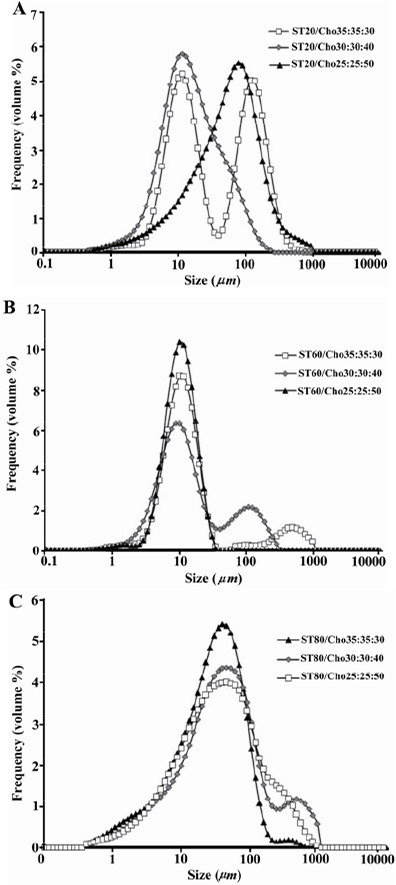
Mean volume diameter (dv) of different niosomal formulations measured by laser light scattering technique are presented in table 2. As shown, by increasing the amount of cholesterol content from 3 to 5 molar ratios the size of vesicles increased from 10.35, 9 and 31.17 to 61.3, 14.4 and 38.47 μm for the Span/Tween 20, Span/Tween 60 and Span/Tween 80 formulation.
Table 2.
Mean volume diameter (dv) (μm) and CPFX encapsulation efficiency percentage of formulations (mean±SD, n=3)
| Formulations | Average size (mean±SD) | Encapsulation efficiency % |
|---|---|---|
| F1 | 10.35±0.3 | 61±5.6 |
| F2 | 13.67±0.61 | 48±4.6 |
| F3 | 61.3±2.56 | 38±6 |
| F4 | 9±0.1 | 74±8.5 |
| F5 | 8.56±0.11 | 67±6.5 |
| F6 | 14.4±0.61 | 63±7 |
| F7 | 31.17±0.55 | 58±4.5 |
| F8 | 41.83±2.97 | 41±3.5 |
| F9 | 38.47±3.96 | 33±3 |
The size distributions of prepared niosomes with different compositions are presented in figure 1 . Size distribution curves of most formulations were as bell-shape patterns indicating log-normal size distributions (Figures 1B and 1C).
Figure 1.

Size distribution of niosomes: A) the effect of cholesterol content on the size distribution of niosomes composed of Span 20/Tween 20/Cholesterol, B) the effect of cholesterol content on the size distribution of niosomes composed of Span 60/Tween 60/ Cholesterol and C) the effect of cholesterol content on the size distribution of niosomes composed of Span 80/Tween 80/Cholesterol.
The morphological observation of niosomes showed that prepared formulations were spherical and homogeneous dispersion. Different shapes and sizes of niosomes were seen in the micrographs, but MLVs were frequently observed (Figure 2A). Morphological studies revealed the size related well with the results of the laser light scattering measurement. In few formulations, trace aggregation was observed (Figure 2C). Moreover, no CPFX crystal was observed.
Figure 2.

Morphological micrographs of niosomes (×400): A) formulation composed of Span 20/Tween 20/Cholesterol; molar ratio (m.r) 35:35:30, B) formulation composed of Span 60/Tween 60/Cholesterol; m.r 35:35:30 and C) formulation composed of Span 80/Tween 80/Cholesterol; m.r 35:35:30.
Drug loading
Encapsulation efficiencies of CPFX in different formulations prepared by remote loading method are presented in table 2. As shown, the encapsulation efficiency of niosomes decreased with increasing cholesterol content from 3 to 5 molar ratios. This effect was more observed at the higher cholesterol ratio.
Drug release
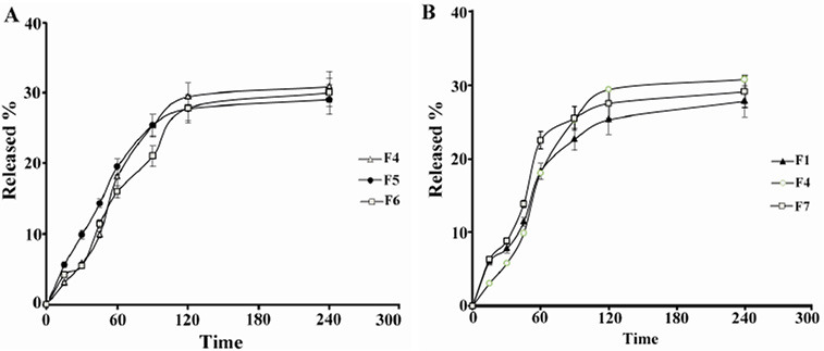
The results of in vitro release of CPFX from niosomes after 4 hr in normal saline at 37°C are shown in figure 3. During 4 hr, 27.8%, 30.8%, 30%, 29% and 29.1of CPFX were released from F1, F4, F5, F6, and F7 niosomes, respectively. R-squared (r2) obtained from linear regression analysis of CPFX release data is presented in table 3. For most of formulations, CPFX release profile better fits with Peppas equation and Higuchi model suggesting the Fickian diffusion release mechanism for CPFX. The release of CPFX from niosomes was a biphasic process. In fast initial phase (first 60 min), around 20% of drug was released, whereas only around 10% of CPFX was released in slow release phase (180 min). The effect of amount of cholesterol on CPFX release profile is shown in figure 3A. A non-significant decrease in the percentage of CPFX released was observed when cholesterol content was increased from 3 to 5 molar ratios (p>0.05). Figure 3B shows the effect of the surfactant type on release profile of CPFX. There were not any significant differences among the overall released amount of CPFX from the different surfactant type niosomes (p>0.05).
Figure 3.

Release of CPFX from niosomes in normal saline at 37°C versus time (mean±SD, n=3). A) effect of cholesterol content and B) effect of surfactant type.
Table 3.
R-squared (r2) obtained from linear regression analysis of CPFX release data which fitted in different release kinetic models
| Formulations | Baker-lonsdale | Higuchi | Hixon-crawell | First order | Peppas | Fickian | Zero order |
|---|---|---|---|---|---|---|---|
| F1 | r2=0.9628 | r2=0.9647 | r2=0.9205 | r2=0.9111 | r2=0.9713 | r2=0.9110 | r2=0.9110 |
| F4 | r2=0.9811 | r2=0.9849 | r2=0.9645 | r2=0.9562 | r2=0.9831 | r2=0.9561 | r2=0.9561 |
| F5 | r2=0.9447 | r2=0.9553 | r2=0.9111 | r2=0.9013 | r2=0.9747 | r2=0.9011 | r2=0.9011 |
| F6 | r2=0.9344 | r2=0.9564 | r2=0.8866 | r2=0.8755 | r2=0.9744 | r2=0.8754 | r2=0.8754 |
| F7 | r2=0.9052 | r2=0.9311 | r2=0.8519 | r2=0.8407 | r2=0.9613 | r2=0.8406 | r2=0.8406 |
In vitro antibacterial activity
MICs (μg/ml) of CPFX-encapsulated, free CPFX, empty niosomes plus free CPFX and empty niosomes against S. aureus, P. aeruginosa, K. pneumonia and E. coli are presented in table 4. The empty niosomes showed no activity against four bacteria tested. The MIC values for noisome encapsulated CPFX were more than free CPFX. The combination of empty niosomes with free CPFX had no additive effect on the antimicrobial activity of CPFX.
Table 4.
MICs (μg/ml) of CPFX-encapsulated, free CPFX, empty niosomes plus free CPFX and empty niosomes against 4 strain of microorganisms (n=3; mean±SD)
| Formulation | Microorganism | |||
|---|---|---|---|---|
| S. aureuse (PTCC 1112) | E. coli (PTCC 1330) | P. aeruginosa (PTCC 1074) | K. pneumonia (PTCC 1053) | |
| Free CPFX | 0.21±0.06 | 0.012±0.00 | 0.5±0.00 | 0.03±0.00 |
| F1 | 0.104±0.03 | 0.006±0.001 | 0.21±0.06 | 0.015±0.00 |
| F4 | 0.104±0.03 | 0.003±0.001 | 0.17±0.06 | 0.015±0.00 |
| F7 | 0.104±0.03 | 0.0057±0.001 | 0.21±0.06 | 0.012±0.003 |
| Empty noisome F1 | -- | -- | -- | -- |
| Empty noisome F4 | -- | -- | -- | -- |
| Empty noisome F7 | -- | -- | -- | -- |
| Empty noisome F1 with CPFX | 0.21±0.06 | 0.012±0.004 | 0.42±0.13 | 0.03±0.00 |
| Empty noisome F4 with CPFX | 0.21±0.06 | 0.012±0.004 | 0.5 ±0.00 | 0.025±0.007 |
| Empty noisome F7 with CPFX | 0.21±0.06 | 0.012±0.004 | 0.5 ±0.00 | 0.03±0.00 |
Discussion
Particle size is an important factor for drug delivery systems which can influence entrapment efficiency and drug release. Our study showed that amount of cholesterol can significantly influence mean diameter of niosomal vesicle. This result is in agreement with the previous studies reporting that increasing the amount of cholesterol resulted in larger vesicles 17,18 . This could be explained based on this fact that cholesterol would be more likely to increase the number of bilayers since it has little effect on the charge at the bilayer surface and interbilayer separation 19. The resultant effect is the forming of larger niosomes. The change in the mean diameter of liquid state surfactants (Span/Tween 20 and Span/Tween 80) vesicles was more significant following the increase in the amount of intercalated cholesterol. This can be related to the flexibility of their bilayers leading to more susceptibility of bilayers to the structural effects of cholesterol.
In addition, Hydrophile-Lipophile Balance (HLB) and length chain of surfactants may also affect the particle size of vesicles. In our study, the combination of a monoalkylsorbitan ester (Span) and polyxylated sorbitan ester (Tween) with the same hydrocarbon chain length was used for the preparation of niosomes. Span/ Tween 20, Span/Tween 60 and Span/Tween 80 mixtures with mean HLB values 12.65, 9.8 and 9. 65, respectively, formed stable niosomes in the presence of cholesterol. Yoshioka et al reported that increasing the HLB value of surfactant results in larger vesicles 20. This is in agreement with results of our study where HLB increment from 9.8 to 12.65 leads to significant increase in mean diameter of vesicles. In the present study, Span/Tween 80 which has an unsaturated and longest alkyl chain (C9=9) resulted in larger vesicles. It may be due to the fact that compared to monolayer types of saturated Span 60, monolayers of unsaturated Span 80 are more expanded and form larger molecular areas 21.
The entrapment efficiency of CPFX in niosomes prepared by remote loading method was relatively high and ranged from 33 to 74%. However, low encapsulation efficiencies of 5–14% were observed using a conventional passive-entrapment method (data not shown). Similar to our results, Oh et al reported an increase in CPFX liposomal encapsulation from 9 to 90% by a remote-loading technique that utilized both pH and potential gradients 22.
Increasing cholesterol content led to reducing the e encapsulated drug amount. This can be related to the amphipathic nature of the drug which may make the drug-bilayer interactions. In addition to entrapment in the hydrophilic compartment by protonation, there is the further possibility of the CPFX molecule being incorporated into the niosome membrane. Hernández-Borrell and Montero 23 exploited the fluorescence properties of CPFX to localize the drug in bilayers by using quenching, anisotropy and binding experiments.
The entrapment of CPFX in solid state, Span/Tween 60, and niosomes was significantly higher than liquid state, Span/Tween 20 and Span/Tween 80, and vesicles (p>0.05). The rigidity of bilayers of Span/Tween 60 containing niosomes and the leaky nature of liquid surfactants bilayers can explain the mentioned difference in CPFX encapsulation ability. Other groups also reported surfactant having the highest phase transition temperature provides the highest encapsulation for the drug 20. Among tested surfactants, Span/Tween 80 had lowest encapsulation efficiency. This finding is consistent with other report 24 in which the lowest colchicine entrapment efficiency of the Span 80 formulation was explained by unsaturated status of Span 80 causing he membrane to be more permeable. Degier et al also reported the introduction of double bonds into the paraffin chains which cause a significant increment of liposomes’ permeability 25.
The rate of drug release from a delivery system is critical and has to be investigated in order to achieve an optional system with desired release characteristics. Furthermore, in vitro release studies are often performed to predict how a delivery system might work in ideal situations, which might give some indication of its in vivo performance. In general, vesicle lamellarity plays a significant role in the retention of entrapped material 26. The usual types of membranes employed are those with porous characteristics, e.g. cellulose acetate or homogeneous permeable polymers such as silicone 27. In the present study, cellulose acetate was used for assessment of drug release from MLVs. Our results indicate initial rapid releases of the drug without detectable lag-time and an equilibrium state or a slower release phase with all formulations. The rapid initial phase may be originated from permeation of free CPFX and desorption of drug from the surface of niosomes and the slower phase related primarily to the diffusion of CPFX through the bilayers. Such effect has also been observed in the release of human insulin 17 and also caffeine 28 from niosomal suspensions. There were not any significant differences among the overall released amount of CPFX from the different niosomal formulations. The better fits with Higuchi model, and Peppas equation in drug release profile indicated the Fickian diffusion. This finding suggests the dominant mechanism is diffusion of CPFX through gel and liquid states bilayers. An investigation on the release rate of the drug revealed that the highest speed of drug delivery is during the first 60 min. This finding shows the main driving force for transporting the drug is the concentration differences between the two compartments of all glass Franz diffusion cells.
Many studies have demonstrated the successful use of vesicular systems including niosomes as ocular drug delivery carriers. Niosomes can provide prolonged and controlled drug action at the corneal surface. Abdelkader et al reported that timolol maleate loaded niosomes showed significantly more sustained reduction of the intra-ocular pressures compared to timolol maleate solution 29. Physicochemical parameters such as size, morphology, physical state of the loaded drug, and drug release profile of the niosomal formulations can have a bearing on the ocular bioavailability. Marsh and Maurice 30 investigated the effect of non-ionic surfactants of different HLB values on corneal permeability in human subjects. They found that Tween 20 and Brij 35 surfactants having HLB values between 16 and 17, were most effective in increasing corneal penetration. Niosome size of greater than 10 μm has been reported to be optimum for ocular delivery. This large size helps providing higher entrapped quantity of drug, better ocular localization and longer stay on the surface of the eye. Additionally, smaller vesicles are less stable due to greater surface tension 31. With respect to sustained drug release profile and size of vesicles, CPFX niosomes prepared in the present study have good potentials for effective ocular delivery of the drug.
CPFX is an antibiotic useful for the treatment of a number of bacterial infections including respiratory, urinary tract, and gastrointestinal infections. In addition, CPFX ophthalmic drop is currently the drug of choice for infection of the eye including conjunctivitis and corneal ulcers. In the current study, antimicrobial activity of CPFX loaded niosomes was assessed by MIC measurement. MICs of niosomal CPFX were lower than those of free drug for all the strains. Similar results were reported for liposomes containing CPFX, meropenem, and gentamicin 32. It was suggested that this increased antibacterial activity is related to the fusional interaction between membrane phospholipids. S. aureus and P. aeruginosa are common organisms responsible for bacterial conjunctivitis. Our result showed that CPFX niosomes had a good effect on S. aureus, and P. aeruginosa and MICs of CPFX niosomes were more than two-fold lower than MICs of free CPFX.
Conclusion
In summary, particle size of nisomes was dependent on the type of surfactant and the amount of cholesterol used in the preparation of the vesicles. Relatively, high drug loading by remote loading method was achieved, and entrapment efficiency was influenced by cholesterol content and phase transition temperature of surfactants. Based on the antibacterial activity, evaluation of niosomal CPFX can be beneficial for designing a new delivery system for this antibiotic. Niosome size of greater than 10 μm has been reported to be suitable for drug administration to eye. According to size of vesicles and sustained drug release profile, our niosomes have good potentials for effective topical delivery of the drug especially ophthalmic ones.
Acknowledgement
This study was financially supported by Isfahan University of Medical Sciences (Grant No. 188097).
References
- 1. Oliphant CM, Green GM. Quinolones: a comprehensive review. Am Fam Physician 2002; 65 (3): 455– 464. [PubMed] [Google Scholar]
- 2. Pham HH, Luo P, Genin F, Dash AK. Synthesis and characterization of hydroxyapatite-ciprofloxacin delivery systems by precipitation and spray drying technique. AAPS PharmSciTech 2002; 3 (1): E1. [DOI] [PMC free article] [PubMed] [Google Scholar]
- 3. Akbari V, Moghim S, Reza Mofid M. Comparison of epothilone and taxol binding in yeast tubulin using molecular modeling. Avicenna J Med Biotechnol 2011; 3 (4): 167– 175. [PMC free article] [PubMed] [Google Scholar]
- 4. Fawaz F, Bonini F, Maugein J, Lagueny A. Ciprofloxacin-loaded polyisobutylcyanoacrylate nanoparticles: pharmacokinetics and in vitro antimicrobial activity. Int J Pharm 1998; 168 (2): 255– 259. [Google Scholar]
- 5. Ke TL, Cagle G, Schlech B, Lorenzetti OJ, Mattern J. Ocular bioavailability of ciprofloxacin in sustained release formulations. J Ocul Pharmacol Ther 2001; 17 (6): 555– 563. [DOI] [PubMed] [Google Scholar]
- 6. Koort JK, Mäkinen TJ, Suokas E, Veiranto M, Jalava J, Knuuti J, et al. Efficacy of ciprofloxacin-releasing bioabsorbable osteoconductive bone defect filler for treatment of experimental osteomyelitis due to Staphylococcus aureus. Antimicrob Agents Chemother 2005; 49 (4): 1502– 1508. [DOI] [PMC free article] [PubMed] [Google Scholar]
- 7. Drulis-Kawa Z, Dorotkiewicz-Jach A. Liposomes as delivery systems for antibiotics. Int J Pharm 2010; 387 (1): 187– 198. [DOI] [PubMed] [Google Scholar]
- 8. Kumar R, Kumar S, Jha SS, Jha AK. Vesicular system-carrier for drug delivery. Der Pharmacia Sinica 2011; 2 (4): 192– 202. [Google Scholar]
- 9. Chen C-S, Yao J, Durst RA. Liposome encapsulation of fluorescent nanoparticles: Quantum dots and silica nanoparticles. J Nanopart Res 2006; 8 (6): 1033– 1038. [Google Scholar]
- 10. Mahale NB, Thakkar PD, Mali RG, Walunj DR, Chaudhari SR. Niosomes: novel sustained release nonionic stable vesicular systems--an overview. Adv Colloid Interface Sci 2012; 183–184: 46– 54. [DOI] [PubMed] [Google Scholar]
- 11. Pardakhty A, Moazeni E. Nano-niosomes in drug, vaccine and gene delivery: a rapid overview. Nanomedicine J 2013; 1 (1): 1– 12. [Google Scholar]
- 12. Abdelkader H, Ismail S, Kamal A, Alany RG. Design and evaluation of controlled-release niosomes and discomes for naltrexone hydrochloride ocular delivery. J Pharm Sci 2011; 100 (5): 1833– 1846. [DOI] [PubMed] [Google Scholar]
- 13. Hasan AA, Madkor H, Wageh S. Formulation and evaluation of metformin hydrochloride-loaded niosomes as controlled release drug delivery system. Drug Deliv 2013; 20 (3–4): 120– 126. [DOI] [PubMed] [Google Scholar]
- 14. Mokhtar M, Sammour OA, Hammad MA, Megrab NA. Effect of some formulation parameters on flurbiprofen encapsulation and release rates of niosomes prepared from proniosomes. Int J Pharm 2008; 361 (1–2): 104– 111. [DOI] [PubMed] [Google Scholar]
- 15. Guinedi AS, Mortada ND, Mansour S, Hathout RM. Preparation and evaluation of reverse-phase evaporation and multilamellar niosomes as ophthalmic carriers of acetazolamide. Int J Pharm 2005; 306 (1–2): 71– 82. [DOI] [PubMed] [Google Scholar]
- 16. Akbari V, Abedi D, Pardakhty A, Sadeghi-Aliabadi H. Ciprofloxacin nano-niosomes for targeting intracellular infections: an in vitro evaluation. J Nanopart Res 2013; 15 (4): 1– 14. [Google Scholar]
- 17. Pardakhty A, Varshosaz J, Rouholamini A. In vitro study of polyoxyethylene alkyl ether niosomes for delivery of insulin. Int J Pharm 2007; 328 (2): 130– 141. [DOI] [PubMed] [Google Scholar]
- 18. Varshosaz J, Pardakhty A, Hajhashemi VI, Najafabadi AR. Development and physical characterization of sorbitan monoester niosomes for insulin oral delivery. Drug Deliv 2003; 10 (4): 251– 262. [DOI] [PubMed] [Google Scholar]
- 19. Cable C. An examination of the effect of surface modifications on the physicochemical and biological properties of non-ionic surfactant vesicles. [dissertation]. [Glasgow]: University of Strathclyde; 1990. [Google Scholar]
- 20. Yoshioka T, Sternberg B, Florence AT. Preparation and properties of vesicles (niosomes) of sorbitan monoesters (Span 20, 40, 60 and 80) and a sorbitan triester (Span 85). Int J pharm 1994; 105 (1): 1– 6. [Google Scholar]
- 21. Peltonen L. The interfacial behaviour of sorbitan surfactant monolayers and the bulk properties of these surfactants as a function of temperature. [Bibliography]. Finland: University of Helsinki; 2001. [Google Scholar]
- 22. Oh YK, Nix DE, Straubinger RM. Formulation and efficacy of liposome-encapsulated antibiotics for therapy of intracellular Mycobacterium avium infection. Antimicrob Agents Chemother 1995; 39 (9): 2104– 2111. [DOI] [PMC free article] [PubMed] [Google Scholar]
- 23. Hernández-Borrell J, Montero MT. Does ciprofloxacin interact with neutral bilayers? An aspect related to its antimicrobial activity. Int J Pharm 2003; 252 (1–2): 149– 157. [DOI] [PubMed] [Google Scholar]
- 24. Hao Y, Zhao F, Li N, Yang Y, Li K. Studies on a high encapsulation of colchicine by a niosome system. Int J Pharm 2002; 244 (1–2): 73– 80. [DOI] [PubMed] [Google Scholar]
- 25. de Gier J, Mandersloot JG, van Deenen LL. Lipid composition and permeability of liposomes. Biochim Biophys Acta 1968; 150 (4): 666– 675. [DOI] [PubMed] [Google Scholar]
- 26. Schilling RJ, Mitra AK. Pharmacodynamics of insulin following intravenous and enteral administrations of porcine-zinc insulin to rats. Pharm Res 1992; 9 (8): 1003– 1009. [DOI] [PubMed] [Google Scholar]
- 27. Dias M, Farinha A, Faustino E, Hadgraft J, Pais J, Toscano C. Topical delivery of caffeine from some commercial formulations. Int J Pharm 1999; 182 (1): 41– 47. [DOI] [PubMed] [Google Scholar]
- 28. Khazaeli P, Pardakhty A, Shoorabi H. Caffeine-loaded niosomes: characterization and in vitro release studies. Drug Deliv 2007; 14 (7): 447– 452. [DOI] [PubMed] [Google Scholar]
- 29. Abdelkader H, Farghaly U, Moharram H. Effects of surfactant type and cholesterol level on niosomes physical properties and in vivo ocular performance using timolol maleate as a model drug. J Pharm Investig 2014; 44 (5): 329– 337. [Google Scholar]
- 30. Marsh RJ, Maurice DM. The influence of non-ionic detergents and other surfactants on human corneal permeability. Exp Eye Res 1971; 11 (1): 43– 48. [DOI] [PubMed] [Google Scholar]
- 31. Sahoo RK, Biswas N, Guha A, Sahoo N, Kuotsu K. Non-ionic surfactant vesicles in ocular delivery: innovative approaches and perspectives. Biomed Res Int 2014; 2014: 263604. [DOI] [PMC free article] [PubMed] [Google Scholar]
- 32. Gubernator J, Drulis-Kawa Z, Dorotkiewicz-Jach A, Doroszkiewicz W, Kozubek A. In vitro antimicrobial activity of liposomes containing ciprofloxacin, meropenem and gentamicin against Gram-negative clinical bacterial strains. Lett Drug Des Discov 2007; 4 (4): 297– 304. [Google Scholar]


