Abstract
Introduction: Metabolic syndrome is a systemic disorder and manifests as a group of conditions including abdominal obesity, dyslipidemia, hypertension and coronary artery disease. The importance of epicardial adipose tissue has been proven through recognition of its contribution to inflammation by pro-inflammatory cytokine discharge. Several investigations have been performed on vitamin D receptors in different tissues. In this study, epicardial adipose tissue thickness (EATT) and the levels of vitamin D were measured and compared with a healthy control group. Material and Methods: 84 patients who had metabolic syndrome without diabetes and 64 healthy individuals were enrolled into the study. In all patients, the EATT was calculated by ecocardiography and the level of serum 25 (OH) vitamin D was measured. Results: It was observed that EATT in patients with metabolic syndrome increases significiantly compared to the healthy control group (P < 0.001). No significant difference between patients and control group was found for the levels of 25 (OH) vitamin D (P = 0.507). There was no correlation between 25 (OH) vitamin D and EATT (P = 0.622). Conclusions: We observed that EATT increased in patients with metabolic syndrome. In contradiction to literature; the levels of 25 (OH) vitamin D was not found to be high in patients with metabolic syndrome. Any significant correlation was not found between EATT and 25 (OH) vitamin D levels. Further studies with a larger patient population are required to assess the relationship.
Keywords: Metabolic syndrome, epicardial adipose tissue thickness, vitamin D
Introduction
Metabolic syndrome is a cluster of risk factors including abdominal obesity, impaired fasting glucose, hypertension and dyslipidemia [1]. Clinical implications include hypercoagulability, obesity, hyperuricemia, osteoporosis, non-alcoholic steatohepatitis, sleep apnea and polycystic ovary syndrome [2]. The incidence of metabolic syndrome is increasing globally and it is a pandemic effecting 20-30% on adult populations [3]. Patients with metabolic syndrome are observed to have myocardial infarction and strokes 3 times more than healthy people. Additionally, these patients are at a higher risk for developing type 2 diabetes and coronary artery disease [4,5].
Epicardial adipose tissue (EAT), is a fatty tissue regarded as a component of visceral fat tissue [6]. EAT thickness increases as visceral fat tissue increases and they are therefore regarded as equivalent. EAT is most frequently found in right ventricle free wall, left ventricle free wall, around the atrium and the adventitia of the coronary artery branches from the surface of the epicardium to the myocardium [7]. The EAT thickness (EATT) varies according to the inflammatory status of the body and the person’s dietary habits. In healthy individuals EAT is protective of vascular functions and also acts as an energy store for the cardium. However, increase of EAT leads to lipolytic, prothrombotic and proinflammatory properties [8]. Recent studies have found significant relationship between EATT and metabolic syndrome [9,10].
The main effect of Vitamin D is on calcium and phosphate homeostasis and bone metabolism. In addition, there are more than 30 tissues with vitamin D receptors (VDR), including endothelium, smooth muscle, myocardium, brain, prostate, breast, colonic and immune system cells [11]. A study in patients with coronary artery disease demonstrated VDR in EAT [12]. An animal study found that vitamin D deficiency leads to hypertrophy of cardiomyocytes and an increase in release of proinflammatory cytokines (TNF-α, IL-6, MCP-1) in EAT [13].
Another animal study found that 2 months after feeding with a vitamin D deficient diet, pancreatic insulin secretion decreased and the animals developed glucose intolerance [14]. Vitamin D deficiency leads to decreased insulin sensitivity, deterioration of beta cell functions, systemic inflammation, glucose intolerance, metabolic syndrome and type 2 diabetes. There is evidence of vitamin D’s effect on these mechanisms [15,16]. There is sufficient data on the relationship between EATT and 25 (OH) vitamin D with metabolic syndrome. However, there is lack of data on the relationship between EATT and 25 (OH) vitamin D. In this study, we measured EATT and 25 (OH) vitamin D levels in patients with isolated metabolic syndrome without diabetes and compared with a control of patients without metabolic syndrome. We also analysed the relationship between 25 (OH) vitamin D and EATT.
Materials and methods
This study included 148 consecutive patients who admitted to Bakirkoy Dr. Sadi Konuk Education and Research Hospital’s Internal Medicine and Cardiology outpatient clinics between December 2013 and April 2014. Of these patients, 84 had metabolic syndrome and 64 were participants with similar age and demographic characteristics forming the control group. The patient group was created from patients with metabolic syndrome who did not have diabetes.
This study was approved by the local ethical committee and informed consent was obtained from all patients.
Exclusion criteria were: patients without metabolic syndrome or with diabetes, patients who did not consent or would not volunteer for the study, patients with one of: mental retardation, psychotic disorders, mood disorders, dementia, delirium and other amnestic disorders; pregnancy, hypothyroidism, hyperthyroidism, acute coronary syndrome, coronary artery disease, congestive heart failure, atrial fibrillation or any coronary rhythm other than sinus rhythm, renal dysfunction, congenital heart disease, myocarditis, pericarditis, cardiomyopathy, valvular heart disease, neoplastic disease, any chronic inflammatory illness, active infection, chronic liver disease, use of medication that includes vitamin D, parathyroid disease, bone metabolism disorders (osteoporosis or osteopenia) or related medication use and menopause.
For all patients included in the study, a form with the following information was collected by the same physician: age, gender, medical history, family history, habits, height, weight, systolic blood pressure (SBP), diastolic blood pressure (DBP), body mass index (BMI), waist circumference (WC) and physical examination findings. All measurements and examinations were performed by the same physician. BMI was calculated according to the “weight (kg)/height (m2)” formula. The homeostatic model assessment of insulin resistance (HOMA-IR) was used to evaluate insulin resistance. The following formula was used: HOMA-IR = (Fasting insulin (µu/ml) × Fasting plasma glucose (mg/dl)/405) [17] Waist circumference (cm) was measured parallel from the exact middle point between the lower border of 12th rib and spina ischiadica major. Limits were determined as 94 cm for men and 80 cm for women in accordance with relevant criteria.
Blood pressure was measured after 20 minutes of resting, both systolic and diastolic. Two measurements were recorded and the mathematical average was taken. Hypertension was defined as ≥ 140 mmHg systolic or ≥ 90 mmHg diastolic pressure in three separate measurements or the use of antihypertensive medication. Diabetes was defined as fasting blood glucose ≥ 126 mg/dL or use of antidiabetic medication. Fasting glucose between 100-126 mg/dL was classified as impaired fasting glucose. Hyperlipidemia was defined as total cholesterole ≥ 200 mg/dL or trigliceride > 150 mg/dL.
The diagnosis of metabolic syndrome was made according to the 2005 International Diabetes Federation (IDF) criteria [18]. Diagnosis was made if the patient had abdominal obesity (waist circumference > 94 cm in men and > 88 cm in females) plus at least two of the following: elevated fasting plasma glucose (≥ 100 mg/dL (5.6 μmol/L)); elevated levels of triglycerides (≥ 150 mg/dL (1.7 μmol/L)); reduced levels of HDL cholesterol [< 40 mg/dL (1.03 μmol/L) in men and 50 mg/dL (1.29 μmol/L) in women)]; elevated blood pressure (systolic blood pressure ≥ 130 mmHg or diastolic blood pressure ≥ 85 mmHg).
Laboratory parameters
Blood for 25 (OH) vitamin D and other laboratory parameters were taken after overnight fasting of 12 hours from the forearm veins into tubes with EDTA. Tubes were centrifuged for 10 minutes at 4000 rpm and stored at -80 degrees centigrade.
Fasting glucose, urea, creatinine, LDL cholesterole, HDL cholesterol, triglycerides, TSH and other biochemical parameters were measured using the Abott Architect C16200 system (Abott Laboratories, IL, USA).
Serum insulin was measured using the Immulite 2000 chemoimmulescense autoanalyse and the “Siemens Healthcare Diagnostics USA” kit. Complete blood count was analysed with the Counter LH 750 autoanalyser (Beckman Counter, CA, USA) and 25 (OH) vitamin D with the Roche 25 (OH) Vitamin D total test, Roche elecsys E 170 Immunoassay.
Determination of epicardial fat tissue thickness
Detailed two dimensional, M-mode and doppler echocardiography was performed in all patients included in this study, by two cardiologist blinded to the patients’ and healthy group’s biochemical results. The echocardiography device used was vivid S-5 (GE Vingmed, Horten, Norway) with a 2.5-3.5 MHz probe. Epicardial adipose tissue was observed as a comparatively echo-free area between the outer border of the myocardium and the visceral layer of the pericardium. EATT was measured on the parasternal longitudinal axis in transverse images, perpendicular to the right ventricular free wall, at the end of diastole. The aortic annulus intraventricular septum was used as anatomical markers for the identification of a measurement perpendicular to the right ventricular free wall [19]. To reduce the margin of error, measurements were made at five consecutive cardiac cycles with the average of all measurements taken as final EATT.
Statistical analysis
Data were analyzed using SPSS 22.0 for Windows software (SPSS Inc, Chicago, IL, USA). Frequency, ratio, mean, minimum, maximum, and standard deviation values were used in the descriptive statistics. The Kolmogorov-Smirnov test was used to control the data distribution. Independent samples t-test and Mann-Whitney U-test were used to analyze quantitative variables. The chi-square test was used to analyze qualitative variables. Spearman’s correlation tests were used for the correlation analysis. The lineer logistic regression analysis was performed to determine the effect levels of the parameters. Standard beta coefficients and 95% confidence intervals (CI) were calculated. Receiver operating curve (ROC) analysis was used to calculate the required cut-off values to distinguish metabolic syndrome patients with maximum sensitivity and specificity. P values <0.05 were considered statistically significant.
Results
Clinical and demographic information of patients and the control group are given in Table 1.
Table 1.
Clinical and demographic characteristics of patients and control groups
| Patients group | Control group | P value | ||||
|---|---|---|---|---|---|---|
|
|
||||||
| Mean ± SD | Mean ± SD | |||||
| Age | 38.5±9.7 | 37.1±9.1 | 0.394 | |||
| Gender | Female | 58 | 69.0% | 52 | 81.3% | 0.092 |
| Male | 26 | 31.0% | 12 | 18.8% | ||
| BMI (kg/m2) | 34.2±6.4 | 25.1±4.2 | 0.0001 | |||
| Waist circumference (cm) | 107.9±14.4 | 85.1±11.4 | 0.0001 | |||
| SBP (mmHg) | 132.9±20.9 | 109.8±13.5 | 0.0001 | |||
| DBP (mmHg) | 82.0±12.4 | 70.7±9.6 | 0.0001 | |||
| Hypertension | 49 | 58.3% | 1 | 1.6% | 0.0001 | |
| Hyperlipidemia | 11 | 13.1% | 1 | 1.6% | 0.011 | |
| Smoking | 29 | 34.5% | 19 | 29.7% | 0.534 | |
BMI: Body mas index, SBP: Systolic blood pressure, DBP: Diastolic blood pressure.
While there was no statistical difference between gender and smoking between the groups (P > 0.05), there was a statistically significant difference between patients and the control group for BMI, waist circumference, SBP, DBP (P = 0.0001) and hyperlipidemia (P = 0.011).
Laboratory parameters for patients and control group are shown in Table 2.
Table 2.
Comparison of patients and control group biochemical and other parameters
| Patients group | Control group | P value | |
|---|---|---|---|
|
| |||
| Mean ± SD | Mean ± SD | ||
| Glucose (mg/dl) | 95.6±11.1 | 84.6±10.2 | 0.0001 |
| Total cholesterol | 188.1±38.7 | 173.8±35.2 | 0.006 |
| LDL-C | 110.5±30.4 | 101.4±32.2 | 0.017 |
| HDL-C | 44.2±12.5 | 51.7±11.8 | 0.0001 |
| Triglyceride (mg/dl) | 176.0±81.1 | 91.7±47.0 | 0.0001 |
| Creatinine (mg/dl) | 0.7±0.2 | 0.7±0.1 | 0.849 |
| Insulin (µu/ml) | 19.3±10.2 | 7.2±3.9 | 0.0001 |
| HOMA-IR | 4.5±2.5 | 1.4±0.8 | 0.0001 |
| EATT (cm) | 0.6±0.2 | 0.4±0.1 | 0.0001 |
| Vitamin D (ng/ml) | 11.8±8.2 | 13.4±10.3 | 0.507 |
LDL-C: Low density lipoprotein cholesterol, HDL-C: High density lipoprotein cholesterol, HOMA-IR: Homeostatic model assessment, EATT: Epicardial adipose tissue thickness.
While there was no difference between patients and the control group in creatinine and vitamin D levels (P > 0,05) there was a statistical difference for glucose, HDL-C, triglyceride, insulin, HOMA IR, EATT (P = 0.0001), total cholesterole (P = 0.006) and LDL-C (P = 0.017).
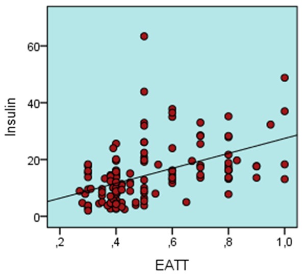
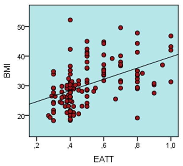
Correlation performed between EATT and other parameters are shown in Table 3. A negative correlation was found between EATT and HDL-C while there was a positive correlation between EATT and age, BMI, waist circumference, TG, SBP, DBP, insulin and HOMA-IR (P < 0.005). No significant correlation was found between vitamin D and age, BMI, waist circumference, TG, HDL-C, SBP, DBP, insulin or HOMA-IR (P > 0.05). Lineer regression analysis was calculated for parameters that correlated with EATT (age, height, weight circumference, SBP, DBP, TG, insulin, HOMA-IR and vitamin D) and a reduced model was determined. Insulin (Figure 1) and BMI (Figure 2) was demonstrated to have a significant independent effect (P < 0.05) on EATT (Table 4).
Table 3.
The correlation analysis between the epicardial adipose tissue thickness with other parameters
| EATT | r value | P value |
|---|---|---|
| Age (year) | 0.204 | 0.018 |
| BMI (kg/m2) | 0.557 | 0.0001 |
| Waist circumference (cm) | 0.528 | 0.0001 |
| Glucose | 0.166 | 0.055 |
| Total cholesterol | 0.049 | 0.575 |
| LDL-C | 0.041 | 0.638 |
| HDL-C | -0.232 | 0.007 |
| Triglyceride (mg/dl) | 0.278 | 0.001 |
| SBP (mmHg) | 0.422 | 0.0001 |
| DBP (mmHg) | 0.402 | 0.0001 |
| Vitamin D (ng/ml) | -0.043 | 0.622 |
| Insulin | 0.528 | 0.0001 |
| HOMA-IR | 0.524 | 0.0001 |
EATT: Epicardial adipose tissue thickness, BMI: Body mass index, LDL-C: Low density lipoprotein cholesterol, HDL-C: High density lipoprotein cholesterol, SBP: Systolic blood pressure, DBP: Diastolic blood pressure, HOMA-IR Homeostatic model assessment.
Figure 1.

Correlation between insulin and epicardial adipose tissue thickness.
Figure 2.

Correlation between BMI and epicardiac adipose tissue thickness.
Table 4.
Lineer regression analysis results for comparison between epicardial adipose tissue thickness and insulin, ALT and BMI
| Multivariate Model | |||||
|---|---|---|---|---|---|
|
| |||||
| Non-Standardised Coefficient | Standardised Coefficient | ||||
| t | P | ||||
|
|
|||||
| B | S.H | Beta | |||
| Insulin | -0.15 | 0.03 | 0.36 | 04.06 | 0.0001 |
| BMI | 0.0001 | 0.0001 | 0.35 | 04.10 | 0.0001 |
BMI: Body mass index.
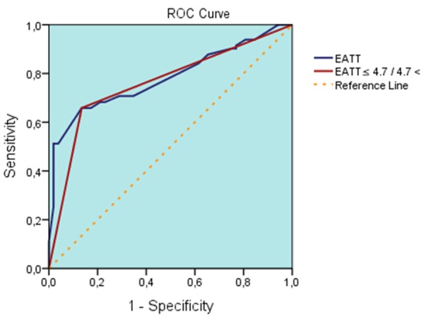
Receiver operating characteristics (ROC) curves, specificity, sensitivity, negative predictive value (NPV) and positive predictive value (PPV) were calculated for the EATT value. EATT prediction values > 4.7 mm were associated with the diagnosis of MeS with a sensitivity of 61%, a specificity of 87%, a positive predictive value of 89%, and a negative predictive value of 62% (area under the curve: 0.782 (0.705-0.859), (P = 0.0001) (Figure 3).
Figure 3.

ROC curve of epicardiac adipose tissue thickness in patients with metabolic syndrome.
Discussion
Our study demonstrated significant differences of EATT in patient with metabolic syndrome versus health controls. Correlation was found between EATT and components of metabolic syndrome. This correlation was positive for BMI, waist circumference, TG, SBP, DBP, insulin, HOMA-IR and negative for HDL-C. However, there was no significant difference between 25 (OH) vitamin D levels between the groups and no correlation was found between 25 (OH) and any component of metabolic syndrome. Also, no significant correlation was found between 25 (OH) vitamin D and EATT.
Metabolic syndrome is evaluated by its main diagnostic criteria: obesity and waist circumference measurements. However, waist circumference has been shown to be inaccurate for the determination of visceral adiposity. EAT is a component of visceral adipose tissue and is a reliable method for the determination of intraabdominal visceral adiposity. Studies have found a significant correlation between EATT and body mass index (BMI), waist circumference, visceral adipose tissue and metabolic syndrome. [9,20,21]. Our study also found significantly increased EATT in the patient group. TNF-α secreted by proinflammatory cytokines and adiposites of EAT lead to insulin resistance and lipolysis through autocrine mechanisms and also prevent the production of adinopectine [9,22,23].
A study on patients with high insulin levels and severe insulin resistance found substantially high levels of adinopectin [24]. When compared to subcutaneous adipose tissue, EAT secretes more chemokines and cytokines in patients with coronary artery disease [25]. These chemokines and cytokines are the precursors of inflammation. Inflammation plays a major role in the development of coronary artery disease, DM and metabolic syndrome. Consequently, the various effects of EAT plays a role in the development of metabolic syndrome. This could explain the correlation between EATT and metabolic syndrome. Also, the measurement of EATT as a component of visceral adipose tissue could be a method of providing information on the development of metabolic syndrome [26].
There is a paucity of data regarding the relationship between EATT and vitamin D. A study on patients with coronary artery disease demonstrated vitamin D receptors in EAT [12]. One animal study found an increase in hypertrophy of cardiomyocytes and secretion of proinflammatory cytokines (TNF-α, IL-6, MCP-1) from EAT in vitamin D deficiency [13]. In the light of this information, we studied the correlation between EATT and 25 (OH) vitamin D but was not demonstrate any significant relationship. This may be due to vitamin D defciency in both the patient group and control group.
In the single-variable linear regression analysis with EATT as the dependent variable and other parameters as independent variables, a significant correlation was found between EATT and BMI, insulin (P = 0.0001). Visceral obesity plays an important role in the development of metabolic syndrome. Our study demonstrated a positive correlation between EATT and BMI in patients with metabolic syndrome, supporting the fact that EAT is an important component of visceral adipose tissue.
The relationship between vitamin D and insulin resistance plus metabolic syndrome has been under discussion in recent years. The most important mechanism has been shown to be the positive effect on pancreatic β cells. Many studies have found a positive relationship between insulin sensitivity and 25 (OH) vitamin D. It has been concluded that deficiency of vitamin D adversely effects β cells leading to insulin resistance and metabolic syndrome [27-29]. Vitamin D also accelerates the conversion of proinsulin to insulin. A study performed on rats found that vitamin D deficiency leads to suppression of insulin production and that replacement therapy in vitamin D deficient rats lead to an increase in fasting insulin production [30,31]. In our study we were unable to demonstrate a significant difference between the vitamin D levels in patients with metabolic syndrome versus healthy controls. This could be due to the relatively small number of patients in this study. We also had a broad range of exclusion criteria. Patients with DM and coronary artery disease were excluded and only patients with isolated metabolic syndrome were included in this study. This led to the exclusion of many patients. The homogenity of our patient group may have lead to our inability to demonstrate a link with 25 (OH) vitamin D. Also, both the patient group and control group were found to be deficient in vitamin D. This could be due to the study being performed during the winter months. As previously known, vitamin D levels are highly dependent on patients’ environment, clothing style, feeding habits and sociocultural characteristics. A study showed that vitamin D deficiency is common among adult Turkish population [32]. Another study found that vitamin D deficiency was more frequent in persons migrating from Turkey to Europe when compared european population [33].
As a result, no statistically significant correlation was found between metabolic syndrome and vitamin D levels. EATT was found to be higher in patients with metabolic syndrome and was associated with insulin resistance. No relationship was noted between EATT and vitamin D in this pilot study. So further detailed studies are need to certain this.
Disclosure of conflict of interest
None.
References
- 1.Grundy SM, Cleeman JI, Daniels SR, Donato KA, Eckel RH, Franklin BA, Gordon DJ, Krauss RM, Savage PJ, Smith SC Jr, Spertus JA, Costa F, American Heart A, National Heart L, Blood I. Diagnosis and management of the metabolic syndrome: an American Heart Association/National Heart, Lung, and Blood Institute Scientific Statement. Circulation. 2005;112:2735–2752. doi: 10.1161/CIRCULATIONAHA.105.169404. [DOI] [PubMed] [Google Scholar]
- 2.Grundy SM. Hypertriglyceridemia, atherogenic dyslipidemia, and the metabolic syndrome. Am J Cardiol. 1998;81:18B–25B. doi: 10.1016/s0002-9149(98)00033-2. [DOI] [PubMed] [Google Scholar]
- 3.Zimmet P, Magliano D, Matsuzawa Y, Alberti G, Shaw J. The metabolic syndrome: a global public health problem and a new definition. J Atheroscler Thromb. 2005;12:295–300. doi: 10.5551/jat.12.295. [DOI] [PubMed] [Google Scholar]
- 4.Stern MP, Williams K, Gonzalez-Villalpando C, Hunt KJ, Haffner SM. Does the metabolic syndrome improve identification of individuals at risk of type 2 diabetes and/or cardiovascular disease? Diabetes Care. 2004;27:2676–2681. doi: 10.2337/diacare.27.11.2676. [DOI] [PubMed] [Google Scholar]
- 5.Kaur J. A comprehensive review on metabolic syndrome. Cardiol Res Pract. 2014;2014:943162. doi: 10.1155/2014/943162. [DOI] [PMC free article] [PubMed] [Google Scholar] [Retracted]
- 6.Paolisso G, Gualdiero P, Manzella D, Rizzo MR, Tagliamonte MR, Gambardella A, Verza M, Gentile S, Varricchio M, D’Onofrio F. Association of fasting plasma free fatty acid concentration and frequency of ventricular premature complexes in nonischemic non-insulin-dependent diabetic patients. Am J Cardiol. 1997;80:932–937. doi: 10.1016/s0002-9149(97)00548-1. [DOI] [PubMed] [Google Scholar]
- 7.Silver M, Silver M. Cardiovascular Pathology. 3rd edition. Philadelphia, PA: Churchill Livingstone; 2001. Examination of the Heart and of Cardiovascular Specimens in Surgical Pathology; pp. 8–9. [Google Scholar]
- 8.Iozzo P. Myocardial, perivascular, and epicardial fat. Diabetes Care. 2011;34(Suppl 2):S371–379. doi: 10.2337/dc11-s250. [DOI] [PMC free article] [PubMed] [Google Scholar]
- 9.Rabkin SW. The relationship between epicardial fat and indices of obesity and the metabolic syndrome: a systematic review and meta-analysis. Metab Syndr Relat Disord. 2014;12:31–42. doi: 10.1089/met.2013.0107. [DOI] [PubMed] [Google Scholar]
- 10.Pierdomenico SD, Pierdomenico AM, Cuccurullo F, Iacobellis G. Meta-analysis of the relation of echocardiographic epicardial adipose tissue thickness and the metabolic syndrome. Am J Cardiol. 2013;111:73–78. doi: 10.1016/j.amjcard.2012.08.044. [DOI] [PubMed] [Google Scholar]
- 11.Deluca HF, Cantorna MT. Vitamin D: its role and uses in immunology. FASEB J. 2001;15:2579–2585. doi: 10.1096/fj.01-0433rev. [DOI] [PubMed] [Google Scholar]
- 12.Romanelli MC, Vianello E, Briganti S, Malavazos A, Dozio E. Expression of vitamin D receptor and 1α-hydroxylase in epicardial fat: association to plasma 25-hydroxyvitamin D level in coronary artery disease patients (1139.8) The FASEB Journal. 2014;28:1138–1139. [Google Scholar]
- 13.Gupta GK, Agrawal T, DelCore MG, Mohiuddin SM, Agrawal DK. Vitamin D deficiency induces cardiac hypertrophy and inflammation in epicardial adipose tissue in hypercholesterolemic swine. Exp Mol Pathol. 2012;93:82–90. doi: 10.1016/j.yexmp.2012.04.006. [DOI] [PMC free article] [PubMed] [Google Scholar]
- 14.Nyomba BL, Bouillon R, De Moor P. Influence of vitamin D status on insulin secretion and glucose tolerance in the rabbit. Endocrinology. 1984;115:191–197. doi: 10.1210/endo-115-1-191. [DOI] [PubMed] [Google Scholar]
- 15.Weyer C, Bogardus C, Mott DM, Pratley RE. The natural history of insulin secretory dysfunction and insulin resistance in the pathogenesis of type 2 diabetes mellitus. J Clin Invest. 1999;104:787–794. doi: 10.1172/JCI7231. [DOI] [PMC free article] [PubMed] [Google Scholar]
- 16.Hu FB, Meigs JB, Li TY, Rifai N, Manson JE. Inflammatory markers and risk of developing type 2 diabetes in women. Diabetes. 2004;53:693–700. doi: 10.2337/diabetes.53.3.693. [DOI] [PubMed] [Google Scholar]
- 17.Matthews DR, Hosker JP, Rudenski AS, Naylor BA, Treacher DF, Turner RC. Homeostasis model assessment: insulin resistance and beta-cell function from fasting plasma glucose and insulin concentrations in man. Diabetologia. 1985;28:412–419. doi: 10.1007/BF00280883. [DOI] [PubMed] [Google Scholar]
- 18.Alberti KG, Zimmet P, Shaw J IDF Epidemiology Task Force Consensus Group. The metabolic syndrome--a new worldwide definition. Lancet. 2005;366:1059–1062. doi: 10.1016/S0140-6736(05)67402-8. [DOI] [PubMed] [Google Scholar]
- 19.Bertaso AG, Bertol D, Duncan BB, Foppa M. Epicardial fat: definition, measurements and systematic review of main outcomes. Arq Bras Cardiol. 2013;101:e18–28. doi: 10.5935/abc.20130138. [DOI] [PMC free article] [PubMed] [Google Scholar]
- 20.Iacobellis G, Corradi D, Sharma AM. Epicardial adipose tissue: anatomic, biomolecular and clinical relationships with the heart. Nat Clin Pract Cardiovasc Med. 2005;2:536–543. doi: 10.1038/ncpcardio0319. [DOI] [PubMed] [Google Scholar]
- 21.Lopez-Jimenez F, Sochor O. Epicardial fat, metabolic dysregulation, and cardiovascular risk: putting things together. Rev Esp Cardiol (Engl Ed) 2014;67:425–427. doi: 10.1016/j.rec.2014.01.011. [DOI] [PubMed] [Google Scholar]
- 22.Hotamisligil GS, Arner P, Caro JF, Atkinson RL, Spiegelman BM. Increased adipose tissue expression of tumor necrosis factor-alpha in human obesity and insulin resistance. J Clin Invest. 1995;95:2409–2415. doi: 10.1172/JCI117936. [DOI] [PMC free article] [PubMed] [Google Scholar]
- 23.Bruun JM, Lihn AS, Verdich C, Pedersen SB, Toubro S, Astrup A, Richelsen B. Regulation of adiponectin by adipose tissue-derived cytokines: in vivo and in vitro investigations in humans. Am J Physiol Endocrinol Metab. 2003;285:E527–533. doi: 10.1152/ajpendo.00110.2003. [DOI] [PubMed] [Google Scholar]
- 24.Semple RK, Soos MA, Luan J, Mitchell CS, Wilson JC, Gurnell M, Cochran EK, Gorden P, Chatterjee VK, Wareham NJ, O’Rahilly S. Elevated plasma adiponectin in humans with genetically defective insulin receptors. J Clin Endocrinol Metab. 2006;91:3219–3223. doi: 10.1210/jc.2006-0166. [DOI] [PubMed] [Google Scholar]
- 25.Mazurek T, Zhang L, Zalewski A, Mannion JD, Diehl JT, Arafat H, Sarov-Blat L, O’Brien S, Keiper EA, Johnson AG, Martin J, Goldstein BJ, Shi Y. Human epicardial adipose tissue is a source of inflammatory mediators. Circulation. 2003;108:2460–2466. doi: 10.1161/01.CIR.0000099542.57313.C5. [DOI] [PubMed] [Google Scholar]
- 26.Ahn SG, Lim HS, Joe DY, Kang SJ, Choi BJ, Choi SY, Yoon MH, Hwang GS, Tahk SJ, Shin JH. Relationship of epicardial adipose tissue by echocardiography to coronary artery disease. Heart. 2008;94:e7. doi: 10.1136/hrt.2007.118471. [DOI] [PubMed] [Google Scholar]
- 27.Chiu KC, Chu A, Go VL, Saad MF. Hypovitaminosis D is associated with insulin resistance and beta cell dysfunction. Am J Clin Nutr. 2004;79:820–825. doi: 10.1093/ajcn/79.5.820. [DOI] [PubMed] [Google Scholar]
- 28.Boucher BJ, Mannan N, Noonan K, Hales CN, Evans SJ. Glucose intolerance and impairment of insulin secretion in relation to vitamin D deficiency in east London Asians. Diabetologia. 1995;38:1239–1245. doi: 10.1007/BF00422375. [DOI] [PubMed] [Google Scholar]
- 29.Arunabh S, Pollack S, Yeh J, Aloia JF. Body fat content and 25-hydroxyvitamin D levels in healthy women. J Clin Endocrinol Metab. 2003;88:157–161. doi: 10.1210/jc.2002-020978. [DOI] [PubMed] [Google Scholar]
- 30.Ayesha I, Bala TS, Reddy CV, Raghuramulu N. Vitamin D deficiency reduces insulin secretion and turnover in rats. Diabetes Nutr Metab. 2001;14:78–84. [PubMed] [Google Scholar]
- 31.Clark SA, Stumpf WE, Sar M. Effect of 1,25 dihydroxyvitamin D3 on insulin secretion. Diabetes. 1981;30:382–386. doi: 10.2337/diab.30.5.382. [DOI] [PubMed] [Google Scholar]
- 32.Satman I, Ozbey N, Boztepe H, Kalaca S, Omer B, Tanakol R, Tutuncu Y, Genc S, Alagol F. Prevalence and correlates of vitamin D deficiency in Turkish adults. 2013 [Google Scholar]
- 33.van der Meer IM, Middelkoop BJ, Boeke AJ, Lips P. Prevalence of vitamin D deficiency among Turkish, Moroccan, Indian and sub-Sahara African populations in Europe and their countries of origin: an overview. Osteoporos Int. 2011;22:1009–1021. doi: 10.1007/s00198-010-1279-1. [DOI] [PMC free article] [PubMed] [Google Scholar]


