Abstract
Objective: Gastric carcinoma is a malignant tumor that responds poorly to both chemotherapy and radiation therapy. In our study, we investigated the anti-cancer effect of honokiol, an active component isolated and purified from the Magnolia officinalis, in human gastric carcinoma MGC-803 cell line. Methods: The cell viability was detected by the CCK8 assay. The cell apoptosis and cell cycle arrest were assessed by flow cytometer. The protein expression of cell cycle regulators and tumor suppressors were analyzed by western blotting. Results: Treatment of human gastric carcinoma cells with honokiol induced cell death in a dose-and time-dependent manner by using CCK8 assay. Consistent with the CCK8 assay, the flow cytometry results showed that the proportion of apoptosis cells had gained when the cells were exposed to honokiol. Moreover, Cyclin B1, CDC2 and cdc25C were downregulated, and the expression of p-CDC2 and p-cdc25c was significantly upregulated upon honokiol treatment. P53 and p21 were significantly upregulated by honokiol treatment. Treatment of MGC-803 cells with honokiol significantly increased the pro-apoptotic Bax level and decreased the anti-apoptotic Bcl-2 level. Conclusions: These results confirmed that honokiol could induce apoptosis and cell cycle arrest, the underlying molecular mechanisms, at least partially, through activation p53 signaling and downregulation CDC2/cdc25C expression.
Keywords: Honokiol, gastric carcinoma, CDC2/cdc25C, p53, cell cycle arrest
Introduction
The bark and/or seed cones of the Magnolia tree have been used in traditional herbal medicines in China. Individual constituents of Magnolia have been reported by many investigators to have anti-cancer effects [1]. Honokiol, a small molecular weight natural product isolated and purified from the Magnolia officinalis, has been shown to possess potent anti-oxidation [2], anti-inflammatory [3], ameliorate body fat accumulation and insulin resistance [4], anti-neoplastic [5,6] and anti-angiogenic properties [7-9]. Functional studies reveal that honokiol can induce cell apoptosis in human chondrosarcoma cells in vitro and reduce tumor volume in vivo [8]. Moreover, honokiol significantly inhibit cyclosporine A-induced and Ras-mediated survival of renal cancer cells through the down-regulations of vascular endothelial growth factor (VEGF) and cytoprotective enzyme HO-1 [10]. Interestingly, honokiol analogs show much higher growth inhibitory activity in A549 human lung cancer cells and significant increase of cell population in the G0/G1 phase [11]. The study further suggests that honokiol combined with paclitaxel or gemcitabine synergistically inhibits the proliferation and induces cell apoptosis in human cancer cell modol [12,13]. However, the pharmacological functions of honokiol are rarely conducted in gastric carcinoma growth and anti-cancer efficacy.
Cell cycle control is the major regulatory mechanisms of cell growth, and activation of p53, a tumor suppressor protein, is involved in the regulation of cell cycle arrest and apoptosis [6]. Many chemotherapeutic drugs or Chinese Herbal Medicine arrest the cell cycle and subsequently induce cell death [14-16]. The phosphorylation of cell division cycle 2 (CDC2) and cdc25C, cycle regulatory proteins, are involved in arresting effect of gastric carcinoma cells on the cell cycle at G2/M phase [15,17]. CDC2 is always overexpressed in malignant carcinoma cells and is correlated with chemosensitivity [18]. Knockdown of CDC2 expression inhibits proliferation, enhances apoptosis, and increases chemosensitivity to temozolomide in glioblastoma cells [18]. In human gastric carcinoma BGC-823 cells, oroxylin A-treated can downregulate the expression of cyclin-dependent kinase 7 (CDK7), which is responsible for the low expression of cyclin B1 and CDC2 [19]. Similarly, gambogic acid-induced G2/M phase cell-cycle arrest via disturbing CDK7-mediated phosphorylation of CDC2/p34 in human gastric carcinoma BGC-823 cells [20]. Moreover, tanshinone IIA inhibits gastric carcinoma AGS cells proliferation through activation p53 signaling and suppression CDC2 and cyclin B1 expression [17,21]. In colorectal cancer cells, p53 can modulate the honokiol-induced apoptosis [6,22]. These findings indicate that cell-cycle regulatory proteins CDC2 and cdc25C and p53 signaling play an evident role to control the proliferation of carcinoma cells. However, honokiol inhibitions cell-cycle progression in human gastric carcinoma cells is unknown.
Gastric carcinoma is one of the leading causes and deaths in the world [23]. Development of colorectal cancer prevention and therapy by Chinese Herbal Medicine is highly desired. In this study, the role of p53 and CDC2/cdc25C on the regulation of honokiol-induced apoptosis in the human gastric carcinoma cells was investigated. Honokiol inhibited CDC2/cdc25C expression in human gastric carcinoma cells; furthermore, honokiol arrested the cell cycle and subsequently induced cell death. In contrast, the expression of p53 was upregulated in honokiol-induced apoptosis. These data suggested that honokiol might be an effective adjuvant therapy drug for human gastric carcinoma.
Materials and methods
Cell culture
The MGC-803 gastric carcinoma cells were obtained from the Chinese Academy of Sciences (Institute of Shanghai Cell Biology and Chinese Type Culture Collection, China), and maintained in DMEM (Dulbecco’s modified Eagle’s medium; Invitrogen), supplemented with 10% fetal bovine serum (FBS) (HyClone, Logan, UT) at 37°C in a humidified, 5% CO2, 95% air atmosphere. The medium was replenished every day. Confluent cells were treated with various concentrations of honokiol (0-40 μM).
Cell viability detection by CCK8
The MGC-803 gastric carcinoma cells (1.0 × 10⁴/well) were plated and treated in 96-well plates (three wells per group) with honokiol (0-40 mg/mL) for 24 or 48, respectively. 10 μL of CCK8 (Dojindo, Kumamoto, Japan) was added to the cells, and the viability of the cells was measured at 490 nm using an ELISA reader (BioTek, Winooski, VT, USA) according to the manufacturer’s instructions.
Quantification of apoptosis by flow cytometry
Apoptosis was assessed using annexin V, a protein that binds to phosphatidylserine (PS) residues which are exposed on the cell surface of apoptotic cells. Cells were treated with vehicle or honokiol for indicated time intervals. After treatment, cells were washed twice with PBS (pH = 7.4), and re-suspended in staining buffer containing 1 μg/ml PI and 0.025 μg/ml annexin V-FITC. Double-labeling was performed at room temperature for 10 min in the dark before the flow cytometric analysis. MGC-803 gastric carcinoma cells were immediately analyzed using FACScan and the Cellquest program. Quantitative assessment of apoptotic cells was also assessed by the terminal deoxynucleotidyl transferase-mediated deoxyuridine triphosphate nick end labeling (TUNEL) method, which examines DNA-strand breaks during apoptosis by using BD ApoAlertTM DNA Fragmentation Assay Kit. Briefly, MGC-803 gastric carcinoma cells were incubated with honokiol. The MGC-803 gastric carcinoma cells were trypsinized, fixed with 4% paraformaldehyde, and permeabilized with 0.1% Triton-X-100 in 0.1% sodiumcitrate. After being washed, MGC-803 gastric carcinoma cells were incubated with the reaction mixture for 60 min at 37°C. The stained cells were then analyzed with flow cytometer (FC500, Beckman Coulter, FL, USA).
Cell cycle assays
MGC-803 gastric carcinoma cells (1.0 × 10⁶/well) were plated and treated in 6-well plates (three wells per group) with vehicle or honokiol (5 or 10 μM) for 48 h. After treatment with honokiol, the cells were harvested and subjected to the following assays. For the cell cycle assay, the cells were washed twice with ice cold PBS, fixed in 70% ethanol at 4°C overnight, incubated with 10 mg/mL Rnase A (Sigma-Aldrich) at 37°C for 30 min, and then incubated with 50 mg/mL propidium iodide (Sigma-Aldrich). Cell cycle distribution was assessed by flow cytometry (FC500, Beckman Coulter, FL, USA).
Western blotting
The MGC-803 gastric carcinoma cells were homogenized and extracted in NP-40 buffer, followed by 5-10 min boiling and centrifugation to obtain the supernatant. Samples containing 50 μg of protein were separated on 10% SDS-PAGE gel, transferred to nitrocellulose membranes (Bio-Rad Laboratories, Hercules, CA, USA). After saturation with 5% (w/v) non-fat dry milk in TBS and 0.1% (w/v) Tween 20 (TBST), the membranes were incubated with the following antibodies: Bax, Bcl-2, CDC2, P-CDC2, cdc25C, P-cdc25C, p53 and p21 (Santa Cruz Biotechnoogy, CA, USA), at dilutions ranging from 1:500 to 1:2,000 at 4°C over-night. After three washes with TBST, membranes were incubated with secondary immunoglobulins (Igs) conjugated to IRDye 800CW Infrared Dye (LI-COR), including donkey anti-goat IgG and donkey anti-mouse IgG at a dilution of 1:10,000-1:20,000. After 1 hour incubation at 37°C, membranes were washed three times with TBST. Blots were visualized by the Odyssey Infrared Imaging System (LI-COR Biotechnology). Signals were densitometrically assessed (Odyssey Application Software version 3.0) and normalized to the β-actin signals to correct for unequal loading using the mouse monoclonal anti-β-actin antibody (Bioworld Technology, USA).
Statistical analysis
The data from these experiments were reported as mean ± standard errors of mean (SEM) for each group. All statistical analyses were performed by using PRISM version 4.0 (GraphPad). Inter-group differences were analyzed by one-way ANOVA, and followed by Tukey’s multiple comparison test as a post test to compare the group means if overall P < 0.05. Differences with P value of < 0.05 were considered statistically significant.
Results
Cell growth inhibition
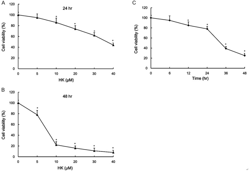
Human gastric carcinoma MGC-803 cell viability was measured when cells were exposed to various concentrations of honokiol (0-40 μM) for 24 (Figure 1A) and 48 h (Figure 1B). MGC-803 cell were growth inhibited with honokiol. As shown in Figure 1A, the concentrations at which honokiol inhibited MGC-803 cell growth by 50% (IC50) was 30 μM for 24 h. The IC50 was 7.5 μM when the cells were exposed to honokiol for 48 h (Figure 1B). Treatment of gastric carcinoma cells with honokiol induced cell growth inhibition in a dose-dependent manner by using CCK8 assay. To evaluate the time-dependent effect of honokiol on the cell viability, the MGC-803 cells were exposed to 10 μM honokiol for various times. As shown in Figure 1C, the cell viability was significantly decreased with increasing durations.
Figure 1.

Effect of honokiol on the cell viability. The cell viability was examined by CCK8 assay when the human gastric carcinoma MGC-803 cells were incubated with various concentrations of honokiol (0-40 μM) for 24 h (A) and 48 h (B). Human neuroglioma cells were incubated with honokiol (10 μM) for 0, 6, 12, 24, 36 and 48 h, and the cell viability was examined by CCK8 assay (C). Values are expressed as mean ± SEM, n = 3 in each group. *P < 0.05 versus control group.
Effects of honokiol on cell apoptosis and cell cycle arrest
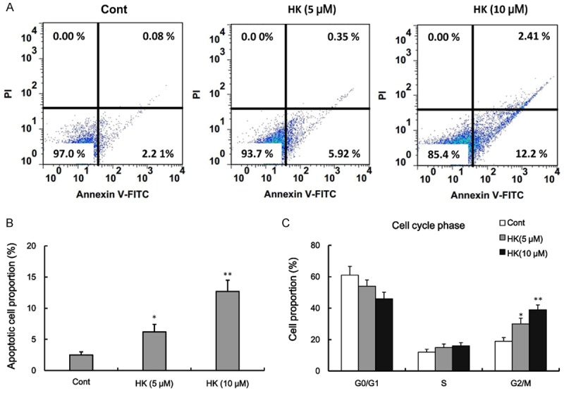
We next investigated whether honokiol induced cell death through an apoptotic mechanism. Annexin V-PI double-labeling was used for the detection of PS externalization, a hallmark of early phase of apoptosis. Consistent with the CCK8 assay, the results showed that growth inhibition was accompanied with an increase in apoptotic cells, as determined by flow cytometry (Figure 2A and 2B). The proportion of apoptosis cells had gained after honokiol treatment as compared with control group (Figure 2A and 2B). To gain insights into the mechanism of the antiproliferative activity of honokiol, its effect on cell-cycle distribution was determined via a flow cytometry assay. As shown in Figure 2C, human gastric carcinoma cells were exposed to honokiol for 48 h, which resulted in an accumulation of cells in G2/Mphase. These results suggested that the effects of honokiol suppressed human gastric carcinoma cell proliferation, at least in part, through delay in the G2/M transition.
Figure 2.

Effect of honokiol on cell apoptosis and cell cycle arrest. Human gastric carcinoma cells were treated with vehicle or honokol (5 or 10 μM) for 48 h, the percentage of apoptotic cells was also analyzed by flow cytometric analysis of annexin V/PI double staining (A) and bar graphs represent the percentage of apoptotic cells (B). The percentage of cell cycle phase was analyzed by flow cytometry analysis after cells exposure to honokiol for 48 h (C). Values are expressed as mean ± SEM, n = 3 in each group. *P < 0.05, **P < 0.01 versus control group.
Effect of honokiol on the cell cycle regulated protein
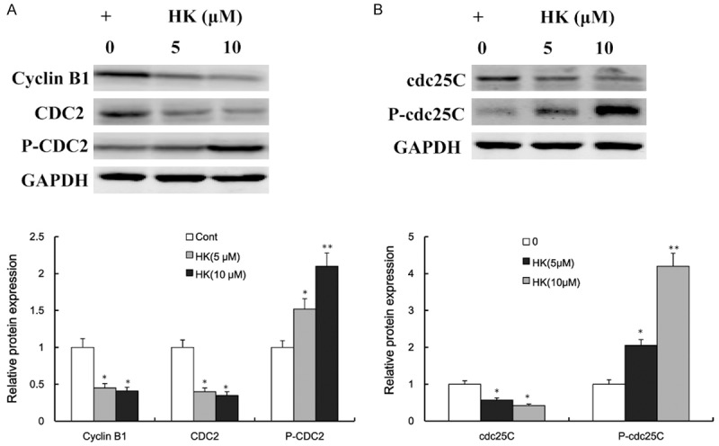
To evaluate the potential molecular mechanism by which honokiol causes a G2/M arrest, we analyzed the steady-state levels of proteins involved in the G2/M checkpoint. The results found that Cyclin B1, CDC2 and cdc25C were downregulated upon honokiol treatment in human gastric carcinoma cells (Figure 3A and 3B). However, we found that the expression of p-CDC2 and p-cdc25c was significantly upregulated when the gastric carcinoma cells were exposed to honokiol (Figure 3A and 3B).
Figure 3.

Effects of honokiol on G2/M checkpoint proteins. Human gastric carcinoma cells were treated with vehicle or honokol (5 or 10 μM) for 48 h, and the expression levels of Cyclin B1, CDC2 and p-CDC2 were determined by western blotting and densitometric analyses (A). The expression levels of cdc25C and p-cdc25C were determined by western blotting and densitometric analyses (B). Values are expressed as mean ± SEM, n = 3 in each group. *P < 0.05, **P < 0.01 versus control group.
Effect of honokiol on p53, p21, BAX and Bcl-2
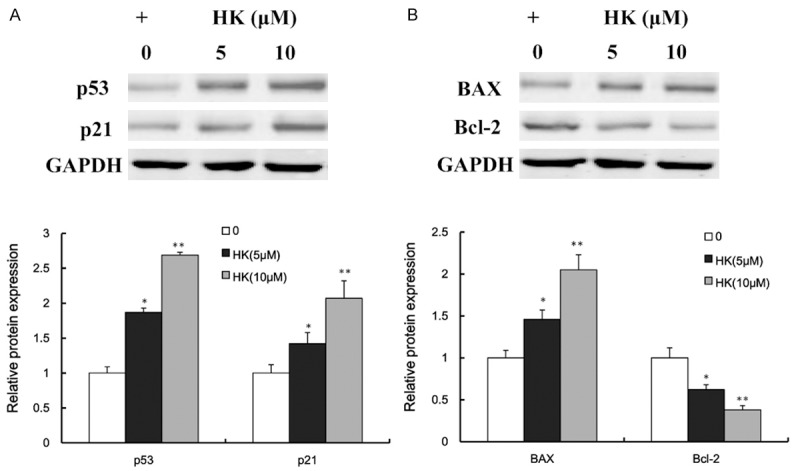
Significant changes in the protein levels of tumor suppressors were observed in human gastric carcinoma cells with honokiol-treated. As shown in Figure 4A, p53 and p21 were significantly upregulated by honokiol treatment. Moreover, the apoptotic response was further investigated by measuring apoptosis-related proteins expression. Treatment of MGC-803 cells with honokiol significantly increased the pro-apoptotic Bax level and decreased the anti-apoptotic Bcl-2 level (Figure 4B). These results indicated that honokiol might induce cell death through activation tumor suppressors signaling pathway.
Figure 4.

Effects of honokiol on tumor suppressors and apoptosis-related proteins. Human gastric carcinoma cells were treated with vehicle or honokol (5 or 10 μM) for 48 h, and the expression levels of p53 and p21 were determined by western blotting and densitometric analyses (A). The expression levels of BAX and Bcl-2 were determined by western blotting and densitometric analyses (B). Values are expressed as mean ± SEM, n = 3 in each group. *P < 0.05, **P < 0.01 versus control group.
Discussion
In this study, we investigated the anti-cancer mechanism of honokiol in human gastric carcinoma MGC-803 cells. We demonstrated that there might be correlated between p53 signaling with cell apoptosis progression. Honokiol could induce gastric carcinoma cells apoptosis, and the underlying mechanism was mediated, at least partially, through down-regulation of CDC2/cdc25C and up-regulation of p53. There are mostly sparse reports of the anticancer activity of honokiol on human tumor, especially on human gastric carcinoma. Previous studies have shown that honokiol can suppress tumor growth, such as human glioblastoma [5], colorectal cancer [6], hepatocellular carcinoma [24] and breast cancer [25]. However, honokiol derivatives show no significant cytotoxic effects in any of the three tested cell lines, CCRF-CEM leukemia cells, U251 glioblastoma and HCT-116 colon cancer cells, at a test concentration of 10 μM [26]. These studies indicate that more research is needed to understand the cytotoxicity and the tumor inhibition of honokiol or honokiol derivatives.
In the present study, we undertook a comprehensive and integrative approach to explore the cytotoxicity and the tumor inhibition of honokiol in human gastric carcinoma MGC-803 cells. We demonstrated that honokiol induced cell death in a dose-and time-dependent manner, and the underlying molecular mechanisms might be correlated with cell cycle arrest. The anti-cancer mechanisms of honokiol in this study were similar to the other tumor cells model [6,24,27]. Honokiol influenced cell-cycle progression, induced G2/M arrest and apoptosis accompanied with p53 signaling activation. It is known that cell cycle dysregulation is a hallmark of tumor cells. Regulation of proteins that mediate critical events of the cell cycle may be a useful antitumor target [17]. CDC2 is the cyclin-dependent kinase responsible for the entry and exit from G2 and mitosis. CDC2 interacts with cyclin B1, and activation of the CDC2/cyclin B1 complex is required for the transition from G2 to M phase of the cell cycle [28]. Cdc25C, a protein tyrosine phosphorylation in cell cycle control, allows progression to mitosis when the CDC2/cyclin B1 complexes are formed [29]. Our results indicated that honokiol induced cell cycle arrest through suppression the expression of CDC2, cyclin B1 and cdc25C. Moreover, honokiol induces cell cycle arrest and apoptosis in acute myeloid leukemia and human malignant pleural mesothelioma cells [30,31]. Intriguingly, honokiol induces G1 cell cycle arrest by reducing the expression of cyclins and CDK, and honokio-induced apoptosis is associated with activation of caspase3 and caspase9 in adult T-cell leukemia [32].
On the other hand, the tumor suppressor gene p53 plays a critical role in the regulation of cell cycle along with the induction of apoptosis and regulates the inhibition of cell growth [33]. In response to honokiol, p53 accumulates due to posttranslational modification, resulting in cell cycle arrest and apoptosis [22]. In human colorectal cell line RKO, honokiol induces apoptosis through p53-independent pathway targeting to activate caspase cascade [22]. Interestingly, honokiol decreased anti-apoptotic survivin protein and gene expression and increased total p53 and the phosphorylated p53 proteins at Ser15 and Ser46 [6]. Together, these studies indicate that the existence of survivin and p53 can modulate the honokiol-induced apoptosis. In our study, human gastric carcinoma cell exposure to honokiol could upregulate the expression of p53 and p21. In an orthotopic model, honokiol suppresses gastric tumor growth and peritoneal dissemination [34], and the same results are detected in nu/nu mice [35].
In conclusion, honokiol could induce apoptosis and cell cycle arrest in human gastric carcinoma MGC-803 cell line, the underlying molecular mechanisms, at least partially, through activation p53 signaling and downregulation CDC2/cdc25C expression. In view of the results of this experiment, it seemed reasonable to highlight the possibility of honokiol in the clinical treatment of gastric carcinoma.
Disclosure of conflict of interest
None.
References
- 1.Lee YJ, Lee YM, Lee CK, Jung JK, Han SB, Hong JT. Therapeutic applications of compounds in the Magnolia family. Pharmacol Ther. 2011;130:157–176. doi: 10.1016/j.pharmthera.2011.01.010. [DOI] [PubMed] [Google Scholar]
- 2.Liou KT, Shen YC, Chen CF, Tsao CM, Tsai SK. Honokiol protects rat brain from focal cerebral ischemia-reperfusion injury by inhibiting neutrophil infiltration and reactive oxygen species production. Brain Res. 2003;992:159–166. doi: 10.1016/j.brainres.2003.08.026. [DOI] [PubMed] [Google Scholar]
- 3.Kim KR, Park KK, Chun KS, Chung WY. Honokiol inhibits the progression of collagen-induced arthritis by reducing levels of pro-inflammatory cytokines and matrix metalloproteinases and blocking oxidative tissue damage. J Pharmacol Sci. 2010;114:69–78. doi: 10.1254/jphs.10070fp. [DOI] [PubMed] [Google Scholar]
- 4.Kim YJ, Choi MS, Cha BY, Woo JT, Park YB, Kim SR, Jung UJ. Long-term supplementation of honokiol and magnolol ameliorates body fat accumulation, insulin resistance, and adipose inflammation in high-fat fed mice. Mol Nutr Food Res. 2013;57:1988–1998. doi: 10.1002/mnfr.201300113. [DOI] [PubMed] [Google Scholar]
- 5.Liang WZ, Chou CT, Chang HT, Cheng JS, Kuo DH, Ko KC, Chiang NN, Wu RF, Shieh P, Jan CR. The mechanism of honokiol-induced intracellular Ca(2+) rises and apoptosis in human glioblastoma cells. Chem Biol Interact. 2014;221:13–23. doi: 10.1016/j.cbi.2014.07.012. [DOI] [PubMed] [Google Scholar]
- 6.Lai YJ, Lin CI, Wang CL, Chao JI. Expression of survivin and p53 modulates honokiol-induced apoptosis in colorectal cancer cells. J Cell Biochem. 2014;115:1888–1899. doi: 10.1002/jcb.24858. [DOI] [PubMed] [Google Scholar]
- 7.Bai X, Cerimele F, Ushio-Fukai M, Waqas M, Campbell PM, Govindarajan B, Der CJ, Battle T, Frank DA, Ye K, Murad E, Dubiel W, Soff G, Arbiser JL. Honokiol, a small molecular weight natural product, inhibits angiogenesis in vitro and tumor growth in vivo. J Biol Chem. 2003;278:35501–35507. doi: 10.1074/jbc.M302967200. [DOI] [PubMed] [Google Scholar]
- 8.Chen YJ, Wu CL, Liu JF, Fong YC, Hsu SF, Li TM, Su YC, Liu SH, Tang CH. Honokiol induces cell apoptosis in human chondrosarcoma cells through mitochondrial dysfunction and endoplasmic reticulum stress. Cancer Lett. 2010;291:20–30. doi: 10.1016/j.canlet.2009.08.032. [DOI] [PubMed] [Google Scholar]
- 9.Vavilala DT, Ponnaluri VK, Kanjilal D, Mukherji M. Evaluation of anti-HIF and anti-angiogenic properties of honokiol for the treatment of ocular neovascular diseases. PLoS One. 2014;9:e113717. doi: 10.1371/journal.pone.0113717. [DOI] [PMC free article] [PubMed] [Google Scholar]
- 10.Banerjee P, Basu A, Arbiser JL, Pal S. The natural product honokiol inhibits calcineurin inhibitor-induced and Ras-mediated tumor promoting pathways. Cancer Lett. 2013;338:292–299. doi: 10.1016/j.canlet.2013.05.036. [DOI] [PMC free article] [PubMed] [Google Scholar]
- 11.Lin JM, Prakasha Gowda AS, Sharma AK, Amin S. In vitro growth inhibition of human cancer cells by novel honokiol analogs. Bioorg Med Chem. 2012;20:3202–3211. doi: 10.1016/j.bmc.2012.03.062. [DOI] [PMC free article] [PubMed] [Google Scholar]
- 12.Wang X, Beitler JJ, Wang H, Lee MJ, Huang W, Koenig L, Nannapaneni S, Amin AR, Bonner M, Shin HJ, Chen ZG, Arbiser JL, Shin DM. Honokiol enhances paclitaxel efficacy in multi-drug resistant human cancer model through the induction of apoptosis. PLoS One. 2014;9:e86369. doi: 10.1371/journal.pone.0086369. [DOI] [PMC free article] [PubMed] [Google Scholar]
- 13.Zhang MW, Xu XJ, Fan JX, Hung YX, Ye YB, Wang J, Guo KY. [Honokiol combined with Gemcitabine synergistically inhibits the proliferation of human Burkitt lymphoma cells and induces their apoptosis] . Zhongguo Shi Yan Xue Ye Xue Za Zhi. 2014;22:93–98. doi: 10.7534/j.issn.1009-2137.2014.01.019. [DOI] [PubMed] [Google Scholar]
- 14.Lee SM, Kwon JI, Choi YH, Eom HS, Chi GY. Induction of G2/M arrest and apoptosis by water extract of Strychni Semen in human gastric carcinoma AGS cells. Phytother Res. 2008;22:752–758. doi: 10.1002/ptr.2355. [DOI] [PubMed] [Google Scholar]
- 15.Yunlan L, Juan Z, Qingshan L. Antitumor activity of di-n-butyl-(2,6-difluorobenzohydroxamato) tin (IV) against human gastric carcinoma SGC-7901 cells via G2/M cell cycle arrest and cell apoptosis. PLoS One. 2014;9:e90793. doi: 10.1371/journal.pone.0090793. [DOI] [PMC free article] [PubMed] [Google Scholar] [Retracted]
- 16.Li Y, Wang X, Cheng S, Du J, Deng Z, Zhang Y, Liu Q, Gao J, Cheng B, Ling C. Diosgenin induces G2/M cell cycle arrest and apoptosis in human hepatocellular carcinoma cells. Oncol Rep. 2015;33:693–698. doi: 10.3892/or.2014.3629. [DOI] [PubMed] [Google Scholar]
- 17.Su CC. Tanshinone IIA inhibits gastric carcinoma AGS cells through increasing p-p38, p-JNK and p53 but reducing p-ERK, CDC2 and cyclin B1 expression. Anticancer Res. 2014;34:7097–7110. [PubMed] [Google Scholar]
- 18.Zhou B, Bu G, Zhou Y, Zhao Y, Li W, Li M. Knockdown of CDC2 expression inhibits proliferation, enhances apoptosis, and increases chemosensitivity to temozolomide in glioblastoma cells. Med Oncol. 2015;32:378. doi: 10.1007/s12032-014-0378-9. [DOI] [PubMed] [Google Scholar]
- 19.Yang Y, Hu Y, Gu HY, Lu N, Liu W, Qi Q, Zhao L, Wang XT, You QD, Guo QL. Oroxylin A induces G2/M phase cell-cycle arrest via inhibiting Cdk7-mediated expression of Cdc2/p34 in human gastric carcinoma BGC-823 cells. J Pharm Pharmacol. 2008;60:1459–1463. doi: 10.1211/jpp/60.11.0006. [DOI] [PubMed] [Google Scholar]
- 20.Yu J, Guo QL, You QD, Zhao L, Gu HY, Yang Y, Zhang HW, Tan Z, Wang X. Gambogic acid-induced G2/M phase cell-cycle arrest via disturbing CDK7-mediated phosphorylation of CDC2/p34 in human gastric carcinoma BGC-823 cells. Carcinogenesis. 2007;28:632–638. doi: 10.1093/carcin/bgl168. [DOI] [PubMed] [Google Scholar]
- 21.Su CC. Tanshinone IIA inhibits human gastric carcinoma AGS cell growth by decreasing BiP, TCTP, Mcl1 and BclxL and increasing Bax and CHOP protein expression. Int J Mol Med. 2014;34:1661–1668. doi: 10.3892/ijmm.2014.1949. [DOI] [PubMed] [Google Scholar]
- 22.Wang T, Chen F, Chen Z, Wu YF, Xu XL, Zheng S, Hu X. Honokiol induces apoptosis through p53-independent pathway in human colorectal cell line RKO. World J Gastroenterol. 2004;10:2205–2208. doi: 10.3748/wjg.v10.i15.2205. [DOI] [PMC free article] [PubMed] [Google Scholar]
- 23.Cao AL, Tang QF, Zhou WC, Qiu YY, Hu SJ, Yin PH. Ras/ERK signaling pathway is involved in curcumin-induced cell cycle arrest and apoptosis in human gastric carcinoma AGS cells. J Asian Nat Prod Res. 2015;17:56–63. doi: 10.1080/10286020.2014.951923. [DOI] [PubMed] [Google Scholar]
- 24.Han LL, Xie LP, Li LH, Zhang XW, Zhang RQ, Wang HZ. Reactive oxygen species production and Bax/Bcl-2 regulation in honokiol-induced apoptosis in human hepatocellular carcinoma SMMC-7721 cells. Environ Toxicol Pharmacol. 2009;28:97–103. doi: 10.1016/j.etap.2009.03.005. [DOI] [PubMed] [Google Scholar]
- 25.Alonso-Castro AJ, Dominguez F, Garcia-Regalado A, Gonzalez-Sanchez I, Cerbon MA, Garcia-Carranca A. Magnolia dealbata seeds extract exert cytotoxic and chemopreventive effects on MDA-MB231 breast cancer cells. Pharm Biol. 2014;52:621–627. doi: 10.3109/13880209.2013.859160. [DOI] [PubMed] [Google Scholar]
- 26.Bernaskova M, Kretschmer N, Schuehly W, Huefner A, Weis R, Bauer R. Synthesis of tetrahydrohonokiol derivates and their evaluation for cytotoxic activity against CCRF-CEM leukemia, U251 glioblastoma and HCT-116 colon cancer cells. Molecules. 2014;19:1223–1237. doi: 10.3390/molecules19011223. [DOI] [PMC free article] [PubMed] [Google Scholar]
- 27.Arora S, Bhardwaj A, Srivastava SK, Singh S, McClellan S, Wang B, Singh AP. Honokiol arrests cell cycle, induces apoptosis, and potentiates the cytotoxic effect of gemcitabine in human pancreatic cancer cells. PLoS One. 2011;6:e21573. doi: 10.1371/journal.pone.0021573. [DOI] [PMC free article] [PubMed] [Google Scholar]
- 28.Lew DJ, Kornbluth S. Regulatory roles of cyclin dependent kinase phosphorylation in cell cycle control. Curr Opin Cell Biol. 1996;8:795–804. doi: 10.1016/s0955-0674(96)80080-9. [DOI] [PubMed] [Google Scholar]
- 29.Taylor WR, Stark GR. Regulation of the G2/M transition by p53. Oncogene. 2001;20:1803–1815. doi: 10.1038/sj.onc.1204252. [DOI] [PubMed] [Google Scholar]
- 30.Li HY, Ye HG, Chen CQ, Yin LH, Wu JB, He LC, Gao SM. Honokiol induces cell cycle arrest and apoptosis via inhibiting class I histone deacetylases in acute myeloid leukemia. J Cell Biochem. 2015;116:287–298. doi: 10.1002/jcb.24967. [DOI] [PubMed] [Google Scholar]
- 31.Chae JI, Jeon YJ, Shim JH. Downregulation of Sp1 is involved in honokiol-induced cell cycle arrest and apoptosis in human malignant pleural mesothelioma cells. Oncol Rep. 2013;29:2318–2324. doi: 10.3892/or.2013.2353. [DOI] [PubMed] [Google Scholar]
- 32.Ishikawa C, Arbiser JL, Mori N. Honokiol induces cell cycle arrest and apoptosis via inhibition of survival signals in adult T-cell leukemia. Biochim Biophys Acta. 2012;1820:879–887. doi: 10.1016/j.bbagen.2012.03.009. [DOI] [PubMed] [Google Scholar]
- 33.Liebermann DA, Hoffman B, Steinman RA. Molecular controls of growth arrest and apoptosis: p53-dependent and independent pathways. Oncogene. 1995;11:199–210. [PubMed] [Google Scholar]
- 34.Pan HC, Lai DW, Lan KH, Shen CC, Wu SM, Chiu CS, Wang KB, Sheu ML. Honokiol thwarts gastric tumor growth and peritoneal dissemination by inhibiting Tpl2 in an orthotopic model. Carcinogenesis. 2013;34:2568–2579. doi: 10.1093/carcin/bgt243. [DOI] [PubMed] [Google Scholar]
- 35.Liu SH, Wang KB, Lan KH, Lee WJ, Pan HC, Wu SM, Peng YC, Chen YC, Shen CC, Cheng HC, Liao KK, Sheu ML. Calpain/SHP-1 interaction by honokiol dampening peritoneal dissemination of gastric cancer in nu/nu mice. PLoS One. 2012;7:e43711. doi: 10.1371/journal.pone.0043711. [DOI] [PMC free article] [PubMed] [Google Scholar]


