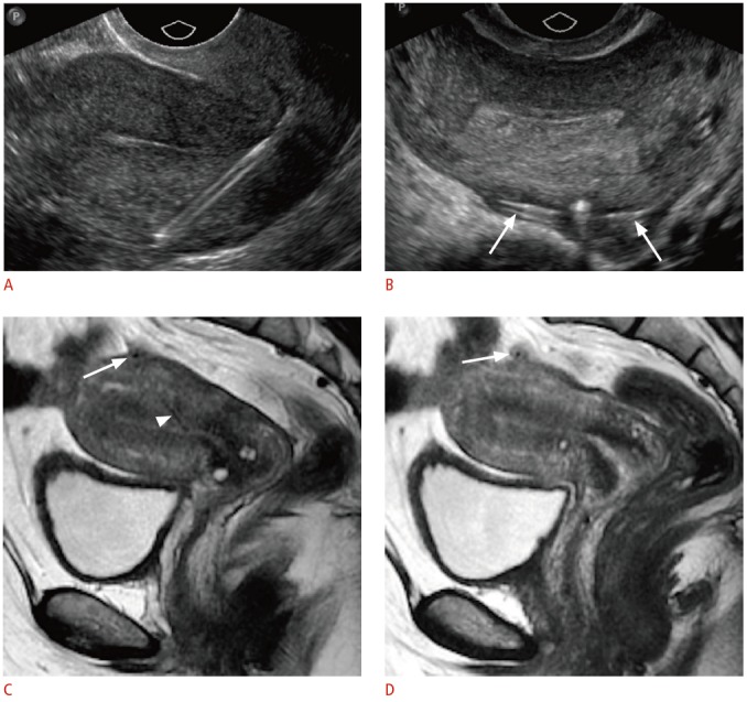
Fig. 11. Partially perforated intrauterine device (IUD) in a 43-year-old female with shortened retrieval strings.

A. Sagittal transvaginal sonogram shows the stem extending through the myometrium of the posterior wall. B. Transverse transvaginal sonogram shows the arms extending outside the serosa (arrows). C, D. Midline (C) and left lateral (D) sagittal magnetic resonance images demonstrate correlated findings of the low-signal IUD coursing through the posterior myometrium (arrowhead) with the arms extending through the serosa (arrows).
