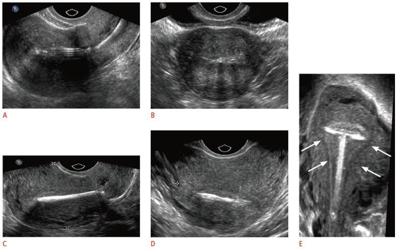
Fig. 3. Transvaginal ultrasonographic appearance of T-shaped intrauterine devices (IUDs).

A, B. Two-dimensional (2D) sagittal (A) and transverse (B) sonograms show hyperechoic levonorgestrel-releasing IUD in the endometrial cavity. C, D. 2D sagittal (C) and transverse (D) sonograms show the bright echo of the copper IUD with marked posterior shadowing. E. Three-dimensional coronal reformatted sonogram demonstrates the properly positioned copper IUD within the endometrial cavity (arrows).
