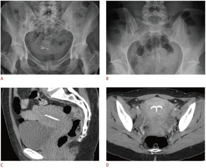
Fig. 4. Radiographic and computed tomographic (CT) appearance of the T-shaped intrauterine device (IUD).

A, B. For the copper IUD in a retroverted uterus (A) and levonorgestrel-releasing IUD in an anteverted uterus (B), pelvic radiographs alone are inadequate for precise localization in relation to the uterine cavity due to normal variations in uterine position. C, D. Coronal (C) and sagittal (D) noncontrast-enhanced CT images demonstrate a radiodense IUD properly positioned within the uterine fundus.
