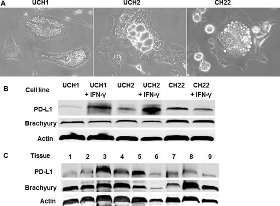
Figure 1. PD-L1 expression in chordoma.

(A) Cell morphology and growth characteristics of chordoma cell lines. UCH1, UCH2, and CH22 cells exhibited round nuclei with clear vacuolated cytoplasm. (B) PD-L1 expression in chordoma cell lines. PD-L1 protein expressions were induced 16-fold and 4-fold by IFN-γ in UCH1 and UCH2 cell lines, respectively. (C) PD-L1 expression in chordoma tissues. Relative expressions of PD-L1 were present in 9 chordoma specimens. PD-L1 expression was evaluated from total protein by western blot and absolute expression of PD-L1 was normalized to β-actin. Three of nine samples were found to have high expression.
