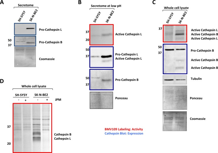
Figure 3. Cathepsin expression and activity of secretomes and cells.
(A) Western blot analysis for cathepsin abundance in the neuroblastoma secretome samples. Higher abundance of pro-cathepsins B and L is detected in the secretome of SK-N-BE2 compared to SH-SY5Y (B) BMV109 probe based cathepsin activity analysis in secretomes. Under low pH conditions (5.5), the active form of cathepsin L is present in higher abundance in the secretome of SK-N-BE2 compared to SH-SY5Y. (C) Probe based cathepsin activity and expression analysis in whole cell lysates. SK-N-BE2 whole cell lysates exhibit higher levels of active cathepsins B and L as compared to SH-SY5Y whole cell lysates. (D) Cathepsin activity in the whole cell lysates in the presence and absence of cathepsin inhibitor JPM. JPM treatment leads to ablation of cathepsin activity in SK-N-BE2 cells.

