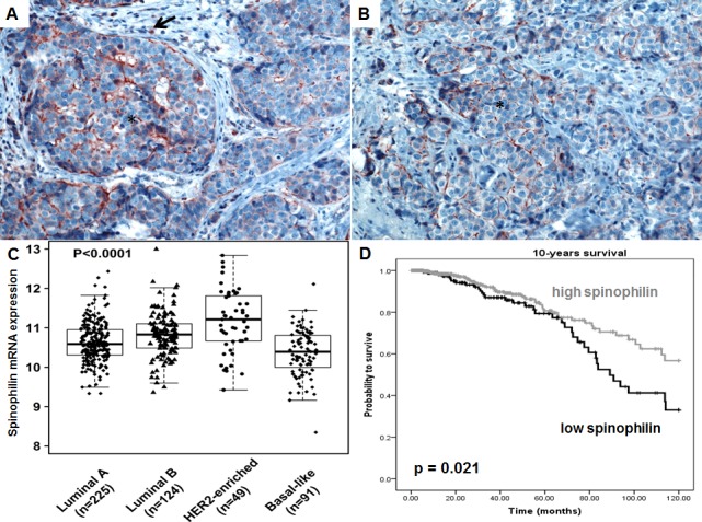
Figure 1. Spinophilin expression in breast cancer tissue and different molecular subtypes.

(A-B) A strong, membranous staining pattern could be observed in breast cancer cells (*) in tissue slides of breast cancer patients. Surrounding inflammatory cells are also positively stained (arrow). (C) Analysis of 489 breast cancer patients of the TCGA data set indicates that basal-like breast cancer subtype exhibit the lowest spinophilin expression. (D) In 921 breast cancer patients, a low spinophilin level is significantly associated with poor survival.
