Abstract
Patient: Female, 31
Final Diagnosis: Hyperinfection syndrome due to Strongyloides stercoralis
Symptoms: Abdominal pain • shortness of breath
Medication: Prednisone
Clinical Procedure: Bronchoscopy with BAL
Specialty: Pulmonology
Objective:
Unusual clinical course
Background:
Strongyloides stercoralis (SS) is a parasite seen in certain parts of the USA and in people from other endemic areas. In these patients steroids might precipitate or exacerbate asthma. Apart from worsening of asthma, serious complications like hyperinfection syndrome and even death can occur in these patients if treated with steroids. Treatment is either ivermectin or albendazole based on severity of the disease. Clinicians have to be very careful when prescribing steroids in patients presenting with an exacerbation of asthma from areas endemic for Strongyloides stercoralis.
Case Report:
A young woman with history of asthma presented with complaints of nausea, vomiting, abdominal pain, wheezing, and dry cough. Physical examination revealed diffuse expiratory wheezing and mild diffuse abdominal pain without rebound or guarding. Laboratory results showed leukocytosis with eosinophilia. Stool studies showed Strongyloides stercoralis. Imaging revealed ground-glass opacities in the right upper and lower lobe along with an infiltrate in the lingular lobe on the left side. Bronchoscopy showed Strongyloides stercoralis. The patient was diagnosed with hyperinfection syndrome due to Strongyloides stercoralis most probably exacerbated by prednisone given for her asthma. Steroids were then discontinued and the patient was started on ivermectin. The patient improved with treatment. Repeat stool examination was negative for Strongyloides stercoralis.
Conclusions:
Clinicians have to be very careful when prescribing steroids in patients presenting with an exacerbation of asthma who are from areas endemic for Strongyloides stercoralis and should test for it (preferably with serology test) before starting treatment.
MeSH Keywords: Asthma, Prednisone, Strongyloides
Background
Corticosteroids are the cornerstone of management for asthma, and usually the response to treatment is excellent. However, steroids might precipitate or exacerbate asthma, which could be life-threatening, in patients infected with a parasite – Strongyloides stercoralis (SS) seen in certain parts of the USA and in people from other endemic areas. Most patients with SS infection of are asymptomatic with eosinophilia, thus a good history and high index of suspicion are needed to help prevent this catastrophic complication. We report a case in which timely diagnosis may have saved a young woman from a poor outcome.
Case Report
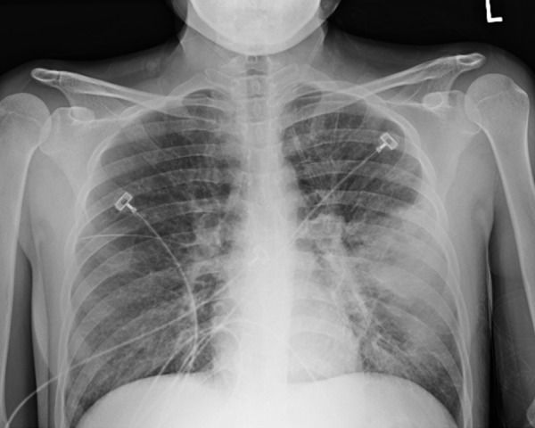
A 31-year-old Hispanic woman with history of asthma, who emigrated from Mexico at age 14, presented with complaints of nausea, vomiting, abdominal pain, wheezing, and dry cough. She had recently been treated for asthma exacerbation with prednisone and was still taking it. She denied any occupational exposure, pets, smoking, and recent travel. Vital signs were: temperature 101°F, pulse 106/min, blood pressure 112/72 mmHg, and respiratory rate 22/min. Systemic examination revealed diffuse expiratory wheezing and mild diffuse abdominal pain without rebound or guarding. Laboratory results showed leukocytosis (11 900/mm3) with eosinophilia (32.2%), negative drug screen, and negative rapid HIV test. Stool studies showed SS. Chest X-ray showed opacity in the left lung (Figure 1). CT chest revealed ground-glass opacities in the right upper and lower lobe along with an infiltrate in the lingular lobe on the left side (Figure 2). Blood cultures were positive for Escherichia coli. The patient was diagnosed with hyperinfection syndrome due to SS, most probably exacerbated by prednisone given for her asthma. Steroids were then discontinued, and the patient was started on ivermectin, broad-spectrum antibiotics, and intensive bronchodilator therapy. Human T-cell lymphotropic virus (HTLV) I/II quantitative Ab were negative. Bronchoscopy with bronchoalveolar lavage (BAL) was done to rule out other infectious causes of the lung infiltrate. The cytological examination of BAL showed larvae of SS (Figure 3A, 3B). The patient was started on ivermectin along with antibiotics for her E. coli bacteremia. Ivermectin was given until her repeat stool examination was negative for SS for 2 weeks (she received it for a total of 17 days). She improved with treatment.
Figure 1.

Chest X-ray showing left lung opacity.
Figure 2.

CT chest showing left lingular lobe infiltrate.
Figure 3.

(A, B) BAL showing larvae of Strongyloides stercoralis.
Discussion
Strongyloidiasis is an infection caused by SS. This disease can be seen in the southeastern United States and in immigrants from endemic areas. Patients may not experience prominent symptoms but when they do they most commonly involve the gastrointestinal, cutaneous, and /or pulmonary systems.
Pulmonary symptoms may include dry cough, shortness of breath, wheezing, and hemoptysis. Features suggestive of recurrent bacterial pneumonia are common in these patients. They may also develop asthma, which paradoxically worsens with corticosteroid use [1,2]. Apart from worsening of asthma, serious complications and death can occur in these patients treated with steroids [3,4]. Gram-negative sepsis is also commonly seen in patients with SS. Hyperinfection syndrome is a severe form of SS, occurring most probably due to repeated autoinfection leading to increased parasite burden and sometimes precipitated by steroid therapy [4]. It has a mortality rate of from 10% to over 80%.
Diagnosis is based on active investigation of the parasite by identification of the larvae in the stool, and a high index of suspicion should be maintained, especially in immunocompromised patients. Standard stool examination is very insensitive for detecting SS. Three specialized tests are recommended: Baermann concentration technique, the Harada-Mori filter paper technique, and a modified agar plate method [6,7]. The most sensitive test (>80%) for diagnosis of strongyloides is enzyme-linked immunosorbent assay (ELISA), which detects immunoglobin G (IgG) to filariform larvae [8].
Treatment of limited disease is with ivermectin or albendazole. Disseminated disease or hyperinfection syndrome may require discontinuation of immunosuppressive therapy and prolonged use of ivermectin alone or in combination with albendazole until the patient shows clinical improvement and has negative stool studies for at least 2 weeks (1 autoinfection cycle) [9,10]. Data on treatment methods are limited to case reports or case series. Use of albendazole alone may have difficulty in clearing the infection and its efficacy has been shown to be lower than that of ivermectin [11].
Conclusions
Due to increased risk of developing disseminated disease or hyperinfection syndrome, clinicians have to be very careful when prescribing steroids in patients presenting with an exacerbation of asthma from areas endemic for SS. We suggest that clinician should test for SS (preferably with serology test) before starting treatment.
References:
- 1.Sen P, Gil C, Estrellas B, et al. Corticosteroid-induced asthma: a manifestation of limited hyperinfection syndrome due to Strongyloides stercoralis. South Med J. 1995;88(9):923–27. [PubMed] [Google Scholar]
- 2.Wehner JH, Kirsch CM, Kagawa FT, et al. The prevalence and response to therapy of Strongyloides stercoralis in patients with asthma from endemic areas. Chest. 1994;106(3):762–66. doi: 10.1378/chest.106.3.762. [DOI] [PubMed] [Google Scholar]
- 3.Newberry AM, Williams DN, Stauffer WM, et al. Strongyloides hyperinfection presenting as acute respiratory failure and gram-negative sepsis. Chest. 2005;128(5):3681–84. doi: 10.1378/chest.128.5.3681. [DOI] [PMC free article] [PubMed] [Google Scholar]
- 4.Rodriguez EA, Abraham T, Williams FK. Severe strongyloidiasis with negative serology after corticosteroids treatment. Am J Case Rep. 2015;16:95–98. doi: 10.12659/AJCR.892759. [DOI] [PMC free article] [PubMed] [Google Scholar]
- 5.Chu E, Whitlock WL, Dietrich RA. Pulmonary hyperinfection syndrome with Strongyloides stercoralis. Chest. 1990;97(6):1475–77. doi: 10.1378/chest.97.6.1475. [DOI] [PubMed] [Google Scholar]
- 6.Rosenblatt JE. Clinical importance of adequately performed stool ova and parasite examinations. Clin Infect Dis. 2006;42:979–80. doi: 10.1086/500943. [DOI] [PubMed] [Google Scholar]
- 7.Hirata T, Nakamura H, Kinjo N, et al. Increased detection rate of Strongyloides stercoralis by repeated stool examinations using the agar plate culture method. Am J Trop Med Hyg. 2007;77:683–84. [PubMed] [Google Scholar]
- 8.Caroll SM, Karthigasu KT, Grove DI. Serodiagnosis of human strongyloidosis by an enzyme- linked immunosorbent assay. Trans R Soc Trop Med Hyg. 1981;75:706–9. doi: 10.1016/0035-9203(81)90156-5. [DOI] [PubMed] [Google Scholar]
- 9.Segarra-Newnham M. Manifestations, diagnosis, and treatment of Strongyloides stercoralis infection. Ann Pharmacother. 2007;41(12):1992–2001. doi: 10.1345/aph.1K302. [DOI] [PubMed] [Google Scholar]
- 10.Pornsuriyasak P, Niticharoenpong K, Sakaibunnan A. Disseminated strongyloidiasis successfully treated with extended duration ivermectin combined with albendazole: a case report of intractable strongyloidiasis. Southeast Asian J Trop Med Public Health. 2004;35:531–34. [PubMed] [Google Scholar]
- 11.Muennig O, Pallin D, Challah C, et al. The cost effectiveness of ivermectin vs. albendazole in the presumptive treatment of strongyloidiasis in immigrants to the United States. Epidemiol Infect. 2004;132:1055–63. doi: 10.1017/s0950268804003000. [DOI] [PMC free article] [PubMed] [Google Scholar]


