
Fig. 6.
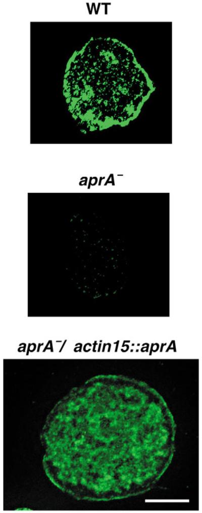
The subcellular distribution of AprA. Cells from preparations similar to those shown in Fig. 3B were imaged with a Zeiss Axioplan II deconvolution microscope. Scale bar: 5 μm.
Official websites use .gov
A
.gov website belongs to an official
government organization in the United States.
Secure .gov websites use HTTPS
A lock (
) or https:// means you've safely
connected to the .gov website. Share sensitive
information only on official, secure websites.
The subcellular distribution of AprA. Cells from preparations similar to those shown in Fig. 3B were imaged with a Zeiss Axioplan II deconvolution microscope. Scale bar: 5 μm.