

Highlights
-
•
Ectopic splenic tissue can be either congenital (accessory spleen) or acquired (splenosis).
-
•
Accessory spleen (AS) is commonly located near the spleen’s hilum and in the pancreas tail.
-
•
Pelvic AS is a very rare entity.
-
•
AS is generally determined incidentally during radiological investigation or surgery.
-
•
Pelvic AS may be considered in differential diagnosis of adnexal masses.
Keywords: Adnexal mass, Accessory spleen, Histopathology
Abstract
Introduction
Accessory Spleen (AS) is a very rare entity and usually near the spleen’s hilum and in the tail of the pancreas. Pelvis reported as an atypical and a rare localization. AS may be formed during embryonic life, they rise from the left side of the dorsal mesogastrium as a result of imperfect fusion of separate splenic masses.
Presentation of case
We report a case of an AS presenting as an left adnexal mass in a middle-aged woman. Transvaginal ultrasonography and magnetic resonance imaging (MRI) revealed a left adnexial mass. Laparatomy was performed, and histological examination revealed that resected mass was splenic tissue.
Discussion
An AS is an incidental finding of no clinical significance in most patients. AS are generally determined during radiological investigations or during open or laparoscopic surgeries. When, the AS settle in the adnexal area; the differential diagnosis could include the causes of adnexal masses like enlarged lymph nodes, subserous fibroid, ovarian tumors, organized hematoma, tuboovarian abscess.
Conclusion
Althought pelvic accessory spleen is a rare condition, should be considered in the differential diagnosis of adnexal masses.
1. Introduction
Accessory spleen (AS) may develop during the sixth week of embryogenesis following the deposition of spleen cells along the path from the midline, usually occurring on the left side. An AS is commonly located near the spleen’s hilum and in the pancreas tail [1], and is generally asymptomatic and diagnosed incidentally during laparotomy or radiological examination performed for other reasons [2]. Adnexal masses typically originate from the genital tract; however, extrapelvic organs are rarely found in this location. Herein, we present the sonographic and histopathologic findings in the case of a woman with left lower quadrant pain whose final diagnosis was AS in the pelvis. Although extremely rare, this possibility must be considered in the differential diagnosis of pelvic masses.
2. Case report
A 43-year-old woman was presented to our university hospital with recurrent left lower abdominal pain of 6 months duration. There was no history of abdominal trauma. She had a history of subtotal hysterectomy and unilateral salphingo-oopherectomy for uterine myoma 9 years prior. She did not know the details of her operation and did not remember which ovary had been removed. Physical and abdominal examination findings were normal. Upon vaginal examination, a solid mass approximately 4 cm in diameter was palpable in the left adnexa. Laboratory evaluation was normal, including erythrocyte sedimentation rate and C-reactive protein levels. Transvaginal ultrasound examination was performed with a 7.0 MHz vaginal transducer (Voluson 730, GE Healthcare, USA). Sonography revealed a left adnexial mass 46.1 × 31.9 mm in diameter showing a homogeneously isoechoic pattern. The mass was solid and well circumscribed, and pathologic blood flow was not detected in Doppler ultrasonography. The cervix and right ovary were normal. Pelvic magnetic resonance imaging (MRI) demonstrated a 4.5 × 4 × 4 cm solid mass in the left adnexa, but did not provide additional information (Fig. 1). CA125, CA15-3, CA19-9, CEA, and AFP values were within normal limits. A laparotomy was performed under general anesthesia, during which inspection of the pelvis revealed a solid, firm, oval, left-sided tumor with a smooth surface, approximately 4 cm in diameter. The tumor was located in the left pararectal space, adjacent to the obturator fossa. Several lymph nodes and considerable lipomatous tissue had been surrounded by the mass. There was also adhesion to and infiltration of the surrounding structures. The right salpinx, ovary, and cervix were normal on inspection. The resected mass was histopathologically confirmed as splenic tissue. Macroscopic analyses showed a pinkish, clear, 5.5 × 5 × 5 cm mass with a smooth surface (Fig. 2). Microscopic analyses showed normal splenic red and white pulp components in a 5:1 ratio, including lymphoid follicles with germinal center formation (Fig. 3). The specimen had a thick capsule with smooth muscle elements. An upper abdominal sonography was performed, confirming that the spleen had a normal localization and size. The patient’s symptoms were completely relieved following surgery and the postoperative period was uneventful.
Fig. 1.

MRI examination. A solid mass (white arrow) at the left of the pelvis is detected in a T1 weighted coronal MR image. The mass is slightly hyperintense to adjacent muscles. The surrounding fat tissue is heterogeneous and the lymph node (short arrow) is seen adjacent to the mass. The rectum (star) is displaced to the right.
Fig. 2.
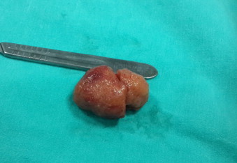
Macroscopic appearance of the mass. Spherical, firm, and pinkish mass, 5.5 × 5 × 5 cm.
Fig. 3.

White and red pulp of splenic tissue (Hematoxylin & Eosin, ×10).
3. Discussion
The spleen is a large encapsulated mass of vascular and lymphoid tissue situated in the upper left quadrant of the abdominal cavity. Various developmental anomalies of the spleen have been observed, including complete agenesis, multiple spleens or polysplenia, isolated small additional accessory spleens, and persistent lobulation. AS may be formed during embryogenesis, developing from the left side of the dorsal mesogastrium as a result of imperfect fusion of separate splenic masses [3]. The localization varies widely, but the most common locations are the splenic hilum (75%) or the pancreas tail (20%), as well as the greater omentum, along the greater curvature of the stomach, and the small and large intestine mesentery [1–3]. The regions adjacent to the left ovary or left testis and the pouch of Douglas are uncommon localizations. The case presented herein is one with a rare localization of AS, of interest due to its proximity to the obturator fossa and retroperitoneal area.
Ectopic splenic tissue can be either congenital (AS or splenunculi) or acquired (splenosis). Splenosis is the auto-transplantation of splenic tissue during splenectomy or following trauma. The patient discussed herein had no history of trauma or splenectomy; therefore, this may have been a case of a previously undiagnosed AS, missed during hysterectomy due to its deep localization. Congenital AS is seen in 10–30% of all autopsies. Unver Dogan et al. [1] investigated 720 autopsy cases, and AS was found in 6.7% cases, 2 of which were pelvic AS; thus, classifying the pelvis as an atypical localization. Further, the common presentation is of a single AS (85%), although two (14%), and rarely three or more (1%), can also be observed. The size of the AS ranges from microscopic to 4 cm in diameter, as observed in the present case [1,3,4]. AS presents 13 times more frequently in females than males, and the mean age is reported between 20 and 40 years [5]; the present case agreeing with these statistics.
A wandering spleen is a separate entity, resulting from incomplete development of the ligamentous splenic apparatus allowing the spleen to migrate within the abdomen. Case reports in the literature mostly present wandering AS. Vural et al. [6] reported on a wandering AS attached to the greater omentum resected from a 26-year old female. Perin et al. [7] reported on a wandering AS in the pelvis treated laparoscopically. Azar et al. [5] also reported on a pelvic wandering AS in a 44-year-old female and emphasized on its differential diagnosis from adnexal masses.
An AS is an incidental finding of no clinical significance in most patients. AS are generally determined during radiological investigations or during open or laparoscopic surgeries [8]. They are usually asymptomatic and have rarely been reported to present clinically as an abdominal mass related to complications such as torsion, spontaneous rupture, hemorrhage, or cyst formation. Torsion and ischemia of AS can lead to acute abdomen as seen in the main spleen. Cowles and Lazar [9] reported on a torsioned wandering AS in the pelvis. Padilla et al. [2] also reported on an acute abdomen due to spontaneous torsion of the AS. The differential diagnosis of pelvic AS could include adnexal mass features such as enlarged lymph nodes, subserosal fibroid, ovarian tumors, organized hematoma, and tubo-ovarian abscess. Our initial diagnosis was also of an ovarian neoplasia. Owing to fact that, pelvic mass was considered as malignancy before surgery and its deep localization, open surgery was prefered. But laparoscopic approach can also be a choice for the treatment.
4. Conclusions
Despite pelvic AS being a rare, usually asymptomatic, pelvic mass condition, it should be considered in the differential diagnosis of symptomatic pelvic masses.
Conflicts of interest
Authors declare that there is no conflicts of interest.
Funding
None.
Consent
Written informed consent was obtained from the patient for publication of this case report and and accompanying images.
Author contribution
Mine Islimye Taskin, Ertan Adali, Banu Gulec Baser contributed to study design and writing, Erdogan Bulbul contributed to data collection and evaluation of MRI. Engin Uzgoren contributed to histologic analysis.
References
- 1.Unver Dogan N., Uysal I.I., Demirci S., Dogan K.H., Kolcu G. Accessory spleens at autopsy. Clin. Anat. 2011;24(6):757–762. doi: 10.1002/ca.21146. [DOI] [PubMed] [Google Scholar]
- 2.Padilla D., Ramia J.M., Martin J., Pardo R., Cubo T., Hernandez-Calvo J. Acute abdomen due to spontaneous torsion of an accessory spleen. Am. J. Emerg. Med. 1999;17(4):429–430. doi: 10.1016/s0735-6757(99)90103-1. [DOI] [PubMed] [Google Scholar]
- 3.Radu C.C., Muţiu G., Pop O. Accessory spleen. Rom. J. Morphol. Embryol. 2014;55(3 Suppl):1243–1246. [PubMed] [Google Scholar]
- 4.Halpert B., Gyorkey F. Lesions observed in accessory spleens of 311 patients. Am. J. Clin. Pathol. 1959;32(2):165–168. doi: 10.1093/ajcp/32.2.165. [DOI] [PubMed] [Google Scholar]
- 5.Azar G.B., Awwad J.T., Mufarrij I.K. Accessory spleen presenting as adnexal mass. Acta Obstet. Gynecol. Scand. 1993;72(7):587–588. doi: 10.3109/00016349309058171. [DOI] [PubMed] [Google Scholar]
- 6.Vural M., Kacar S., Koşar U., Altin L. Symptomatic wandering accessory spleen in the pelvis: sonographic findings. J. Clin. Ultrasound. 1999;27(9):534–536. doi: 10.1002/(sici)1097-0096(199911/12)27:9<534::aid-jcu8>3.0.co;2-x. [DOI] [PubMed] [Google Scholar]
- 7.Perin A., Cola R., Favretti F. Accessory wandering spleen: report of a case of laparoscopic approach in an asymptomatic patient. Int. J. Surg. Case Rep. 2014;5(12):887–889. doi: 10.1016/j.ijscr.2014.10.045. [DOI] [PMC free article] [PubMed] [Google Scholar]
- 8.Yee L.F., Carvajal S.H., de Lorimier A.A., Mulvihill S.J. Laparoscopic splenectomy. The initial experience at University of California, San Francisco. Arch. Surg. 1995;130:874–879. doi: 10.1001/archsurg.1995.01430080076012. [DOI] [PubMed] [Google Scholar]
- 9.Cowles R.A., Lazar E.L. Symptomatic pelvic accessory spleen. J. Am. Surg. 2007;194(2):225–226. doi: 10.1016/j.amjsurg.2006.11.023. [DOI] [PubMed] [Google Scholar]