Abstract
Mammalian prions, transmissible agents causing lethal neurodegenerative diseases, are composed of assemblies of misfolded cellular prion protein (PrP) 1. A novel PrP variant, G127V, was under positive evolutionary selection during the epidemic of kuru, an acquired prion disease epidemic of the Fore population in Papua New Guinea, and appeared to provide strong protection against disease in the heterozygous state2. We have now investigated the protective role of this variant and its interaction with the common worldwide M129V PrP polymorphism; V127 was seen exclusively on a M129 PRNP allele. Here we demonstrate that transgenic mice expressing both variant and wild type human PrP are completely resistant to both kuru and classical CJD prions (which are closely similar) but can be infected with variant CJD prions, a human prion strain resulting from exposure to BSE prions to which the Fore were not exposed. Remarkably however, mice expressing only PrP V127 were completely resistant to all prion strains demonstrating a different molecular mechanism to M129V, which provides its relative protection against classical CJD and kuru in the heterozygous state. Indeed this single amino acid substitution (G→V) at a residue invariant in vertebrate evolution is as protective as deletion of the protein. Further study in transgenic mice expressing different ratios of variant and wild type PrP indicates that not only is PrP V127 completely refractory to prion conversion, but acts as a potent dose-dependent inhibitor of wild type prion propagation.
Prions cause fatal neurodegenerative conditions such as scrapie in sheep, bovine spongiform encephalopathy (BSE) in cattle and Creutzfeldt-Jakob disease (CJD) in humans1. The fundamental molecular process, seeded propagation of assemblies of misfolded host protein, is increasingly recognised as being of importance in all the major human neurodegenerative diseases3. There is a common polymorphism, present worldwide, in the substrate protein in human prion disease, human prion protein (PrP), where either methionine (M) or valine (V) is present at residue 129. MV heterozygosity provides relative protection against acquired, sporadic, and some inherited prion diseases4-6 and may have been selected during the evolution of modern humans by ancestral prion disease epidemics7. This protective effect is thought to relate to inhibition of homotypic protein-protein interactions5, 8 although residue 129 also influences the propagation of particular prion strains via conformational selection9, 10. Heterozygosity at another polymorphism, E219K, also provides resistance to CJD in Japan11.
Kuru was a devastating epidemic prion disease transmitted by endocannibalism and restricted to a remote area in Papua New Guinea (PNG). We reported a novel PrP variant (G127V) amongst unaffected individuals which appeared be a resistance factor selected by the epidemic and unique to this region2. Given the proximity to residue 129, we considered that it may have a similar action to M129V, blocking homotypic interactions and exerting its protective effect only in the heterozygous state. No PRNP codon 127VV homozygotes were identified in the kuru-exposed population and V127 was always seen on an M129 allele2. We therefore generated multiple lines of transgenic mice, expressing only human PrP (HuPrP) on a congenic FVB/N Prnpo/o background, to investigate whether G127V was indeed protective and whether this protection was dependent on heterozygosity or was an intrinsic property of the variant protein. Additionally we investigated its interaction with the residue 129 polymorphism.
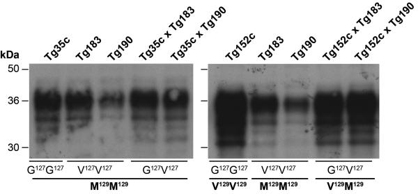
Two lines of transgenic mice homozygous for HuPrP V127 were studied: Tg(HuPrP V127M129/V127M129 Prnpo/o)-183 (V127M129 Tg183) and Tg(HuPrP V127M129/V127M129 Prnpo/o)-190 (V127M129 Tg190). PrP expression levels in homozygotes as compared to pooled normal human brain were 2-fold for V127M129 Tg183 and 1-fold for V127M129 Tg190. G127V heterozygous mice were derived by crossing these lines with FVB-congenic versions of Tg35 mice homozygous for HuPrP G127M129 12-15 or Tg152 mice homozygous for HuPrP G127V129 15-19 designated Tg(HuPrP G127M129/G127M129 Prnpo/o)-35c (G127M129 Tg35c) and Tg(HuPrP G127V129/G127V129 Prnpo/o)-152c (G127V129 Tg152c) respectively. G127M129 Tg35c and G127V129 Tg152c express wild type huPrP at 2- and 6-times respectively as compared to pooled normal human brain (table 1). Extended Data Fig.1 shows relative PrPC expression levels in all transgenic mice used in this study.
Table 1. Transgenic mice expressing G127 and V127 human PrP.
| Transgenic mice | PRNP Codon 127-129 genotype | Total HuPrP* expression level (x) | WT† G127 allele expression level (x) | Variant‡ V127 allele expression level (x) | PrP Ratio G127 : V127 |
|---|---|---|---|---|---|
| Parental lines: | |||||
| Tg35c | G127M129/G127M129 | 2 | 2 | - | - |
| Tg152c | G127V129/G127V129 | 6 | 6 | - | - |
| Tg183 | V127M129/V127M129 | 2 | - | 2 | - |
| Tg190 | V127M129/V127M129 | 1 | - | 1 | - |
| F1 crosses: | |||||
| Tg35c × Tg183 | G127M129/V127M129 | 2 | 1 | 1 | 1 : 1 |
| Tg35c × Tg190 | G127M129/V127M129 | 1.5 | 1 | 0.5 | 2 : 1 |
| Tg152c × Tg183 | G127V129/V127M129 | 4 | 3 | 1 | 3 : 1 |
| Tg152c × Tg190 | G127V129/V127M129 | 3.5 | 3 | 0.5 | 6 : 1 |
| Tg152c +/o (Hemi)§ | G127V129 | 3 | 3 | - | - |
WT = wild type
PrP expression level is relative to pooled 10% (w/v) normal human brain homogenate
Wild type alleles are either G127M129 or G127V129
Variant allele is V127M129
Generated by crossing Tg152c with FVB/PrP-null mice
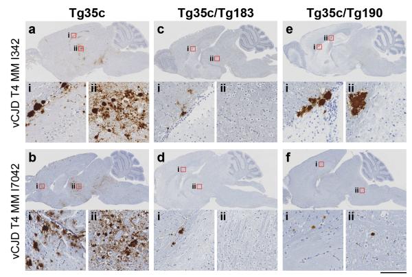
All transgenic lines were then challenged by intracerebral prion inoculation from well-characterised and previously transmitted human prion disease cases including all three PRNP codon 129 genotypes and comprising four cases of kuru, 12 cases of classical CJD and two cases of variant CJD (vCJD). Heterozygous HuPrP G127M129/V127M129 mice (Tg35c × Tg183 and Tg35c × Tg190) (table 1), having the genotype associated with disease resistance in the kuru-exposed human population, proved completely resistant to all four kuru isolates (which included all three PRNP codon 129 genotypes and two molecular strain types) (Fig. 1a and table 2a) while mice expressing wild type HuPrP G127M129/G127M129 (Tg35c) (Fig. 1a, table 2a) or G127V129/G127V129 (Tg152c) (table 2b) were fully susceptible with 100% attack rates. This is consistent with the population genetic data suggesting kuru resistance of G127V individuals. We have previously reported that prion strains seen in kuru brain are indistinguishable from those seen in classical CJD patients19. Similarly, none of four classical CJD isolates from patients of PRNP genotype 129MM transmitted to either line of G127M129/V127M129 mice (table 2a and Fig. 1b), while all four transmitted uniformly to Tg35c mice expressing wild type huPrP (Fig. 1b and table 2a). Remarkably, however, occasional G127M129/V127M129 mice developed clinical disease, and a larger number showed evidence of subclinical infection (positive PrP immunohistochemistry and/or western blot for PrPSc), on challenge with vCJD prions (table 2d and Extended Data Fig. 2). vCJD is a novel BSE-derived prion strain17, 20, 21 to which the population in the kuru-affected area of PNG were not exposed.
Figure 1. Transmission rates of human prions to transgenic mice homo- or heterozygous for human PrP V127.
Transgenic mice were intracerebrally inoculated with brain homogenate from patients with kuru (a) or classical CJD (b). Codon 127 and 129 PRNP genotypes of the recipient mice are shown (G, glycine, M, methionine, V, valine). G127M129 is a wild type human allele, and the V127M129 allele is seen only in humans from the kuru-exposed population of Papua New Guinea. Attack rate reports the total of clinically affected and sub-clinically infected mice as a proportion of the number of inoculated mice after prolonged (>600 days) post-inoculation periods. Primary prion transmission data are reported in tables 2 and 3.
Table 2. Transmission of human prions to transgenic mice heterozygous for human PrP V127.
| Inoculum |
Transmission data |
|||||||
|---|---|---|---|---|---|---|---|---|
| Aetiology | Source Code | Human PrPSc type* | Attack rate† | Incubation period, (days ± SEM) or (days p.i) | Attack rate† | Incubation period, (days ± SEM) or (days p.i) | Attack rate† | Incubation period, (days ± SEM) or (days p.i) |
|
| ||||||||
| A | Tg35c G127M129/G127M129 |
Tg35c × Tg183 G127M129/V127M129 |
Tg35c × Tg190 G127M129/V127M129 |
|||||
| kuru | I516 | T3 VV | 14/14 | 509 ± 56 (3) | 0/11 | >518-605 | 0/13 | >517-605 |
| kuru | I520 | T3 VV | 12/12 | 558 ± 4 (4) | 0/14 | >525-622 | 0/14 | >460-622 |
| kuru | I10336 | T3 MV | 13/13 | 454 ± 14 (10) | 0/14 | >522-606 | 0/11 | >504-607 |
| kuru | I518 | T2 MM | 11/11 | 493 ± 32 (7) | 0/12 | >545-620 | 0/13 | >544-603 |
| iCJD (GH) | I035 | T1 MM | 10/10 | 221 ± 3 (10) | 0/10 | >462-603 | 0/8 | >506-602 |
| sCJD | I11058 | T1 MM | 8/8 | 231 ± 3 (8) | 0/8 | >467-605 | 0/7 | >487-605 |
| iCJD (DM) | I026 | T2 MM | 10/10 | 256 ± 9 (8) | 0/8 | >507-601 | 0/7 | >520-602 |
| sCJD | I7040 | T2 MM | 10/10 | 233 ± 3 (10) | 0/8 | >582-604 | 0/7 | >600-604 |
| B | Tg152c G127V129/G127V129 |
Tg152c × Tg183 G127V129/V127M129 |
Tg152c × Tg190 G127V129/V127M129 |
|||||
|---|---|---|---|---|---|---|---|---|
| kuru | I516 | T3 VV | 9/9 | 218 ± 1 (6) | 0/13 | >447-608 | 9/15 | 543, 592 |
| kuru | I520 | T3 VV | 9/9 | 196 ± 7 (7) | 0/18 | >491-616 | 13/13 | 498 ± 17 (5) |
| kuru | I10336 | T3 MV | 15/15 | 212 ± 3 (11) | 15/15 | 456 ± 3 (15) | 15/15 | 316 ± 4 (14) |
| kuru | I518 | T2 MM | 11/11 | 211 ± 4 (8) | 3/11 | >494-620 | 13/14 | 468 ± 13 (9) |
| sCJD | I280 | T2 VV | 8/8 | 203 ± 5 (4) | 0/4 | >600-602 | 10/10 | 559 ± 14 (8) |
| sCJD | I278 | T2 VV | 6/6 | 236 ± 8 (6) | 0/8 | >424-602 | 8/9 | 582 |
| sCJD | I284 | T2 MV | 9/9 | 338 ± 4 (8) | 0/9 | >410-602 | 5/7 | >545-608 |
| sCJD | I1478 | T2 MV | 10/10 | 248 ± 8 (8) | 0/6 | >451-608 | 2/7 | >482-603 |
| sCJD | I7394 | T3 VV | 9/9 | 213 ± 1 (8) | 7/7 | >537-677 | 10/10 | 427 ± 8 (10) |
| sCJD | I764 | T3 MV | 10/10 | 219 ± 5 (7) | 9/10 | >368-588 | 10/10 | 461 ± 7 (9) |
| iCJD (GH) | I2651 | T3 VV | 8/8 | 200 ± 3 (6) | 0/6 | >567-607 | 7/7 | 538 ± 2 (5) |
| iCJD (GH) | I020 | T3 MV | 9/9 | 211 ± 2 (6) | 7/7 | >489-602 | 10/10 | 399 ± 8 (9) |
| C | Tg152c × Tg183 G127V129/V127M129 |
Tg152c × Tg190 G127V129/V127M129 |
Tg152c+/o (Hemi) G127V129 |
|||||
|---|---|---|---|---|---|---|---|---|
| iCJD (GH) | I020 | T3 MV | 7/7 | >489-602 | 10/10 | 399 ± 8‡ (9) | 14/14 | 252 ± 3‡ (14) |
| sCJD | I7394 | T3 VV | 7/7 | >537-677 | 10/10 | 427 ± 8‡ (10) | 12/12 | 245 ± 5‡ (12) |
| kuru | I10336 | T3 MV | 15/15 | 456 ± 3 (15) | 15/15 | 316 ± 4‡ (14) | 15/15 | 222 ± 2‡ (15) |
| D | Tg35c G127M129/G127M129 |
Tg35c × Tg183 G127M129/V127M129 |
Tg35c × Tg190 G127M129/V127M129 |
|||||
|---|---|---|---|---|---|---|---|---|
| vCJD | I342 | T4 MM | 15/15 | >426-603 | 9/15 | 467, 556 | 17/18 | 419±17 (3) |
| vCJD | I7042 | T4 MM | 14/14 | 559, 561 | 4/13 | >496-607 | 10/12 | 596 |
iCJD, iatrogenic CJD; sCJD, sporadic CJD; GH, growth hormone; DM, dura mater
According to classification of Hill et al.28
Attack rate is defined as the total number of both clinically affected and sub-clinically infected mice as a proportion of the total number of inoculated mice. Sub-clinical prion infection was assessed by immunoblotting and/or immunohistochemical examination of brain. Incubation periods are reported for clinically affected mice in days; where n ≥ 3 the mean ± SEM is reported with the number of mice contributing to the mean shown in parentheses, otherwise individual incubation times are given. In groups where no clinical transmission of prion disease was observed, the attack rate represents subclinical infection only and the interval between inoculation and death (from either senescence, culling due to inter-current illness or termination of the experiment) is reported as >x-y days.
Mean incubation periods for all 3 isolates are significantly lower in Tg152c hemizygotes than in Tg190 × Tg152c heterozygotes (P<0.0001; two-tailed unpaired t-test).
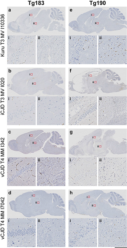
The key question with respect to whether the protective effect of V127 is via a similar mechanism to the M129V polymorphism was addressed by challenge of mice homozygous for HuPrP V127M129. While mice homozygous for wild type HuPrP G127M129 or G127V129 are both highly susceptible to CJD prions, remarkably mice homozygous for HuPrP V127M129 were completely resistant to all 18 human prion disease isolates, including vCJD prions, with no clinical transmissions or evidence of subclinical infection (Fig. 1a and b, table 3 and Extended Data Fig. 3).
Table 3. Transmission of human prions to transgenic mice homozygous for human PrP V127.
| Inoculum |
Transmission data |
|||||
|---|---|---|---|---|---|---|
| Aetiology | Source Code | Human PrPSc type* | Attack rate† | Incubation period, (days p.i) | Attack rate† | Incubation period, (days p.i) |
|
Tg183
V127M129/V127M129 |
Tg190
V127M129/V127M129 |
|||||
| kuru | I516 | T3 VV | 0/19 | >488-604 | 0/22 | >391-609 |
| kuru | I520 | T3 VV | 0/19 | >391-609 | 0/19 | >370-609 |
| kuru | I10336 | T3 MV | 0/21 | >405-607 | 0/24 | >463-617 |
| kuru | I518 | T2 MM | 0/12 | >439-600 | 0/13 | >432-603 |
| vCJD | I342 | T4 MM | 0/8 | >553-605 | 0/10 | >506-604 |
| vCJD | I7042 | T4 MM | 0/11 | >522-609 | 0/14 | >446-607 |
| iCJD (GH) | I035 | T1 MM | 0/10 | >434-622 | 0/10 | >450-602 |
| sCJD | I11058 | T1 MM | 0/8 | >411-609 | 0/9 | >454-612 |
| iCJD (DM) | I026 | T2 MM | 0/9 | >381-602 | 0/8 | >516-602 |
| sCJD | I7040 | T2 MM | 0/8 | >524-601 | 0/6 | >564-600 |
| sCJD | I280 | T2 VV | 0/9 | >532-602 | 0/9 | >425-601 |
| sCJD | I278 | T2 VV | 0/7 | >530-602 | 0/7 | >417-601 |
| sCJD | I284 | T2 MV | 0/6 | >549-603 | 0/6 | >466-609 |
| sCJD | I1478 | T2 MV | 0/7 | >517-602 | 0/7 | >418-617 |
| sCJD | I7394 | T3 VV | 0/8 | >500-600 | 0/8 | >600-603 |
| sCJD | I764 | T3 MV | 0/10 | >447-602 | 0/8 | >484-607 |
| iCJD (GH) | I2651 | T3 VV | 0/7 | >563-606 | 0/7 | >487-599 |
| iCJD (GH) | I020 | T3 MV | 0/6 | >546-603 | 0/6 | >572-600 |
(days p.i), days post inoculation; vCJD, variant CJD; sCJD, sporadic CJD; iCJD, iatrogenic CJD; GH, growth hormone; DM, dura mater
According to classification of Hill et al.28
Attack rate is defined as the total of clinically affected and sub-clinically infected mice as a proportion of the number of inoculated mice. Sub-clinical prion infection was assessed by immunoblotting and/or immunohistochemical examination of brain. As no clinical transmission of prion disease was observed the interval between inoculation and death (from either senescence, culling due to inter-current illness or termination of the experiment) is reported as >x-y days.
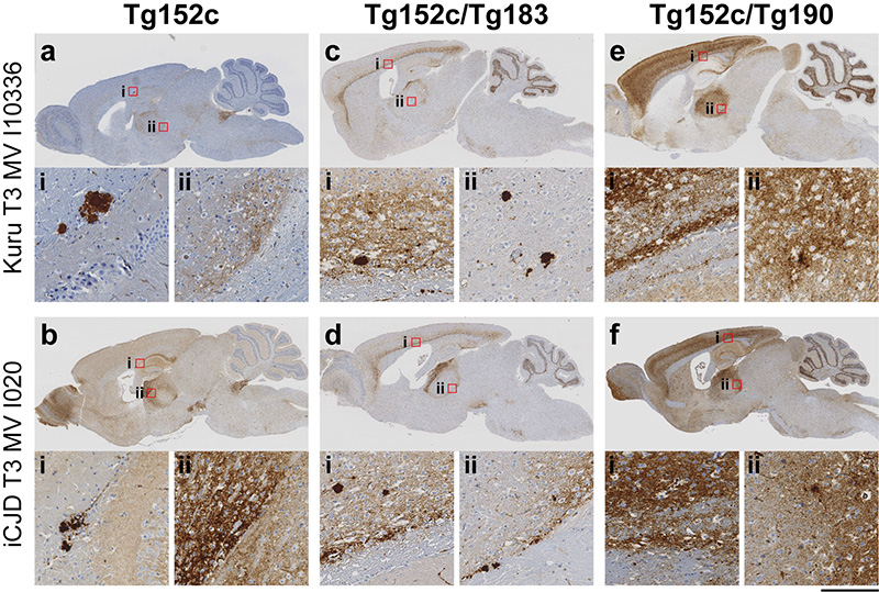
We also challenged mice heterozygous at both residues 127 and 129 of HuPrP (G127V129/V127M129) produced by crossing Tg183 or Tg190 mice with Tg152c mice (tables 1 and 2B). The higher level of expression of HuPrP V129 in Tg152c mice as compared to the other lines meant that there was a marked difference in the ratio of expression of the variant (V127M129) and wild type (G127V129) HuPrP in these crosses (table 1). While Tg183 and Tg35c had closely similar levels of expression of V127M129 and G127M129 HuPrP respectively, such that the Tg35c × Tg183 cross closely modelled the human PRNP G127V genotype, the expression ratios of wild type G127V129 to variant V127M129 HuPrP were approximately 6:1 and 3:1 for the Tg152c × Tg190 and Tg152c × Tg183 crosses respectively (table 1, Extended Data Fig.1 and 4). Interestingly, in these crosses some transmissions of kuru and classical CJD prions were seen (table 2b, Extended Data Fig. 4 and 5). In the Tg152c × Tg183 crosses, with a 3-fold excess of wild type PrP, subclinical infections were seen with four out of the 12 isolates used, and one kuru isolate (I10336) was associated with clinical disease (table 2b). However with the Tg152c × Tg190 crosses, with a 6-fold excess of wild type PrP, transmissions were seen from all isolates, the majority resulting in clinical disease (table 2b).
To further investigate the effect of HuPrP V127 on prion propagation, we compared hemizygous Tg(HuPrP G127V129 Prnpo/o)-152c mice with the Tg152c × Tg183 and Tg152c × Tg190 crosses (table 2c). The hemizygous line was challenged with two classical CJD and one kuru isolate that had resulted in a 100% attack rate in both crosses. Again, 100% attack rates were seen for all three isolates but the incubation periods were significantly shorter (P< 0.0001; table 2c) in the absence of HuPrP V127 than in either cross. These data, together with comparison of the two crosses themselves (table 2b), are consistent with a dominant negative effect of HuPrP V127 expression. Although dominant negative inhibition has been reported for other natural polymorphic residues of mammalian PrP in transgenic mice22-24 none have shown complete prevention of prion conversion when expressed at a 1:1 ratio with wild type PrP. Dominant negative effects on yeast prion propagation have also been reported25.
Our transgenic modelling of the HuPrP G127V polymorphism demonstrates that it confers strong protection against prion disease in the heterozygous state. However, most importantly, the molecular basis of this effect is clearly distinct from that proposed for the well-established HuPrP M129V polymorphism which is protective against developing sporadic CJD only in the heterozygous state: inhibition of homotypic protein-protein interactions during the process of prion propagation. Here we demonstrate that HuPrP V127 is intrinsically resistant to prion conversion and indeed capable of inhibiting propagation of wild type prions in a dose-dependent manner. Mice expressing only HuPrP V127 appear as resistant to prion disease as PrP null mice26 and understanding the structural basis of this effect may therefore provide critical insight into the molecular mechanism of mammalian prion propagation.
At its height when first recognised by Western medicine in 1957, kuru was a devastating epidemic largely affecting women and children and killing up to 2% of the population annually in some villages. Indeed some villages became largely devoid of young women of childbearing age. While collapse of the Fore population was prevented by cessation of endocannibalism in the late 1950s which interrupted the route of transmission and led to a gradual decline in incidence27, our data suggest that if transmission had continued the epicentre of the affected region might have been repopulated with kuru-resistant individuals as a population genetic response to the epidemic. Understanding the structural basis of why HuPrP V127 is unable to propagate prions of multiple strain types may provide key insights into prion propagation and the development of rational therapeutics.
Methods
Ethics Statement
Storage and biochemical analyses of post-mortem human brain samples and transmission studies to mice were performed with written informed consent from patients with capacity to give consent. Where patients were unable to give informed consent, assent was obtained from their relatives in accordance with UK legislation and Codes of Practice. Samples were stored and used in accordance with the Human Tissue Authority Codes of Practice and in line with the requirements of the Human Tissue Authority licence held by UCL Institute of Neurology. This study was performed with approval from the Medical Research Advisory Committee of the Government of Papua New Guinea, the National Hospital for Neurology and Neurosurgery and the UCL Institute of Neurology Joint Research Ethics Committee (now National Research Ethics Service Committee, London – Queen Square) - REC references: 03/N036, 03/N038 and 03/N133. Work with mice was performed under approval and licence granted by the UK Home Office (Animals (Scientific Procedures) Act 1986); Project Licence number 70/6454 which conformed to University College London institutional and ARRIVE guidelines (www.nc3rs.org.uk/ARRIVE/).
Generation of transgenic mice
The 759bp human PrP ORF was amplified by PCR with pfu polymerase from genomic DNA prepared from the brain of a patient with the 127V polymorphism on the 129M allele, using forward primer 5′-GTCGACCAGTCATTATGGCGAACCTT-3′ and reverse primer 5′-CTCGAGAAGACCTTCCTCATCCCACT-3′. Restriction sites Sal I and Xho I (underlined) were introduced in the forward and reverse primers respectively for cloning. The blunt ended PCR fragment generated by pfu polymerase was subcloned into Sma I digested pSP72 vector and sequenced to ensure that no spurious alterations had been introduced by the PCR and to confirm the presence of valine-127 and methionine-129 polymorphisms matching the patient DNA template. The amplified human PrP ORF with the confirmed polymorphisms was then isolated by Sal I and Xho I digestion. Subsequent subcloning into the Sal I site of the cosmid vector SHaCosTt, packaging and preparation of high quality DNA of the Not I transgene insert was as previously reported12. Microinjection of the purified Not I DNA fragment was carried out according to standard protocol into single cell eggs of Prnp null mice which had been backcrossed onto an FVB/N genetic background. Genotyping was performed by PCR and PrP expression levels estimated by western blot analysis as previously reported 12, 29. Two homozygous lines were established for variant HuPrP V127M129 described as Tg(HuPrP V127M129/V127M129 Prnpo/o)-183 (V127M129 Tg183) and Tg(HuPrP V127M129/V127M129 Prnpo/o)-190 (V127M129 Tg190), with transgene expression levels of 2 and 1 × respectively, that of pooled 10% (w/v) normal human brain homogenate (table 1). FVB-congenic versions of Tg35 mice homozygous for HuPrP G127M129 12-15 and Tg152 mice homozygous for HuPrP G127V129 15-19 were used as wild type human PrP-expressing controls, designated Tg(HuPrP G127M129/G127M129 Prnpo/o)-35c (G127M129 Tg35c) and Tg(HuPrP G127V129/G127V129 Prnpo/o)-152c (G127V129 Tg152c) respectively. The PRNP codon 127-129 genotypes and relative expression levels of wild type and variant PrP in the parental transgenic lines and in the F1 crosses are shown in table 1.
Transmission Studies
Strict bio-safety protocols were followed. Inocula were prepared, using disposable equipment for each inoculum, in a microbiological containment level 3 laboratory and inoculations performed within a class 1 microbiological safety cabinet as described previously12, 19. Ten mice per group of control G127M129 Tg35c and G127V129 Tg152c lines and 15 per group of newly generated V127M129 Tg183, V127M129 Tg190 lines and their respective crosses with Tg35c and Tg152c, were inoculated (see below) with human brain homogenates from neuropathologically confirmed patients comprising, four kuru cases, eight sporadic CJD cases, four iatrogenic CJD cases and two cases of vCJD. The genotype of each mouse was confirmed by PCR of ear punch DNA prior to inclusion and all mice were uniquely identified by sub-cutaneous transponders. Disposable cages were used and all cage lids and water bottles were also uniquely identified by transponder and remained with each cage of mice throughout the incubation period. Care of the mice was according to institutional guidelines. Mice (female, aged 6-8 weeks) were randomly assigned to experimental groups and anaesthetised with a mixture of halothane and O2, and intracerebrally inoculated into the right parietal lobe with 30 μl of a 1% (w/v) brain homogenate prepared in Dulbecco’s phosphate buffered saline lacking Ca2+ or Mg2+ ions (D-PBS). All mice were thereafter examined daily for early indicators of clinical prion disease including piloerection, sustained erect ears, intermittent generalised tremor, unsustained hunched posture, rigid tail, mild loss of coordination,and clasping hind legs when lifted by the tail. Definite diagnosis of clinical prion disease (triggering experimental end point) was reached if mice exhibited any two early indicator signs in addition to one confirmatory sign, or any two confirmatory signs. The confirmatory signs included ataxia,impairment of righting reflex, dragging of hind limbs, sustained hunched posture, or significant abnormal breathing. Mice were killed (by CO2 asphyxiation) if they exhibited any signs of distress or once a diagnosis of prion disease was established. At post-mortem brains from inoculated mice were removed, divided sagittally with half frozen and half fixed in 10% buffered formol saline. Subsequent immunohistochemical or biochemical investigations were performed blind to sample provenance.
Neuropathology and Immunohistochemistry
Fixed brain was immersed in 98% formic acid for 1 hour and paraffin wax embedded. Serial sections of 4 μm thickness were pre-treated by boiling for 10 min in a low ionic strength buffer (2.1 mM Tris, 1.3 mM EDTA, 1.1 mM sodium citrate, pH 7.8) before exposure to 98% formic acid for 5 min. Abnormal PrP accumulation was examined using anti-PrP monoclonal antibody ICSM 35 (D-Gen Ltd, London) on a Ventana automated immunohistochemical staining machine (Ventana Medical Systems Inc., Tucson, Arizona) using proprietary secondary detection reagents (Ventana Medical Systems Inc) before development with 3′3 diaminobenzedine tetrachloride as the chromogen30. Harris haematoxylin and eosin staining was done by conventional methods. Appropriate controls were used throughout.
Immunoblotting
Preparation of brain homogenates (10% (w/v) in D-PBS), proteinase K digestion (75 μg/ml for 1 h at 37°C) and subsequent immunoblotting was performed as described previously28, 30. Blots were probed with anti-PrP monoclonal antibody ICSM 35 (D-Gen Ltd, London) in conjunction with an anti-mouse IgG-alkaline phosphatase conjugate and development in chemiluminescent substrate (CDP-Star; Tropix Inc). Primary screening of brain homogenates was performed blind to sample identity.
Extended Data
Extended Data Figure 1. Immunoblots showing relative PrPC expression levels in transgenic mice.
The provenance of each brain sample is designated above each lane and molecular markers are indicated on the left. The PRNP codon 127 (G, glycine, V, valine) or codon 129 (M, methionine, V, valine) genotypes of the transgenic mice are designated below. Transgenic mouse brains were analysed by enhanced chemiluminescence without proteinase-K digestion and equal amounts of total protein loaded in each well and probed with anti-PrP monoclonal antibody ICSM 35. Wild type human PrP expression levels of G127M129/G127M129 Tg35c and G127V129/G127V129 Tg152c mice are 2- and 6-fold higher respectively, than seen in 10 % (w/v) pooled human brain homogenate. Homozygous V127M129 Tg183 and Tg190 mice have 2-fold higher or equivalent PrP expression levels respectively compared to 10 % (w/v) pooled human brain homogenate.
Extended Data Figure 2. Immunohistochemical analysis of transgenic mouse brain following challenge with vCJD prions.
All mice were intracerebrally challenged with the same vCJD prion isolates (I342 and I7042). Abnormal PrP deposition in fixed post-mortem brain from affected mice was detected using anti-PrP monoclonal antibody ICSM 35. (a-b) Homozygous Tg35c mice (expressing G127M129/G127M129 wild type PrP only) show intense and widespread PrP plaque deposits. Magnified areas show (ai) hippocampus, (bi) frontal cortex and (aii and bii) thalamus. In contrast, heterozygous G127M129/V127M129 Tg35c/Tg183 mice expressing equivalent levels of G127M129 and V127M129 PrP (c-d) show only weak PrP deposition in the corpus callosum (ci and di) with no abnormal PrP deposition detected in other brain areas, for example, in the thalamus (cii and dii). Heterozygous G127M129/V127M129 Tg35c/Tg190 mice which express a lower level of V127M129 PrP relative to wild type G127M129 PrP (e-f) show greater levels of PrP deposition than seen in Tg35c/Tg183 mice following challenge with the same vCJD prion isolates, (ei, eii and fi) corpus callosum; (fii) pons. Scale bar, upper panels (a-f) 2 mm; magnified panels, 100 μm.
Extended Data Figure 3. Immunohistochemical analysis of homozygous PRNP V127M129/V127M129 transgenic mouse brain following challenge with human prions.
Mice were intracerebrally challenged with kuru, classical and variant CJD prions. Following prolonged (>600 days) post inoculation periods, abnormal PrP deposition in fixed post-mortem brain was examined using anti-PrP monoclonal antibody ICSM 35. (a-d) V127M129/V127M129 Tg183 mice with 2-fold overexpression of V127M129 PrP. (E-H) V127M129/V127M129 Tg190 mice expressing endogenous levels of V127M129 PrP. Red square boxes in the main panels (a-h) define the area of magnified images of hippocampus (i) and thalamus (ii) shown in the lower panels. Scale bar, a-h, 2 mm; magnified panels i and ii, 100 μm. The lack of detection of abnormal PrP deposition in brain indicates that the mice are not subclinically infected with prions.
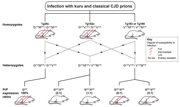
Extended Data Figure 4. Schematic diagram summarising transmissions of kuru and classical CJD prions to transgenic mice expressing human PrP.
G127M129/G127M129 Tg35c mice or G127V129/G127V129 Tg152c mice are homozygous for wild type human PrP alleles and are fully susceptible to kuru and classical CJD prions. V127M129/V127M129 Tg183 or V127M129/V127M129 Tg190 transgenic mice are homozygous for the variant V127M129 allele found only in humans from the kuru-exposed population of Papua New Guinea and are entirely resistant to infection with kuru and classical CJD prions. The levels of PrP expression in the brain of these homozygous transgenic mice relative to a pooled human brain homogenate are 1× (Tg190 mice) 2× (Tg35c and Tg183 mice) and 6× (Tg152c mice). Generation of F1 mice through inter-breeding the various homozygous lines produces different combinations of the various human PrP alleles leading to differences in the relative expression levels of the various prion proteins in brain. The PrP expression ratios from the two PrP alleles in the crosses are shown in parentheses above the cartoon mice. Full, intermediate or low susceptibility of the mice to infection with kuru and classical CJD prions is indicated by three, two or one diagonal red bar, respectively, drawn across the mice. Mice with no red bar are entirely resistant to infection with kuru and classical CJD prions.
Extended Data Figure 5. Immunohistochemical analysis of homozygous PRNP G127V129/G127V129 and heterozygous G127V129/V127M129 transgenic mouse brain following challenge with human prions.
Mice were intracerebrally challenged with kuru and classical CJD prions and abnormal PrP deposition in fixed post-mortem brain from affected mice was examined using anti-PrP monoclonal antibody ICSM 35. Red square boxes labelled i and ii in panels a-f mark brain areas that are magnified and displayed below; (i) cortex and (ii) thalamus. (a-b) Wild type G127V129/G127V129 Tg152c mice. (c-d) Heterozygous G127V129/V127M129 Tg152c/Tg183 mice. (e-f) Heterozygous G127V129/V127M129 Tg152c/190 mice Scale bar, a-f, 2 mm; magnified panels i and ii, 100 μm. The detection of abnormal PrP deposition in brain indicates that the mice are infected with prions.
Acknowledgments
This work was funded by the UK Medical Research Council. We thank all staff at our Biological Services Facility for animal care, Catherine O’Malley and Sarah Lyall for technical support with histology, and Richard Newton and Ray Young for preparation of figures. We thank patients and families for consent to use human tissues. We thank UK neurological and neuropathology colleagues for providing brain tissue from CJD patients. Some of this work was undertaken at University College London Hospitals/University College London, which received a proportion of funding from the Department of Health National Institute for Health Research Biomedical Research Centres funding scheme. We thank the Fore communities and Professor Peter Siba and colleagues at the Papua New Guinea Institute of Medical Research for their long term support. We thank the late Carleton Gajdusek, the late Joseph Gibbs, Story Landis, and their associates from the Laboratory of Central Nervous System Studies of the National Institutes of Health, Bethesda, USA for archiving and sharing kuru samples.
Footnotes
Competing financial interest
JC is a Director and JC and JDFW are shareholders of D-Gen Limited, an academic spin-out company in the field of prion diagnosis, decontamination and therapy. D-Gen supplied the ICSM35 antibody used in this study.
References
- 1.Collinge J. Prion diseases of humans and animals: their causes and molecular basis. Annu. Rev. Neurosci. 2001;24:519–550. doi: 10.1146/annurev.neuro.24.1.519. [DOI] [PubMed] [Google Scholar]
- 2.Mead S, et al. A novel protective prion protein variant that colocalizes with kuru exposure. N Engl J Med. 2009;361:2056–2065. doi: 10.1056/NEJMoa0809716. [DOI] [PubMed] [Google Scholar]
- 3.Jucker M, Walker LC. Self-propagation of pathogenic protein aggregates in neurodegenerative diseases. Nature. 2013;501:45–51. doi: 10.1038/nature12481. [DOI] [PMC free article] [PubMed] [Google Scholar]
- 4.Collinge J, Palmer MS, Dryden AJ. Genetic predisposition to iatrogenic Creutzfeldt-Jakob disease. Lancet. 1991;337:1441–1442. doi: 10.1016/0140-6736(91)93128-v. [DOI] [PubMed] [Google Scholar]
- 5.Palmer MS, Dryden AJ, Hughes JT, Collinge J. Homozygous prion protein genotype predisposes to sporadic Creutzfeldt-Jakob disease. Nature. 1991;352:340–342. doi: 10.1038/352340a0. [DOI] [PubMed] [Google Scholar]
- 6.Collinge J. Molecular neurology of prion disease. J Neurol Neurosurg Psychiatry. 2005;76:906–919. doi: 10.1136/jnnp.2004.048660. [DOI] [PMC free article] [PubMed] [Google Scholar]
- 7.Mead S, et al. Balancing selection at the prion protein gene consistent with prehistoric kuru-like epidemics. Science. 2003;300:640–643. doi: 10.1126/science.1083320. [DOI] [PubMed] [Google Scholar]
- 8.Antonyuk SV, et al. Crystal structure of human prion protein bound to a therapeutic antibody. Proc. Natl. Acad. Sci. USA. 2009;106:2554–2558. doi: 10.1073/pnas.0809170106. [DOI] [PMC free article] [PubMed] [Google Scholar]
- 9.Collinge J. Variant Creutzfeldt-Jakob disease. Lancet. 1999;354:317–323. doi: 10.1016/S0140-6736(99)05128-4. [DOI] [PubMed] [Google Scholar]
- 10.Collinge J, Clarke A. A general model of prion strains and their pathogenicity. Science. 2007;318:930–936. doi: 10.1126/science.1138718. [DOI] [PubMed] [Google Scholar]
- 11.Shibuya S, Higuchi J, Shin RW, Tateishi J, Kitamoto T. Protective prion protein polymorphisms against sporadic Creutzfeldt-Jakob disease. Lancet. 1998;351:419. doi: 10.1016/S0140-6736(05)78358-6. [DOI] [PubMed] [Google Scholar]
- 12.Asante EA, et al. BSE prions propagate as either variant CJD-like or sporadic CJD-like prion strains in transgenic mice expressing human prion protein. EMBO J. 2002;21:6358–6366. doi: 10.1093/emboj/cdf653. [DOI] [PMC free article] [PubMed] [Google Scholar]
- 13.Asante EA, Li YG, Gowland I, Jefferys JG, Collinge J. Pathogenic human prion protein rescues PrP null phenotype in transgenic mice. Neurosci. Lett. 2004;360:33–36. doi: 10.1016/j.neulet.2004.01.049. [DOI] [PubMed] [Google Scholar]
- 14.Wadsworth JD, et al. Human prion protein with valine 129 prevents expression of variant CJD phenotype. Science. 2004;306:1793–1796. doi: 10.1126/science.1103932. [DOI] [PubMed] [Google Scholar]
- 15.Wadsworth JD, Asante EA, Collinge J. Contribution of transgenic models to understanding human prion disease. Neuropathol Appl Neurobiol. 2010;36:576–597. doi: 10.1111/j.1365-2990.2010.01129.x. [DOI] [PMC free article] [PubMed] [Google Scholar]
- 16.Collinge J, et al. Unaltered susceptibility to BSE in transgenic mice expressing human prion protein. Nature. 1995;378:779–783. doi: 10.1038/378779a0. [DOI] [PubMed] [Google Scholar]
- 17.Hill AF, et al. The same prion strain causes vCJD and BSE. Nature. 1997;389:448–450. doi: 10.1038/38925. [DOI] [PubMed] [Google Scholar]
- 18.Asante EA, et al. Dissociation of pathological and molecular phenotype of variant Creutzfeldt-Jakob disease in transgenic human prion protein 129 heterozygous mice. Proc. Natl. Acad. Sci. USA. 2006;103:10759–10764. doi: 10.1073/pnas.0604292103. [DOI] [PMC free article] [PubMed] [Google Scholar]
- 19.Wadsworth JD, et al. Kuru prions and sporadic Creutzfeldt-Jakob disease prions have equivalent transmission properties in transgenic and wild-type mice. Proc. Natl. Acad. Sci. USA. 2008;105:3885–3890. doi: 10.1073/pnas.0800190105. [DOI] [PMC free article] [PubMed] [Google Scholar]
- 20.Collinge J, Sidle KC, Meads J, Ironside J, Hill AF. Molecular analysis of prion strain variation and the aetiology of ‘new variant’ CJD. Nature. 1996;383:685–690. doi: 10.1038/383685a0. [DOI] [PubMed] [Google Scholar]
- 21.Bruce ME, et al. Transmissions to mice indicate that ‘new variant’ CJD is caused by the BSE agent. Nature. 1997;389:498–501. doi: 10.1038/39057. [DOI] [PubMed] [Google Scholar]
- 22.Perrier V, et al. Dominant-negative inhibition of prion replication in transgenic mice. Proc Natl Acad Sci USA. 2002;99:13079–13084. doi: 10.1073/pnas.182425299. [DOI] [PMC free article] [PubMed] [Google Scholar]
- 23.Hizume M, et al. Human prion protein (PrP) 219K is converted to PrPSc but shows heterozygous inhibition in variant Creutzfeldt-Jakob Disease infection. J Biol Chem. 2009;284:3603–3609. doi: 10.1074/jbc.M809254200. [DOI] [PubMed] [Google Scholar]
- 24.Striebel JF, Race B, Meade-White KD, LaCasse R, Chesebro B. Strain specific resistance to murine scrapie associated with a naturally occurring human prion protein polymorphism at residue 171. PLoS Pathog. 2011;7:e1002275. doi: 10.1371/journal.ppat.1002275. [DOI] [PMC free article] [PubMed] [Google Scholar]
- 25.DiSalvo S, Derdowski A, Pezza JA, Serio TR. Dominant prion mutants induce curing through pathways that promote chaperone-mediated disaggregation. Nat Struct Mol Biol. 2011;18:468–492. doi: 10.1038/nsmb.2031. [DOI] [PMC free article] [PubMed] [Google Scholar]
- 26.Bueler H, et al. Mice devoid of PrP are resistant to scrapie. Cell. 1993;73:1339–1347. doi: 10.1016/0092-8674(93)90360-3. [DOI] [PubMed] [Google Scholar]
- 27.Alpers MP. The epidemiology of kuru: monitoring the epidemic from its peak to its end. Philos Trans R Soc Lond B Biol Sci. 2008;363:3707–3713. doi: 10.1098/rstb.2008.0071. [DOI] [PMC free article] [PubMed] [Google Scholar]
- 28.Hill AF, et al. Molecular classification of sporadic Creutzfeldt-Jakob disease. Brain. 2003;126:1333–1346. doi: 10.1093/brain/awg125. [DOI] [PubMed] [Google Scholar]
- 29.Asante EA, et al. Absence of spontaneous disease and comparative prion susceptibility of transgenic mice expressing mutant human prion proteins. J Gen Virol. 2009;90:546–558. doi: 10.1099/vir.0.007930-0. [DOI] [PMC free article] [PubMed] [Google Scholar]
- 30.Wadsworth JD, et al. Molecular diagnosis of human prion disease. Methods Mol Biol. 2008;459:197–227. doi: 10.1007/978-1-59745-234-2_14. [DOI] [PubMed] [Google Scholar]


