



Abstract
Phosphotransacetylase (Pta), a key enzyme in bacterial metabolism, catalyzes the reversible transfer of an acetyl group from acetyl phosphate to coenzyme A (CoA) to produce acetyl-CoA and Pi. Two classes of Pta have been identified based on the absence (PtaI) or presence (PtaII) of an N-terminal regulatory domain. PtaI has been fairly well studied in bacteria and one genus of archaea; however, only the Escherichia coli and Salmonella enterica PtaII enzymes have been biochemically characterized, and they are allosterically regulated. Here, we describe the first biochemical and kinetic characterization of a eukaryotic Pta from the oomycete Phytophthora ramorum. The two Ptas from P. ramorum, designated PrPtaII1 and PrPtaII2, both belong to class II. PrPtaII1 displayed positive cooperativity for both acetyl phosphate and CoA and is allosterically regulated. We compared the effects of different metabolites on PrPtaII1 and the S. enterica PtaII and found that, although the N-terminal regulatory domains share only 19% identity, both enzymes are inhibited by ATP, NADP, NADH, phosphoenolpyruvate (PEP), and pyruvate in the acetyl-CoA/Pi-forming direction but are differentially regulated by AMP. Phylogenetic analysis of bacterial, archaeal, and eukaryotic sequences identified four subtypes of PtaII based on the presence or absence of the P-loop and DRTGG subdomains within the N-terminal regulatory domain. Although the E. coli, S. enterica, and P. ramorum enzymes all belong to the IIa subclass, our kinetic analysis has indicated that enzymes within a subclass can still display differences in their allosteric regulation.
INTRODUCTION
Acetate production has been studied for many years in bacteria but has received less attention in eukaryotic microbes, even though acetate is produced as an important end product of energy metabolism in yeasts (1–3) and protists (4–6). Four different pathways for production of acetate from acetyl-coenzyme A (CoA) have been identified in eukaryotic microbes (7). ADP-forming acetyl-CoA synthetase (Acs) (EC 6.2.1.13) (equation 1) has been implicated in acetate production in amitochondriate protists and some species of archaea.
| (1) |
Acetate:succinate CoA-transferase (EC 2.8.3.8) (equation 2) is present in kinetoplastids, Trichomonas species, the trematode Fasciola hepatica, the yeast Saccharomyces cerevisiae, and the rumen fungus Neocallimastix sp. strain L2 (8–12).
| (2) |
Acetyl-CoA hydrolase (EC 3.1.2.1) (equation 3) is involved in peroxisomal acetate formation in kinetoplastids (7).
| (3) |
Phosphotransacetylase (Pta) (EC 2.3.1.8) (equation 4) and acetate kinase (Ack) (EC 2.7.2.1) (equation 5) form a pathway for the interconversion of acetate and acetyl-CoA that was previously thought to be limited to bacteria and one genus of archaea but has now been shown to be present in eukaryotes, such as green algae and Phytophthora (13, 14).
| (4) |
| (5) |
The Pta-Ack pathway is best understood in its roles in both acetate production and assimilation in Escherichia coli and other bacteria. The pathway is responsible for the production of acetate during mixed-acid fermentation under hypoxic conditions and in a metabolic overflow mechanism in which acetyl-CoA is diverted from the tricarboxylic acid (TCA) cycle when there is an imbalance between the rapid uptake of glucose and its conversion into products (15). Under high acetate concentrations, this low-affinity pathway can also be used for assimilation of acetate by its conversion to acetyl-CoA (16).
In eukaryotes, the Pta-Ack pathway has been investigated only in the green alga Chlamydomonas, in which two parallel Pta-Ack pathways have been identified (13). Proteomic studies have suggested that the Pat1-Ack2 pathway is localized to mitochondria (note that phosphotransacetylase is designated Pat in Chlamydomonas), and the Pat2-Ack1 pathway is localized to chloroplasts (13, 17). Acetate has been found to be one of the major fermentative products excreted by Chlamydomonas during growth under dark, anoxic conditions, and ACK1, ACK2, PAT1, and PAT2 transcript levels are increased, in agreement with a role for the Pat-Ack pathway in acetate production (18). The ack1 and pat2 mutants were the most vulnerable to anoxia, and strains could not be recovered after a 24-h exposure to anoxia (19). Far less acetate was produced in each of the mutants after imposition of anoxia, yet small amounts of acetate (<20%) were still produced in the ack1-ack2 double mutant, suggesting Pat-Ack is not the only pathway for acetate production in Chlamydomonas (19).
Analysis of bacterial and archaeal Pta sequences revealed two classes (20). PtaI enzymes consist of a single catalytic domain, whereas the PtaII enzymes have an additional N-terminal regulatory domain (20, 21). The Pta from the archaeon Methanosarcina thermophila is the best-studied class I enzyme (22–25), and several structures have been solved (26, 27). A ternary-complex mechanism based on kinetic and structural studies has been proposed for the enzyme (24). Two PtaII enzymes have been characterized, one from Salmonella enterica (SePtaII) and one from E. coli (EcPtaII) (20, 21); however, a structure for a PtaII has not been reported. The N-terminal regulatory domains of EcPtaII and SePtaII contain two recognizable subdomains designated the P-loop and the DRTGG subdomains (21). Truncations of the N-terminal domain of EcPtaII revealed that the P-loop subdomain is required for regulation of the enzyme by NADH, ATP, phosphoenolpyruvate (PEP), and pyruvate and that the DRTGG subdomain is vital for the sigmoidal response that is observed in allosteric enzymes (20, 21).
Here, we report the first biochemical and kinetic investigation of a eukaryotic Pta, the PtaII enzyme from Phytophthora ramorum, a pathogenic oomycete that causes sudden oak death (28). P. ramorum has a single open reading frame (ORF) that encodes Ack and two ORFs that encode class II Ptas (here designated PrPtaII1 and PrPtaII2). Our characterization of PrPtaII1 demonstrates that the enzyme strongly prefers the acetyl-CoA/Pi-forming direction, unlike the S. enterica and E. coli enzymes. PrPtaII1 displays substrate cooperativity for acetyl phosphate and CoA and is allosterically regulated through inhibition by ATP, AMP, NADP, NADH, PEP, and pyruvate. Our phylogenetic analysis of the Pta family, the first reported that includes eukaryotic sequences, indicates there are four different subclasses of PtaII based on differences in the N-terminal regulatory domain. A comparison of the bacterial and eukaryotic enzymes and the phylogenetic diversity suggests that the N-terminal domain and its regulatory role have evolved throughout the domains Bacteria and Eukarya.
MATERIALS AND METHODS
Materials.
Chemicals were purchased from Sigma-Aldrich, VWR, Fisher Scientific, and Gold Biotechnology. Oligonucleotide primers were purchased from Integrated DNA Technologies. A codon-optimized gene encoding P. ramorum PrPtaII1 (JGI 78441; http://genome.jgi-psf.org/Phyra1_1/Phyra1_1.home.html) was synthesized by GenScript and supplied in the E. coli expression vector pET21b, which provides for addition of a C-terminal His tag for use in nickel affinity column purification. Plasmid pPTA69 (kindly provided by Jorge Escalante-Semerena, University of Georgia) encodes S. enterica PtaII with a His6 tag fused to the N terminus of the protein (20).
Phylogenetic analysis of Pta.
BLASTP and TBLASTN (29, 30) were used to search the sequence databases at the National Center for Biotechnology Information (NCBI) (http://www.ncbi.nlm.nih.gov), the Broad Institute (http://www.broadinstitute.org/), and the U.S. Department of Energy Joint Genome Institute (http://genome.jgi-psf.org/) for putative Pta amino acid sequences, using M. thermophila PtaI as the query sequence. Sequences were aligned using ClustalW with the Gonnet protein weight matrix, a gap-opening penalty of 10.0, and a gap extension penalty of 0.2 (31). Phylogenetic analysis of the aligned sequences was performed with the MEGA 5.1 program (32) using the neighbor-joining algorithm with partial deletion estimates. Five hundred bootstrap replicates were executed, and bootstrap values of 75% or greater are shown. The EMBOSS Needle Pairwise Sequence Alignment program (http://www.ebi.ac.uk) was used to determine sequence identity and similarity (33). The presence or absence of the P-loop and DRTGG subdomains in each PtaII sequence was analyzed using the Pfam Protein Families Database (http://pfam.xfam.org/search) with an E value cutoff of E−10 (34).
Heterologous production and purification of Pta.
Recombinant plasmids were introduced into E. coli Rosetta 2(DE3) pLysS, and cells were grown in Luria-Bertani broth with 50 μg/ml ampicillin and 34 μg/ml chloramphenicol at 37°C with shaking at 200 rpm until an optical density at 600 nm (OD600) of 0.6 to 0.8 was reached. Production of recombinant Pta was induced by addition of isopropyl-β-d-thiogalactopyranoside to a final concentration of 1 mM.
Following overnight incubation at ambient temperature, cells were harvested by centrifugation. The cells were resuspended in buffer A (25 mM Tris, 150 mM NaCl, 25 mM imidazole, 10% glycerol, pH 7.5), disrupted by three passages through a chilled French pressure cell at 138 mPa, and centrifuged at 100,000 × g for 90 min at 4°C. The supernatant was applied to a HisTrap HP Ni2+ affinity column (GE Healthcare, Inc.) equilibrated with buffer A. The protein was eluted using a linear gradient from 25 mM to 500 mM imidazole in 25 mM Tris-HCl, 150 mM NaCl, and 10% glycerol (pH 7.5). Fractions with Pta activity were pooled and dialyzed against buffer containing 25 mM Tris-HCl and 10% glycerol (pH 7.5). The recombinant enzyme was determined to be electrophoretically pure by SDS-PAGE. The protein concentration for purified PrPtaII1 was calculated from the absorbance at 280 nm using an extinction coefficient of 36,330 M−1 cm−1.
Activity assays.
PrPtaII1 activity in the acetyl-CoA-forming direction was measured by monitoring the increase in absorbance at 233 nm due to thioester bond formation (ε233 = 5.55 mM−1 cm−1) (20, 21). The reaction mixture contained 50 mM Tris (pH 7.5), 20 mM KCl, 20 mM NH4Cl, and 1 mM dithiothreitol (DTT), and the concentrations of acetyl phosphate and CoA were varied. Activity in the acetyl phosphate-forming direction was measured with two different assays. The thioester assay monitors the decrease in absorbance at 233 nm due to the release of CoA (ε233 = 4.44 mM−1 cm−1) (22), and Ellman's thiol assay monitors the increase in absorbance at 412 nm due to the formation of the thiophenolate anion with DTNB [5′,5-dithiobis(2-nitrobenzoic acid)] (ε233 = 14,150 M−1 cm−1) (20, 21). The reaction mixtures for both assays contained 50 mM Tris (pH 7.5), 20 mM KCl, 20 mM NH4Cl, 1 mM DTT, and 1 mM DTNB, and the concentrations of acetyl-CoA and Pi were varied. The reaction mixtures for all assays were preincubated for 3 min at 37°C. Reactions were initiated by the addition of enzyme and were performed in triplicate.
SePtaII activity in the acetyl-CoA-forming direction was measured by monitoring the increase in absorbance at 233 nm. The reaction mixture contained 50 mM Tris (pH 7.5), 40 mM NH4Cl, 1 mM CoA, and 3 mM acetyl phosphate. Reaction mixtures containing acetyl phosphate and enzyme were preincubated for 1 min at 37°C, and reactions were initiated by the addition of CoA. All reactions were performed in triplicate.
Assays were performed in 96-well plates, and the absorbance was monitored using the Synergy HT Multi-mode Microplate Reader (BioTek Instruments, Inc.). Data are expressed as means ± standard deviations (SD).
Kinetic analysis.
To determine apparent kinetic parameters for PrPtaII1 in the acetyl-CoA-forming direction, the concentration of one substrate was varied while the second substrate was held constant at saturating concentration (determined to be 4 mM for CoA and 5 mM for acetyl phosphate). KaleidaGraph (Synergy Software) was used to fit the data to the Michaelis-Menten equation (equation 6), where V0 is the initial velocity, [S] is the substrate concentration, V is the maximum velocity, and Km is the Michaelis constant.
| (6) |
When acetyl phosphate was varied, the data displayed positive cooperativity when fitted to the Hill equation (equation 7) (35, 36), where K0.5 is the substrate concentration at half-maximal velocity and h is the Hill constant.
| (7) |
Determining the IC50.
Metabolic intermediates, coenzymes, and nucleotide triphosphates were tested as allosteric effectors of PrPtaII1 and SePtaII. Substrate concentrations were held at saturating levels, and effector molecule concentrations were varied from 3 μM to 3 mM. Half-maximal inhibitory concentrations (IC50s) were determined for all PrPtaII1 and SePtaII allosteric inhibitors by measuring the decrease in activity as a function of increasing inhibitor concentration. IC50s were determined using GraphPad Prism 5 (GraphPad Software, Inc.) by fitting the data with a log [inhibitor]-versus-response curve.
Site-directed mutagenesis.
The QuikChange Lightning site-directed mutagenesis kit (Stratagene, Inc.) was used for mutagenesis according to the manufacturer's instructions. The primers used in the alteration of the Gly-300 codon were as follows: 5′PrPTAG300D, CACCTGAAAAAATATAAAGACGACGCGATGATTATCACCAGTGGT; 3′PrPTAG300D, ACCACTGGTGATAATCATCGCGTCGTCTTTATATTTTTTCAGGTG; 5′PrPTAG300A, CACCTGAAAAAATATAAAGACGCGGCGATGATTATCACCAGTGGT; 3′PrPTAG300A, ACCACTGGTGATAATCATCGCCGCGTCTTTATATTTTTTCAGGTG. Alterations were confirmed by sequencing at the Clemson University Genomic Institute (CUGI).
Gel filtration chromatography.
The native molecular mass of recombinant PrPtaII1 was examined by gel filtration chromatography using an Äkta fast protein liquid chromatography (FPLC) system with a Superose 12 column (GE Healthcare). The gel filtration column was calibrated with cytochrome c (12.4 kDa), carbonic anhydrase (29 kDa), albumin (66 kDa), amylase (200 kDa), apoferritin (443 kDa), and thyroglobulin (669 kDa) (Sigma-Aldrich Co.). The column was equilibrated with buffer containing 50 mM Tris and 150 mM KCl (pH 7.5) and developed at a rate of 0.5 ml/min.
RESULTS
Discovery of the different subclasses of PtaII.
The Pta enzyme family was previously divided into two classes (20). PtaI enzymes are approximately 350 amino acids long and consist of only a catalytic domain. PtaII enzymes are approximately twice that size, with a C-terminal catalytic domain and an N-terminal regulatory domain (20, 21). Although Pta was commonly considered to be a bacterial enzyme, sequences have now been identified in the Eukarya (14). To determine how widespread the Pta enzyme family is in this domain, searches of the sequence databases were performed using the M. thermophila PtaI sequence as the query. Pta sequences were identified in a number of eukaryotes, such as green algae, lycophytes, moss, and oomycetes, but were absent in fungi, diatoms, higher plants, and metazoans. Every completed eukaryotic genome that has a Pta sequence also has an ORF encoding a product with identity to Ack, consistent with these enzymes acting as a pathway, as in bacteria. In a phylogeny of Pta sequences constructed based on the catalytic domain, the PtaII sequences form a separate clade (Fig. 1). All of the eukaryotic sequences belong to the PtaII class, except for those from Perkinsus marinus, Emiliana huxleri, and Thecomonas trahens. The P. marinus PtaI is a single-domain enzyme, whereas the E. huxleyi and T. trahens Pta sequences (shown in red in Fig. 1) are multidomain enzymes but are considered to be a PtaI rather than PtaII because the Pta catalytic domain is fused to Ack rather than an N-terminal regulatory domain, as for the PtaII enzymes. The E. huxleyi enzyme has just the Pta and Ack domains, whereas the T. trahens Pta has four domains: a domain with unknown function, an Ack domain, a Pta domain, and a poly(R)-hydroxyalkanoic acid synthase domain.
FIG 1.
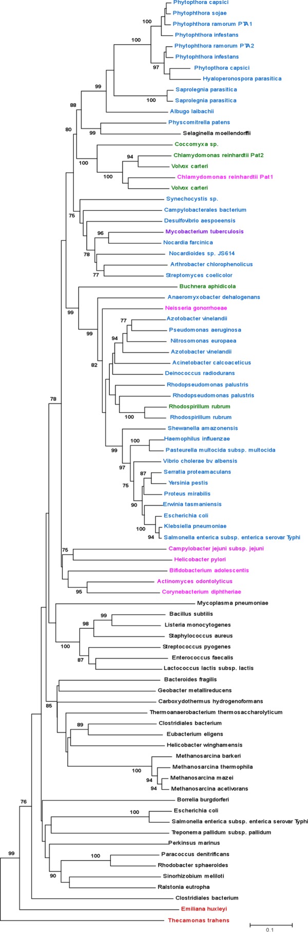
Phylogeny of the PtaI and PtaII family. The phylogenetic tree was constructed based on the sequences of the Pta catalytic domains. PtaI sequences are shown in black, PtaIIa sequences are shown in blue, PtaIIb sequences are shown in purple, PtaIIc sequences are shown in green, and PtaIId sequences are shown in pink. The scale bar indicates the expected number of amino acid replacements per site. Only bootstrap values of 75% or higher are shown.
The N-terminal regulatory domains of EcPtaII and SePtaII contain two recognizable subdomains designated the P-loop and the DRTGG subdomains (21). The P-loop nucleoside triphosphatase (NTPase) subdomain contains a conserved nucleotide triphosphate-binding motif similar to that found in enzymes involved in translation, transcription, intracellular trafficking, membrane transport, and DNA replication and repair (37). The DRTGG subdomain has an unknown function and is named after some of its most conserved residues. This domain is related to the cystathionine-beta-synthase (CBS) domain that exists in both membrane-bound and cytosolic proteins and is known to function in eukaryotes, prokaryotes, and archaea (38).
Although the PtaII N-terminal domains are similar in size, analysis of their sequences has revealed four subclasses based on the presence or absence of the P-loop and DRTGG subdomains (Fig. 2). The PtaIIa N-terminal domain includes both subdomains, whereas the PtaIIb, PtaIIc, and PtaIId subclasses lack one or both subdomains. The majority of PtaII sequences belong to the PtaIIa subclass, with PtaIIb, PtaIIc, and PtaIId sequences distributed infrequently throughout the phylogeny. Surprisingly, the two Chlamydomonas reinhardtii PtaII enzymes belong to two different subclasses, PtaIId and PtaIIc.
FIG 2.
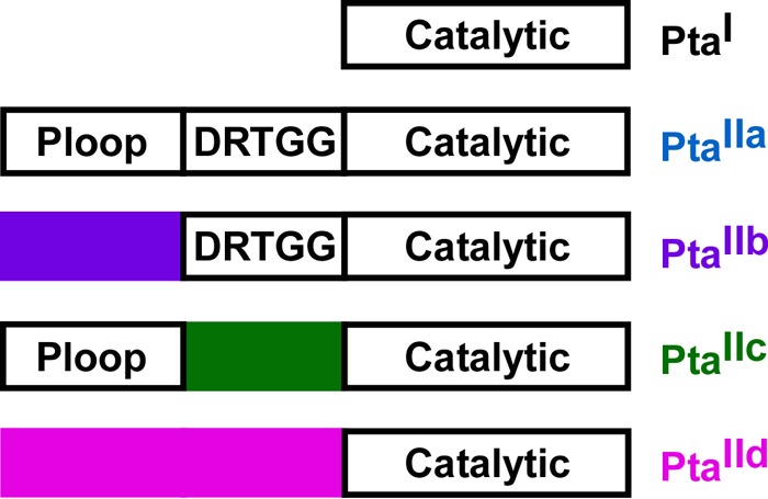
Subdomain structures of the PtaI and PtaII classes. PtaI enzymes have only a catalytic domain; PtaII enzymes have a catalytic domain and an N-terminal domain. Four subclasses of PtaII enzymes have been identified based on the presence or absence of the P-loop and DRTGG subdomains, as shown. The domains are not drawn to scale.
Purification and molecular properties of PrPtaII1.
The genome of P. ramorum (http://genome.jgi-psf.org/Phyra1_1/Phyra1_1.home.html) has two PtaIIa ORFs, designated PtaII1 (protein identifier [ID] 78441) and PtaII2 (protein ID 78440). An E. coli codon-optimized gene encoding PrPtaII1 (GenScript Inc.) was cloned into pET21b (C-terminal His tag), and the recombinant enzyme was produced in E. coli and purified by nickel affinity chromatography to electrophoretic homogeneity. In determining the optimal reaction conditions for PrPtaII1, we tested the requirement for KCl and NH4Cl, as other PtaII enzymes have been shown to have increased activity in the presence of one or both salts (20, 21). Maximum activity was observed in the presence of 20 mM NH4Cl and 20 mM KCl, and both salts are required. Increasing the concentration of either or both salts does not further increase enzyme activity. Optimal activity also requires the presence of 1 mM (final concentration) DTT. PrPtaII1 has the highest activity at 37°C; therefore, all experiments were conducted at 37°C. Due to aggregation issues, the molecular mass of the enzyme could not be determined by gel filtration. Brinsmade and Escalante-Semerena (20) encountered similar discrepancies with the S. enterica Pta.
Kinetic analysis of PrPtaII1.
In determining kinetic parameters for PrPtaII1 in the acetyl-CoA/Pi-forming direction, plots of substrate concentration versus velocity were sigmoidal. Apparent kinetic parameters were determined by fitting the experimental data to the Hill equation (equation 7), in which a Hill constant (h) greater than 1.0 represents positive cooperativity and a Hill constant less than 1.0 represents negative cooperativity (39, 40). PrPtaII1 exhibited positive cooperativity (h = 1.75 ± 0.18) with acetyl phosphate and slight positive cooperativity (h = 1.04 ± 0.05) with CoA (Table 1). The maximum activity observed in the acetyl phosphate/CoA-forming direction is approximately 3-fold lower than that in the acetyl-CoA/Pi-forming direction (8.9 ± 0.5 μmol min−1 mg−1 versus 23.8 ± 0.7 μmol min−1 mg−1, respectively), but kinetics parameters could not be determined in this direction due to an inability to reach saturation with inorganic phosphate.
TABLE 1.
Kinetic parameters for wild-type PrPtaII and the G300D variant in the acetyl-CoA-forming direction
| Enzyme |
K0.5 (mM) |
kcat/K0.5 (s−1 mM−1) |
Hill constant (h) |
|||
|---|---|---|---|---|---|---|
| CoA | AcP | CoA | AcP | CoA | AcP | |
| Wild type | 1.05 ± 0.31 | 1.85 ± 0.14 | 35.1 ± 6.0 | 18.04 ± 1.26 | 1.04 ± 0.05 | 1.75 ± 0.18 |
| G300D | 0.24 ± 0.05 | 1.75 ± 0.10 | 0.32 ± 0.06 | 0.043 ± 0.001 | 1.50 ± 0.05 | 6.50 ± 1.76 |
Allosteric regulation of the PtaIIa enzyme family.
Although the catalytic domains of all Pta enzymes share strong identity (e.g., the catalytic domains of PrPtaII1 and SePtaII share 52.0% identity), the N-terminal domains within a subclass can differ substantially. The PrPtaII1 N-terminal domain shares only 19% identity with the N-terminal domains of the E. coli and S. enterica PtaIIa enzymes, raising the question of whether this eukaryotic Pta is subject to allosteric regulation similar to that of the bacterial PtaII enzymes. We tested nucleotides, coenzymes, and metabolic intermediates from both the glycolytic pathway and the citric acid cycle as effector molecules of PrPtaII1. NAD+, NADH, NADP, and NADPH were potent inhibitors, with NADP having the strongest effect (Fig. 3A). ATP, ADP, AMP, PEP, and pyruvate were also found to inhibit activity (Fig. 3B), but to a lesser extent than the nicotinamide derivatives. Fructose-1,6-bisphosphate (1 mM), oxaloacetate (1 mM), α-ketoglutarate (1.5 mM), and citrate (1.5 mM) had no effect on PrPtaII1 activity.
FIG 3.
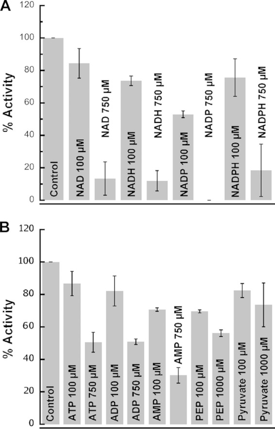
Regulation of PrPtaII1 by allosteric effectors. PrPtaII1 activity in the acetyl-CoA-forming direction was monitored in the absence and presence of allosteric effector molecules. All data were normalized to the control, which represents the enzymatic activity observed in the absence of an effector molecule. (A) The results are displayed as percent activity in the presence of 100 μM and 750 μM NAD+, NADH, NADP, and NADPH. (B) The results are displayed as percent activity in the presence of 100 μM and 750 μM ATP, ADP, and AMP and 100 μM and 1,000 μM PEP and pyruvate. The data are expressed as means ± SD.
Although SePtaII and EcPtaII were shown to be allosterically regulated (20, 21), only ATP, NADH, PEP, and pyruvate were examined for EcPtaII, and only pyruvate and NADH were tested in the acetyl phosphate-forming direction for SePtaII. IC50s were not reported for EcPtaII, and the IC50s for SePtaII were reported only in the acetyl phosphate/CoA-forming direction. To allow direct comparison between the eukaryotic and bacterial enzymes, recombinant SePtaII was produced and purified; the effects of ATP, AMP, NADP, NADH, and PEP on SePtaII enzymatic activity were determined in the acetyl-CoA/Pi-forming direction; and IC50s were determined (Table 2). Our results show that both SePtaII and PrPtaII1 are regulated by the same allosteric effectors, although the potencies differed. Because NADP was the most potent inhibitor of PrPtaII1, we evaluated its effect on substrate affinity. The Km values for CoA were relatively unchanged by the presence of increasing concentrations of NADP, but the Vmax decreased (Fig. 4A), suggesting mixed inhibition by NADP toward CoA. The K0.5 values for acetyl phosphate increase and the cooperativity decreases as the NADP concentration is increased (Fig. 4B), indicating that NADP has a direct effect on the affinity of PrPtaII1 for acetyl phosphate.
TABLE 2.
IC50s for PtaII enzymes in the acetyl-CoA-forming direction
| Effector molecule | IC50 (μM) |
||
|---|---|---|---|
| PrPta | PrPtaG300D | SePta | |
| ATP | 736 ± 6 | 133 ± 8 | 364 ± 19 |
| AMP | 287 ± 4 | 53 ± 2 | NDa |
| NADP | 135 ± 4 | 81 ± 3 | 97 ± 8 |
| NADH | 275 ± 21 | 52 ± 2 | 24 ± 6 |
| PEP | 1,018 ± 128 | 74 ± 4 | 67 ± 3 |
ND, IC50 could not be determined due to the biphasic response.
FIG 4.

Effects of NADP on CoA and acetyl phosphate utilization by PrPtaII1. (A) Enzymatic activity was measured in the acetyl-CoA-forming direction in the presence of 5 mM acetyl phosphate with various concentrations of NADP using the thioester-forming assay. ●, 0 μM NADP; ■, 67.1 μM NADP; ◆, 134.2 μM NADP. (B) Enzymatic activity in the acetyl-CoA-forming direction was measured in the presence of 4 mM CoA with various concentrations of NADP using the thioester-forming assay. ●, 0 μM NADP; ■; 67.1 μM NADP; ◆, 134.2 μM NADP.
For PrPtaII1, activity steadily decreases as the AMP concentration is increased, similar to the behavior observed with other PrPtaII1 effectors. AMP has a much different effect on SePtaII, resulting in activation of the enzyme when present at low concentrations and inhibition at higher concentrations. SePtaII has 28% higher activity in the presence versus absence of 30 μM AMP (Fig. 5); however, as the concentration of AMP was increased to 100 μM, the activity returned to the level observed in the absence of effector. Activity is inhibited further as the AMP concentration is increased, with approximately 50% inhibition in the presence of 1 mM and complete loss of activity at 3 mM AMP. Pyruvate also has an unusual biphasic effect on SePtaII activity (Fig. 5). In the presence of 50 μM pyruvate, activity was 133% that observed in the absence of pyruvate. However, as the pyruvate concentration was increased further to 100 μM, activity began to decrease, and inhibition was observed in the presence of 500 μM pyruvate.
FIG 5.

AMP and pyruvate activate and inhibit SePtaII in the acetyl-CoA-forming direction. SePtaII activity in the acetyl-CoA-forming direction was determined in the presence of increasing concentrations of AMP (■) and pyruvate (●). All data were normalized to the control, which represents the enzymatic activity observed in the absence of an effector molecule. The data are expressed as means ± SD.
Analysis of PrPta1G300D and PrPta1G300A.
Brinsmade and Escalante-Semerena (20), using an S. enterica Acs knockout mutant, screened for Pta variants that supported the growth of the mutant on 10 mM acetate, conditions under which Acs would normally be responsible for acetate activation to acetyl-CoA. They isolated 10 single-amino-acid variants, for which the mutations all mapped to the N-terminal regulatory domain of PtaII. Three of the variants (R252H, G273D, and M294I) displayed altered responses to at least one of the allosteric effectors. Differences in substrate affinities between the wild-type and variant enzymes were minor. A G273D variant had approximately 2-fold-increased Vmax in the acetyl-CoA/Pi-forming direction and 3-fold-increased Vmax in the acetyl phosphate/CoA-forming direction. This variant also showed approximately 3-fold stimulation by pyruvate versus 1.2-fold stimulation for the wild-type enzyme but remained subject to strong inhibition by NADH (20). This Gly residue is conserved in both PrPtaII1 and PrPtaII2, unlike the other two residues identified, which altered the wild-type SePtaII response to allosteric regulators.
To investigate the role of the conserved Gly residue in allosteric regulation of PrPtaII1, we targeted the corresponding residue, Gly300, for alteration. The G300A variant was soluble but was not active in either direction of the reaction. This result suggests either that this alteration resulted in complete loss of activity or that the structural integrity of the protein was compromised but not so severely that it was no longer soluble. Kinetic analysis of the PrPtaII1 G300D variant revealed a nearly 450-fold reduction in the catalytic rate in the acetyl-CoA-forming direction (Table 1), and activity was completely abolished in the acetyl phosphate-forming direction, indicating the importance of this residue in catalysis. A slight decrease in the K0.5 for acetyl phosphate and a 4-fold decrease in the K0.5 for CoA were observed for the G300D variant versus the wild-type enzyme. The kcat/K0.5 value for each substrate decreased significantly, with a 420-fold decrease for acetyl phosphate and a 110-fold decrease observed for CoA (Table 1). This alteration resulted in increased substrate cooperativity for both CoA and acetyl phosphate. The Hill constant for CoA increased by half, and that for acetyl phosphate increased 3.7-fold versus the wild type (Table 1).
We analyzed the effects of several allosteric inhibitors on the PrPtaII1 G300D variant and found that the IC50s for ATP, AMP, and NADH decreased approximately 5-fold versus the wild type, whereas only a 1.6-fold decrease was observed for NADP (Table 2). A nearly 14-fold decrease was observed in the IC50 for PEP (Table 2). Interestingly, nearly 50% inhibition was observed with 50 μM pyruvate, but activity increased as the pyruvate concentration was increased. At 1 mM pyruvate, enzyme activity had reached the uninhibited level, and by 3 mM pyruvate concentration, the activity was 124% that of the control (Fig. 6). The EC50 for pyruvate was determined to be 503 ± 24 μM.
FIG 6.

Alteration of the Gly300 residue in PrPtaII1 changes the effect of pyruvate. Enzyme activity was determined in the presence of increasing amounts of pyruvate in the acetyl-CoA-forming direction for PrPta1 (●) and PrPta1G300D (■). The data are expressed as means ± SD.
DISCUSSION
Phylogenetic analysis of Pta sequences shows that PtaI and PtaII sequences form separate clades (Fig. 1). Notably, nearly all of the eukaryotic enzymes belong to the PtaII class, whereas both PtaI and PtaII enzymes are well represented in bacteria. In fact, E. coli and S. enterica possess both PtaI and PtaII enzymes. Based on these distributions and what is currently known about the roles of Pta in various bacteria and Chlamydomonas, it is difficult to determine what dictates whether an organism is more likely to have a PtaI or a PtaII enzyme.
Our sequence analysis of the regulatory domain of PtaII enzymes has revealed four subclasses based on the presence or absence of the P-loop and DRTGG subdomains. Our phylogeny indicates that the eukaryotic enzymes group together within the PtaII clade and that the eukaryotic PtaIIa sequences are clustered together. Both of the P. ramorum PtaII sequences belong to this subclass and fall within this cluster.
Like P. ramorum, C. reinhardtii and Volvox carteri also have two PtaII enzymes. However, Pat2 from C. reinhardtii lacks the DRTGG subdomain and thus belongs to the PtaIIc subclass, whereas Pat1 lacks identity to either subdomain within the N terminus and is classified as a PtaIId enzyme. The V. carteri PtaII sequences both belong to PtaIIc and are most closely related to the P. ramorum enzymes. The PtaIIa subclass is the most prevalent, followed by PtaIId. The infrequent occurrence of PtaIIb and PtaIIc sequences and their distribution throughout the phylogeny suggest these subclasses arose recently and sporadically through the loss of domains.
Yang et al. (19) have shown that the C. reinhardtii Pta enzymes have different localization, with Pat1 localized to mitochondria and Pat2 localized to chloroplasts. The fact that these two enzymes belong to different subclasses most likely reflects different regulatory requirements in each organelle. The presence of two Pta enzymes in P. ramorum may also indicate different cellular localization. However, the fact that they both belong to the IIa subclass suggests that they are similarly regulated.
It is interesting that although Pta has been identified in the oomycete Phytophthora and in the green alga Chlamydomonas, it has not been identified in other stramenopiles, such as the diatoms. A search of diatom sequences revealed an Ack sequence in Phaeodactylum tricornutum; however, the only gene encoding an Ack partner enzyme present in that genome is that of xylulose 5-phosphate/fructose 6-phosphate phosphoketolase (Xfp), the partner enzyme for Ack in euascomycete and basidiomycete fungi. No ORFS for Ack or Pta have been identified in any other diatom sequences available.
Eukaryotic PtaII versus bacterial PtaII.
Our characterization of PrPtaII1 allowed us to investigate the similarities and differences between bacterial and eukaryotic PtaII enzymes and provides us with a better overall picture of the regulation of this subclass. PrPtaII1, SePtaII, and EcPtaII all display positive cooperativity, but for different substrates. SePtaII and PrPtaII1 display positive cooperativity for acetyl phosphate but little or no cooperativity for CoA, whereas EcPtaII displays positive cooperativity for CoA but not for acetyl phosphate.
As EcPtaII and SePtaII are both allosterically regulated (20, 21), we examined whether PrPtaII1 is also subject to allosteric regulation and if the same metabolites are effectors for all three enzymes. ATP, NADH, PEP, and pyruvate were previously shown to be inhibitors of EcPtaII (21), and we have now shown that they are also inhibitors of SePtaII and PrPtaII1, although the order of potency of the inhibitors differs. We have also found that AMP influences both SePtaII and PrPtaII1 activities, but in different ways. AMP inhibits PrPtaII1 but activates SePtaII at lower concentrations and then inhibits activity at higher concentrations. AMP has a similar effect on glycogen phosphorylase (41). At low concentrations, AMP acts as a strong activator by binding to a site close to the subunit interface and increasing the release of glucose 1-phosphate from glycogen (42, 43). At higher concentrations, AMP binds to the ATP inhibitor site (44, 45) located at the entrance to the channel to the catalytic site (45), resulting in inhibition. By analogy, this may suggest that AMP and ATP bind at separate effector sites on SePtaII.
It is surprising that both Pta enzymes studied here display negative regulation by both NAD(P) and NAD(P)H and ATP, ADP, and AMP. Typically, an enzyme would be expected to respond differently to signals of high versus low cellular energy or the redox indicator pairs rather than to all of them. One possible explanation is that the regulatory domain signals a response to one set of signals (e.g., ATP and NADH) and the C-terminal catalytic domain is regulated by the opposite signals. However, no such regulation of class I Pta enzymes that consist of just the catalytic domain have been reported, so it is difficult to speculate further on this at this time.
The role of the P-loop and DRTGG subdomains in catalysis and allosteric regulation.
Through analysis of EcPtaII truncations, Campos-Bermudez et al. (21) demonstrated that the N-terminal domain is required for maximal catalytic activity and that the P-loop subdomain is required for the regulation of the enzyme by metabolic effector molecules. Using a positive-selection method to identify Pta variants that allow S. enterica growth at low acetate concentrations in an acs mutant, Brinsmade and Escalante-Semerena (20) identified three single-amino-acid variants altered in the N-terminal domain for which regulation by allosteric effectors differs. Of particular interest is the G273D SePtaII variant, which displayed much stronger activation by pyruvate than the wild-type enzyme but similar inhibition by NADH (20). This glycine is located in the DRTGG subdomain and is conserved among all PtaIIa and PtaIIb enzymes, including both enzymes from P. ramorum.
We altered Gly300, the equivalent residue in PrPtaII1, to Ala and Asp to investigate whether the enzyme is regulated similarly to SePtaII. The G300A variant lacked activity in either direction, and the G300D variant had greatly reduced activity in the acetyl-CoA/Pi-forming direction and no activity in the acetyl phosphate/CoA-forming direction. With the exception of pyruvate, each allosteric effector had an effect on the activity of the PrPtaII1 G300D variant similar to that on the wild type, although the IC50s were reduced. Remarkably, pyruvate enhanced the activity of the variant in the acetyl-CoA/Pi-forming direction. However, activity was still very low in the opposite direction. In the corresponding SePtaII variant, this alteration greatly stimulated activity in the acetyl phosphate/CoA-forming direction. This result demonstrates that residue G300 in the DRTGG subdomain of the N-terminal regulatory domain is important across both bacteria and eukaryotes in determining how the enzyme reacts to the presence and absence of pyruvate. In addition, our results suggest that residues within the regulatory domain may also play a catalytic role, since the G300A variant lacks activity and the G300D variant has reduced activity in one direction and activity in the other direction was abolished, both in the absence of any effector molecules.
A possible physiological role for Pta in Phytophthora.
Our knowledge of Pta and its physiological role has largely been limited to bacterial enzymes. The Pta-Ack pathway has been shown to be essential for growth and invasion in several pathogenic bacteria, including Vibrio cholerae (46), uropathogenic E. coli (47), S. enterica (48), and Listeria monocytogenes (49). A eukaryotic Pta has not previously been characterized, and the only report on the physiological role of a nonbacterial Pta is from the green alga Chlamydomonas (19).
An important aspect of adapting to changing environments is the ability to use a variety of carbon sources, including acetate. In addition to having the Ack-Pta pathway utilized by bacteria to activate acetate under high-acetate concentrations, P. ramorum also has the ubiquitous AMP-forming Acs that a number of eukaryotic microbes use for acetate activation. The fact that PrPtaII1 preferentially operates in the acetyl-CoA/Pi-forming direction suggests the physiological role of the Ack-PtaII1 pathway is in utilization of acetate as a carbon source. Perhaps, these pathways function in Phytophthora as in the bacteria E. coli, in which the high-affinity Acs pathway functions in acetate activation at low acetate concentrations and the low-affinity Ack-Pta pathway functions at high acetate concentrations (50, 51). However, there are no reports in the literature investigating the ability of Phytophthora to utilize acetate as a carbon source, leaving this question unanswered.
The presence of a second Pta raises the possibility that the two enzymes are specialized, with PrPtaII2 operating in the acetyl phosphate/CoA-forming direction. Alternatively, PrPtaII2 may catalyze both directions of the reaction, and metabolic effectors may regulate these activities, as well as the activity of PrPtaII1. The two P. ramorum Pta sequences share 82.3% identity overall, with 91% identity between the catalytic domains and 75.6% identity in the N-terminal domains. Thus, specialization in terms of directionality may not be as likely, and the presence of two Pta enzymes may instead reflect localization to different compartments within the cell, as seen in Chlamydomonas.
Concluding remarks.
The PtaII class is much more complex than previously thought, with four subclasses, and PtaIIa enzymes are subject to allosteric regulation by a number of effectors (52, 53). Regulation of enzymes from the other three subclasses and further study of the N-terminal domain are needed. An understanding of the regulation exerted by this domain would be greatly facilitated by having a structure of a PtaII enzyme.
ACKNOWLEDGMENTS
This work was supported by awards from the National Science Foundation (award 0920274) and the South Carolina Experiment Station Project (SC-1700340).
Footnotes
This article is technical contribution 6241 of the Clemson University Experiment Station.
REFERENCES
- 1.Bubb WA, Wright LC, Cagney M, Santangelo RT, Sorrell TC, Kuchel PW. 1999. Heteronuclear NMR studies of metabolites produced by Cryptococcus neoformans in culture media: identification of possible virulence factors. Magn Reson Med 42:442–453. [DOI] [PubMed] [Google Scholar]
- 2.Himmelreich U, Allen C, Dowd S, Malikic R, Shehan BP, Mountford C, Sorrell TC. 2003. Identification of metabolites of importance in the pathogenesis of pulmonary cryptococcoma using nuclear magnetic resonance spectroscopy. Microbes Infect 5:285–290. doi: 10.1016/S1286-4579(03)00028-5. [DOI] [PubMed] [Google Scholar]
- 3.Oura E. 1977. Reaction products of yeast fermentations. Process Biochem 12:19–21. [Google Scholar]
- 4.Mazet M, Morand P, Biran M, Bouyssou G, Courtois P, Daulouede S, Millerioux Y, Franconi JM, Vincendeau P, Moreau P, Bringaud F. 2013. Revisiting the central metabolism of the bloodstream forms of Trypanosoma brucei: production of acetate in the mitochondrion is essential for parasite viability. PLoS Negl Trop Dis 7:e2587. doi: 10.1371/journal.pntd.0002587. [DOI] [PMC free article] [PubMed] [Google Scholar]
- 5.Montalvo FE, Reeves RE, Warren LG. 1971. Aerobic and anaerobic metabolism in Entamoeba histolytica. Exp Parasitol 30:249–256. doi: 10.1016/0014-4894(71)90089-0. [DOI] [PubMed] [Google Scholar]
- 6.Reeves RE, Warren LG, Susskind B, Lo HS. 1977. An energy-conserving pyruvate-to-acetate pathway in Entamoeba histolytica. Pyruvate synthase and a new acetate thiokinase. J Biol Chem 252:726–731. [PubMed] [Google Scholar]
- 7.Tielens AG, van Grinsven KW, Henze K, van Hellemond JJ, Martin W. 2010. Acetate formation in the energy metabolism of parasitic helminths and protists. Int J Parasitol 40:387–397. doi: 10.1016/j.ijpara.2009.12.006. [DOI] [PubMed] [Google Scholar]
- 8.Fleck CB, Brock M. 2009. Re-characterisation of Saccharomyces cerevisiae Ach1p: fungal CoA-transferases are involved in acetic acid detoxification. Fungal Genet Biol 46:473–485. doi: 10.1016/j.fgb.2009.03.004. [DOI] [PubMed] [Google Scholar]
- 9.Marvin-Sikkema FD, Pedro Gomes TM, Grivet JP, Gottschal JC, Prins RA. 1993. Characterization of hydrogenosomes and their role in glucose metabolism of Neocallimastix sp. L2. Arch Microbiol 160:388–396. [DOI] [PubMed] [Google Scholar]
- 10.Saz HJ, deBruyn B, de Mata Z. 1996. Acyl-CoA transferase activities in homogenates of Fasciola hepatica adults. J Parasitol 82:694–696. doi: 10.2307/3283876. [DOI] [PubMed] [Google Scholar]
- 11.Steinbuchel A, Muller M. 1986. Anaerobic pyruvate metabolism of Tritrichomonas foetus and Trichomonas vaginalis hydrogenosomes. Mol Biochem Parasitol 20:57–65. doi: 10.1016/0166-6851(86)90142-8. [DOI] [PubMed] [Google Scholar]
- 12.Van Hellemond JJ, Opperdoes FR, Tielens AG. 1998. Trypanosomatidae produce acetate via a mitochondrial acetate:succinate CoA transferase. Proc Natl Acad Sci U S A 95:3036–3041. doi: 10.1073/pnas.95.6.3036. [DOI] [PMC free article] [PubMed] [Google Scholar]
- 13.Atteia A, van Lis R, Gelius-Dietrich G, Adrait A, Garin J, Joyard J, Rolland N, Martin W. 2006. Pyruvate formate-lyase and a novel route of eukaryotic ATP synthesis in Chlamydomonas mitochondria. J Biol Chem 281:9909–9918. doi: 10.1074/jbc.M507862200. [DOI] [PubMed] [Google Scholar]
- 14.Ingram-Smith C, Martin SR, Smith KS. 2006. Acetate kinase: not just a bacterial enzyme. Trends Microbiol 14:249–253. doi: 10.1016/j.tim.2006.04.001. [DOI] [PubMed] [Google Scholar]
- 15.Akesson M, Karlsson EN, Hagander P, Axelsson JP, Tocaj A. 1999. On-line detection of acetate formation in Escherichia coli cultures using dissolved oxygen responses to feed transients. Biotechnol Bioeng 64:590–598. doi: 10.1002/(SICI)1097-0290(19990905)64:5<590::AID-BIT9>3.0.CO;2-T. [DOI] [PubMed] [Google Scholar]
- 16.Wolfe AJ. 2005. The acetate switch. Microbiol Mol Biol Rev 69:12–50. doi: 10.1128/MMBR.69.1.12-50.2005. [DOI] [PMC free article] [PubMed] [Google Scholar]
- 17.Terashima M, Specht M, Naumann B, Hippler M. 2010. Characterizing the anaerobic response of Chlamydomonas reinhardtii by quantitative proteomics. Mol Cell Proteomics 9:1514–1532. doi: 10.1074/mcp.M900421-MCP200. [DOI] [PMC free article] [PubMed] [Google Scholar]
- 18.Mus F, Dubini A, Seibert M, Posewitz MC, Grossman AR. 2007. Anaerobic acclimation in Chlamydomonas reinhardtii: anoxic gene expression, hydrogenase induction, and metabolic pathways. J Biol Chem 282:25475–25486. doi: 10.1074/jbc.M701415200. [DOI] [PubMed] [Google Scholar]
- 19.Yang W, Catalanotti C, D'Adamo S, Wittkopp TM, Ingram-Smith CJ, Mackinder L, Miller TE, Heuberger AL, Peers G, Smith KS, Jonikas MC, Grossman AR, Posewitz MC. 2014. Alternative acetate production pathways in Chlamydomonas reinhardtii during dark anoxia and the dominant role of chloroplasts in fermentative acetate production. Plant Cell 26:4499–4518. doi: 10.1105/tpc.114.129965. [DOI] [PMC free article] [PubMed] [Google Scholar]
- 20.Brinsmade SR, Escalante-Semerena JC. 2007. In vivo and in vitro analyses of single-amino acid variants of the Salmonella enterica phosphotransacetylase enzyme provide insights into the function of its N-terminal domain. J Biol Chem 282:12629–12640. doi: 10.1074/jbc.M611439200. [DOI] [PubMed] [Google Scholar]
- 21.Campos-Bermudez VA, Bologna FP, Andreo CS, Drincovich MF. 2010. Functional dissection of Escherichia coli phosphotransacetylase structural domains and analysis of key compounds involved in activity regulation. FEBS J 277:1957–1966. doi: 10.1111/j.1742-4658.2010.07617.x. [DOI] [PubMed] [Google Scholar]
- 22.Lawrence SH, Ferry JG. 2006. Steady-state kinetic analysis of phosphotransacetylase from Methanosarcina thermophila. J Bacteriol 188:1155–1158. doi: 10.1128/JB.188.3.1155-1158.2006. [DOI] [PMC free article] [PubMed] [Google Scholar]
- 23.Lundie LL Jr, Ferry JG. 1989. Activation of acetate by Methanosarcina thermophila. Purification and characterization of phosphotransacetylase. J Biol Chem 264:18392–18396. [PubMed] [Google Scholar]
- 24.Lawrence SH, Luther KB, Schindelin H, Ferry JG. 2006. Structural and functional studies suggest a catalytic mechanism for the phosphotransacetylase from Methanosarcina thermophila. J Bacteriol 188:1143–1154. doi: 10.1128/JB.188.3.1143-1154.2006. [DOI] [PMC free article] [PubMed] [Google Scholar]
- 25.Iyer PP, Ferry JG. 2001. Role of arginines in coenzyme A binding and catalysis by the phosphotransacetylase from Methanosarcina thermophila. J Bacteriol 183:4244–4250. doi: 10.1128/JB.183.14.4244-4250.2001. [DOI] [PMC free article] [PubMed] [Google Scholar]
- 26.Iyer PP, Lawrence SH, Luther KB, Rajashankar KR, Yennawar HP, Ferry JG, Schindelin H. 2004. Crystal structure of phosphotransacetylase from the methanogenic archaeon Methanosarcina thermophila. Structure 12:559–567. doi: 10.1016/j.str.2004.03.007. [DOI] [PubMed] [Google Scholar]
- 27.Iyer PP, Lawrence SH, Yennawar HP, Ferry JG. 2003. Expression, purification, crystallization and preliminary X-ray analysis of phosphotransacetylase from Methanosarcina thermophila. Acta Crystallogr D Biol Crystallogr 59:1517–1520. doi: 10.1107/S0907444903013428. [DOI] [PubMed] [Google Scholar]
- 28.Grunwald NJ, Garbelotto M, Goss EM, Heungens K, Prospero S. 2012. Emergence of the sudden oak death pathogen Phytophthora ramorum. Trends Microbiol 20:131–138. doi: 10.1016/j.tim.2011.12.006. [DOI] [PubMed] [Google Scholar]
- 29.Altschul SF, Gish W, Miller W, Myers EW, Lipman DJ. 1990. Basic local alignment search tool. J Mol Biol 215:403–410. doi: 10.1016/S0022-2836(05)80360-2. [DOI] [PubMed] [Google Scholar]
- 30.Altschul SF, Madden TL, Schaffer AA, Zhang J, Zhang Z, Miller W, Lipman DJ. 1997. Gapped BLAST and PSI-BLAST: a new generation of protein database search programs. Nucleic Acids Res 25:3389–3402. doi: 10.1093/nar/25.17.3389. [DOI] [PMC free article] [PubMed] [Google Scholar]
- 31.Thompson JD, Gibson TJ, Plewniak F, Jeanmougin F, Higgins DJ. 1997. The Clustal X windows interface: flexible strategies for multiple sequence alignment aided by quality analysis tools. Nucleic Acids Res 25:4876–4882. doi: 10.1093/nar/25.24.4876. [DOI] [PMC free article] [PubMed] [Google Scholar]
- 32.Tamura K, Peterson D, Peterson N, Stecher G, Nei M, Kumar S. 2011. MEGA5: molecular evolutionary genetics analysis using maximum likelihood, evolutionary distance, and maximum parsimony methods. Mol Biol Evol 28:2731–2739. doi: 10.1093/molbev/msr121. [DOI] [PMC free article] [PubMed] [Google Scholar]
- 33.Rice P, Longden I, Bleasby A. 2000. EMBOSS: the European molecular biology open software suite. Trends Genet 16:276–277. doi: 10.1016/S0168-9525(00)02024-2. [DOI] [PubMed] [Google Scholar]
- 34.Finn RD, Mistry J, Tate J, Coggill P, Heger A, Pollington JE, Gavin OL, Gunasekaran P, Ceric G, Forslund K, Holm L, Sonnhammer EL, Eddy SR, Bateman A. 2010. The Pfam protein families database. Nucleic Acids Res 38:D211-222. doi: 10.1093/nar/gkp985. [DOI] [PMC free article] [PubMed] [Google Scholar]
- 35.Hill AV. 1910. The possible effects of the aggregation of the molecules of haemoglobin on its dissociation curves. J Physiol 40:iv–vii. [Google Scholar]
- 36.Motulsky H. 2004. Fitting Models to Biological Data Using Linear and Nonlinear Regression: A Practical Guide to Curve Fitting. Oxford University Press, Oxford, United Kingdom. [Google Scholar]
- 37.Leipe DD, Koonin EV, Aravind L. 2003. Evolution and classification of P-loop kinases and related proteins. J Mol Biol 333:781–815. doi: 10.1016/j.jmb.2003.08.040. [DOI] [PubMed] [Google Scholar]
- 38.Baykov AA, Tuominen HK, Lahti R. 2011. The CBS domain: a protein module with an emerging prominent role in regulation. ACS Chem Biol 6:1156–1163. doi: 10.1021/cb200231c. [DOI] [PubMed] [Google Scholar]
- 39.Traut T. 2008. Allosteric regulatory enzymes. Springer, New York, NY. doi: 10.1007/978-0-387-72891-9. [DOI] [Google Scholar]
- 40.Copeland RA. 2000. Enzymes: a practical introduction to structure, mechanism, and data analysis, 2nd ed Wiley-VCH, Weinheim, Germany. [Google Scholar]
- 41.Black WJ, Wang JH. 1968. Studies on the allosteric activation of glycogen phosphorylase b by nucleotides. I. Activation of phosphorylase b by inosine monophosphate. Biol Chem 243:5892–5898. [PubMed] [Google Scholar]
- 42.Johnson LN, Stura EA, Wilson KS, Sansom MS, Weber IT. 1979. Nucleotide binding to glycogen phosphorylase b in the crystal. J Mol Biol 134:639–653. doi: 10.1016/0022-2836(79)90371-1. [DOI] [PubMed] [Google Scholar]
- 43.Stura EA, Zanotti G, Babu YS, Sansom MS, Stuart DI, Wilson KS, Johnson LN, Van de Werve G. 1983. Comparison of AMP and NADH binding to glycogen phosphorylase b. J Mol Biol 170:529–565. doi: 10.1016/S0022-2836(83)80160-0. [DOI] [PubMed] [Google Scholar]
- 44.Kasvinsky PJ, Madsen NB, Sygusch J, Fletterick RJ. 1978. The regulation of glycogen phosphorylase alpha by nucleotide derivatives. Kinetic and x-ray crystallographic studies. J Biol Chem 253:3343–3351. [PubMed] [Google Scholar]
- 45.Klinov SV, Kurganov BI. 2001. Combined kinetic mechanism describing activation and inhibition of muscle glycogen phosphorylase b by adenosine 5′-monophosphate. Biophys Chem 92:89–102. doi: 10.1016/S0301-4622(01)00189-2. [DOI] [PubMed] [Google Scholar]
- 46.Chiang SL, Mekalanos JJ. 1998. Use of signature-tagged transposon mutagenesis to identify Vibrio cholerae genes critical for colonization. Mol Microbiol 27:797–805. doi: 10.1046/j.1365-2958.1998.00726.x. [DOI] [PubMed] [Google Scholar]
- 47.Anfora AT, Halladin DK, Haugen BJ, Welch RA. 2008. Uropathogenic Escherichia coli CFT073 is adapted to acetatogenic growth but does not require acetate during murine urinary tract infection. Infect Immun 76:5760–5767. doi: 10.1128/IAI.00618-08. [DOI] [PMC free article] [PubMed] [Google Scholar]
- 48.Kim YR, Brinsmade SR, Yang Z, Escalante-Semerena J, Fierer J. 2006. Mutation of phosphotransacetylase but not isocitrate lyase reduces the virulence of Salmonella enterica serovar Typhimurium in mice. Infect Immun 74:2498–2502. doi: 10.1128/IAI.74.4.2498-2502.2006. [DOI] [PMC free article] [PubMed] [Google Scholar]
- 49.Gueriri I, Bay S, Dubrac S, Cyncynatus C, Msadek T. 2008. The Pta-AckA pathway controlling acetyl phosphate levels and the phosphorylation state of the DegU orphan response regulator both play a role in regulating Listeria monocytogenes motility and chemotaxis. Mol Microbiol 70:1342–1357. doi: 10.1111/j.1365-2958.2008.06496.x. [DOI] [PubMed] [Google Scholar]
- 50.Kumari S, Tishel R, Eisenbach M, Wolfe AJ. 1995. Cloning, characterization, and functional expression of acs, the gene which encodes acetyl coenzyme A synthetase in Escherichia coli. J Bacteriol 177:2878–2886. [DOI] [PMC free article] [PubMed] [Google Scholar]
- 51.Brown TD, Jones-Mortimer MC, Kornberg HL. 1977. The enzymic interconversion of acetate and acetyl-coenzyme A in Escherichia coli. J Gen Microbiol 102:327–336. doi: 10.1099/00221287-102-2-327. [DOI] [PubMed] [Google Scholar]
- 52.Kuhn ML, Zemaitaitis B, Hu LI, Sahu A, Sorensen D, Minasov G, Lima BP, Scholle M, Mrksich M, Anderson WF, Gibson BW, Schilling B, Wolfe AJ. 2014. Structural, kinetic and proteomic characterization of acetyl phosphate-dependent bacterial protein acetylation. PLoS One 9:e94816. doi: 10.1371/journal.pone.0094816. [DOI] [PMC free article] [PubMed] [Google Scholar]
- 53.Weinert BT, Iesmantavicius V, Wagner SA, Scholz C, Gummesson B, Beli P, Nystrom T, Choudhary C. 2013. Acetyl phosphate is a critical determinant of lysine acetylation in E. coli. Mol Cell 51:265–272. doi: 10.1016/j.molcel.2013.06.003. [DOI] [PubMed] [Google Scholar]