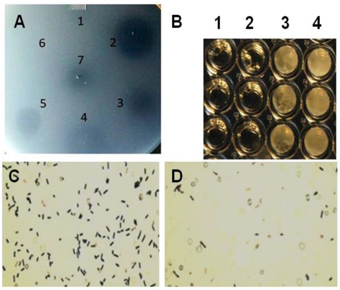
Figure 2.
Plate lysis, minimal inhibitory concentration and Gram-stain of lytic enzyme treated Clostridium perfringens 12917. (A) Plate (spot) lysis assay 10 µL spots: 1. PlyCpAmi (8 μg); 2. PlyGVE2CpCWB (10 μg) and lysozyme (2.5 μg); 3. PlyGVE2CpCWB (20 μg); 4. Lysozyme (5 μg); 5. PlyCP39O (5 μg); 6. Ampicillin (5 μg); and 7. Lower concentration PlyGVE2CpCWB (4 μg); (B) Minimal inhibitory concentration (MIC) for the recombinant PlyGVE2CpCWB protein. A representative MIC assay is illustrated that was determined by serially diluting the endolysin in 1:2 increments using diluent in sterile flat bottom, tissue culture treated, 96-well microtiter plates leaving wells with 100 µL. Well concentrations: 1. 2500 µg/mL, 2. 1250 µg/mL, 3. 625 µg/mL, and 4. 312.5 µg/mL. Buffer alone was used for control; (C) Gram-stain image of C. perfringens 12917 following treatment with PlyGVE2CpCWB protein. The bacterium was untreated (Panel C) or treated (Panel D) with the purified recombinant protein. Gram stain magnification is 1000×, as the 100× oil objective was used with a 10× eyepiece magnification.

