

Abstract
Spinal epidermoid cyst, congenital or acquired, is mainly congenital associated with spinal dysraphism, rarely in isolation. Intramedullary epidermoid cysts (IECs) are rare with less than 60 cases reported so far; isolated variety (i.e., without spinal dysraphism) is still rarer. Complete microsurgical excision is the dictum of surgical treatment. A 14-year-old boy presented with 4-month history of upper backache accompanied with progressive descending paresthesia with paraparesis with early bladder and bowel involvement. His condition deteriorated rapidly making him bedridden. Neurological examination revealed upper thoracic myeloradiculopathy probably of neoplastic origin with sensory localization to D5 spinal level. Digital X-ray revealed no feature suggestive of spinal dysraphism. Contrast magnetic resonance imaging (MRI) characteristics clinched the presumptive diagnosis. Near-total microsurgical excision was done leaving behind a small part of the calcified capsule densely adhered to cord. Histopathological features were confirmative of an epidermoid cyst. Postoperatively, he improved significantly with a gain of motor power sufficient to walk without support within a span of 6 months. Spinal IECs, without any specific clinical presentation, are often diagnosed based upon intraoperative and histopathological findings, however early diagnosis is possible on complete MRI valuation. Complete microsurgical excision, resulting in cessation of clinical progression and remission of symptoms, has to be limited to sub-total or near-total excision if cyst is adherent to cord or its confines.
Keywords: Diffusion weighted imaging, epidermoid, intramedullary, spinal dysraphism, thoracic vertebra
Introduction
An epidermoid cyst is a slow growing indolent rare lesion.[1,2] Most of the spinal epidermoid cysts are subdural and extramedullary. Intramedullary localization is very rare with <60 cases reported so far since the first reporting by Chiari (1833), and very few cases can be diagnosed preoperatively by magnetic resonance imaging (MRI) characteristics.[2,3,4,5,6,7] Most common location is thoracic followed by lumbosacral region.[6,8] Although microsurgical total excision is the treatment of choice, it is often not possible due to adherence to cord.
Case Report
A 14-year-old boy presented to us with a 4-month history of upper backache associated with progressive descending tingling, and numbness (unpleasant, burning in character), and profound progressive weakness in both lower limbs (LLs) making him bedridden, with early bladder, bowel involvement in form of increased frequency of micturition, and constipation. There was no past history of lumbar puncture, spinal trauma or previous spinal surgery. X-ray revealed no feature suggestive of spinal dysraphism [Figure 1]. Examination revealed spastic paraparesis with motor power of 2/5 around all joints of LLs, exaggerated knee and ankle jerks, extensor plantar with loss of all modalities of sensations below T8 segment bilaterally. Upper limbs, general, and systemic examination were normal. MRI of thoracic spine revealed an irregularly marginated intramedullary mass (2 cm × 2 cm × 1.3 cm) at D5 vertebral level, with fusiform expansion of cord and obliteration of anterior and posterior subarachnoid spaces at the same level. The lesion was hypointense on T1-weighted image [Figures 2a and 3a], hyperintense on T2-weighted image [Figures 2b and 3b], with postgadolinium enhancement peripherally [Figures 2c and 3c].
Figure 1.

Digital X-ray of cervical spine and upper thoracic region (anteroposterior, lateral) without any features of spinal dysraphism
Figure 2.
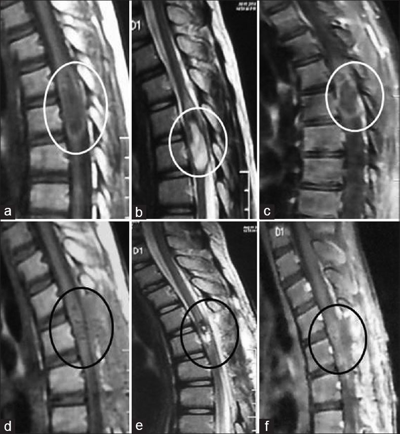
Sagittal magnetic resonance imaging of cervicothoracic spine (a-c) preoperative (T1 hypointense, T2 hyperintense with peripheral wall enhancement with cord compression), and (d-f) postoperative (very minimal residual without cord compression)
Figure 3.

Axial magnetic resonance imaging of cervicothoracic spine (a-c) preoperative (T1 hypointense, T2 hyperintense with peripheral wall enhancement with cord compression), and (d-f) postoperative (very minimal residual without cord compression)
We proceeded for D4–D5 laminectomy aiming for complete microsurgical excision. Dura was tense and bulging. Cord appeared thickened and expanded. Slightly lateral to midline the lesion was bulging and had thinned out the overlying cord. On fine dissection over the most bulging part, a pearly white, avascular, soft, flaky lesion within the cord substance was observed with gushing out of dense, cheese-like suckable material. Near-total excision of the lesion was done leaving behind the part of the capsule calcified and densely adhered to cord antero-medially [Figure 4a and b]. After hemostasis, watertight dural closure was done with wound closure layer by layer. Intraoperative diagnosis of intramedullary epidermoid cyst (IEC) was confirmed on histopathology [Figure 4c and d]. Postoperatively, with physiotherapy, and rehabilitation he had gradual progressive improvement of the preoperative neurological deficits. Follow-up MRI of spine at 1-month revealed very minimal residual [Figures 2d–f and 3d–f]. By 6 months follow-up he regained his power of both LLs to 4+/5 with minimal spasticity, and is now able to walk with support.
Figure 4.

Intraoperative images (a) intramedullary pearly white, avascular, soft, flaky, cheese-like suckable material being expulsed, (b) image after evacuation of cystic contents, and (c and d) histopathological images showing desquamated epithelium surrounded by keratin-producing squamous epithelium suggestive of epidermoid cyst
Discussion
Spinal epidermoid in children constitutes <1% of all central nervous system tumors.[1] Guidetti and Gagliardi reported the incidence as 0.7%,[1,2] whereas Bansal et al., reported the incidence as high as 20.7%.[3] IECs, extremely rare with <60 cases reported so far, mostly affect thoracic (D4–D6, and D11–D12) followed by lumbar region.[2,3,5,6,7,8]
Pathogenesis of spinal epidermoid is either congenital or acquired. Most of spinal epidermoid are congenital because of displacement of ectodermal inclusions during the closure of neural tube. Congenital epidermoid cyst can be associated with other abnormalities, e.g., hemivertebra, dermal sinus, spina bifida, syringomyelia, but may occur in isolation also. Defect of the overlying bone reported in about 10%, is possible but less frequent than in dermoid or some extramedullary epidermoids.[2,4,9,10] Isolated spinal epidermoid may present with either progressive compressive myelopathy, as in our case, or acute onset chemical meningitis (rupture of cyst and spread of cholesterol crystals into cerebral spinal fluid). Acquired epidermoid cyst, due to iatrogenic penetration of skin fragments, have been reported after lumbar puncture or meningomyelocele repair with a latency of years together.[1,3,7,14,15,18]
Slow growth and nonspecific clinical presentation are the reasons for delay in diagnosis. Clinical presentation is usually neurological, but rarely there may be urological involvement.[8] Diagnosis, often based on intraoperative and histological findings, can be established earlier by MRI with a range from 1 to 8 months. Computer tomography scan of an epidermoid cyst is characterized by a low density lesion that does not enhance.[6,7,11,12,19] MRI findings may be quite divergent, because of the disparity of signal intensity secondary to various lipid and protein composition. Amato et al. stated, in patients with spinal dysraphism MRI findings of well-defined, heterogeneous, T1-weighted hypointense lesion without perilesional edema may suggest tumor of developmental origin, e.g., epidermoid cyst, teratoma, or dermoid cyst. In absence of associated dysraphisms, diagnosis might be made by careful and complete MRI study including diffusion weighted imaging (DWI) sequences.[10,16,17,18] MRI characteristics of variable signal intensity between different parts of the same cyst at times, absence of perilesional edema, fairly well defined limits, and peripheral postgadolinium enhancement favors the diagnosis of epidermoid cyst. Margins of the epidermoid cyst may occasionally be “shaggy” because of chronic inflammatory response to squamous tissue “leak” through capsule and variable gliosis along the margin extending into the cord. These features help in differentiating from other intramedullary tumors, e.g., dermoid cyst, ependymoma, astrocytoma, teratoma, and hemangioma.[4,7,17,20] Some authors believe the peripheral enhancement represents normal tissue reaction surrounding tumor, while others consider it as outer tumor wall composed of tumor cells.[2,7,21] We believe it may be due to normal tissue reaction.
The surgical goal is complete excision. Sometimes, capsule is very thin and tightly adherent to the cord or located within its confines, warranting safe maximal resection to avoid damage to cord. Most authors believe in not attempting total removal of capsule in such cases. In most cases, even partial removal resulted in total remission of symptoms.[7,11,12,13] Adherence of capsule to cord anteromedially. Operative complications may include damage to the neurovascular structures, and sphincter disturbances. Aseptic chemical meningitis, unique to epidermoid lasting for weeks, if severe and long-lasting can lead to a “granulomatous” type of arachnoiditis.[13] Plugging of proximal and distal arachnoid space prior to tumor resection and irrigating and washing the site with normal saline prior to dural closure diminishes the chances of spillage of epidermoid and reduces the chances of chemical meningitis.
Histopathologically, epidermoid and dermoid cysts are lined by stratified squamous epithelium supported by an outer layer of collagenous tissue; presence of skin adnexa dictates the diagnosis of dermoid. In epidermoid cyst, progressive desquamation of keratin from epithelial lining toward the interior of cyst produces a soft cheesy white material with histopathology revealing desquamated epithelium surrounded by keratin-producing squamous epithelium.[3,7,11,12,14,22]
The risk of recurrence does exist and depends upon the degree of resection. Gross total removal along with the capsule diminishes the chances of recurrence. Though, radiotherapy has been mentioned as the treatment modality for only one case in the literature, symptomatic recurrences are best treated by surgery.[5,23]
Conclusion
Spinal IECs are rare tumors without any specific clinical presentation. Clinical presentation is usually neurological. Diagnosis is often based on intraoperative and histopathological findings, but possible early on complete MRI profile including DWI sequences. Complete microsurgical excision is the treatment of choice, and results in cessation of clinical progression and remission of symptoms sometimes limited by adherence of cyst to cord or its confines. Maximal safe resection in those cases reduces the morbidity and helps the patients with good long term functional outcome in these slow growing benign conditions.
Footnotes
Source of Support: Nil.
Conflict of Interest: None declared.
References
- 1.Guidetti B, Gagliardi FM. Epidermoid and dermoid cysts. Clinical evaluation and late surgical results. J Neurosurg. 1977;47:12–8. doi: 10.3171/jns.1977.47.1.0012. [DOI] [PubMed] [Google Scholar]
- 2.Penisson-Besnier I, Guy G, Gandon Y. Intramedullary epidermoid cyst evaluated by computed tomographic scan and magnetic resonance imaging: Case report. Neurosurg. 1989;25:955–9. doi: 10.1097/00006123-198912000-00017. [DOI] [PubMed] [Google Scholar]
- 3.Bansal S, Suri A, Borkar SA, Kale SS, Singh M, Mahapatra AK. Management of intramedullary tumors in children: Analysis of 82 operated cases. Childs Nerv Syst. 2012;28:2063–9. doi: 10.1007/s00381-012-1835-4. [DOI] [PubMed] [Google Scholar]
- 4.Roux A, Mercier C, Larbrisseau A, Dube LJ, Dupuis C, Del Carpio R. Intramedullary epidermoid cysts of the spinal cord. Case report. J Neurosurg. 1992;76:528–33. doi: 10.3171/jns.1992.76.3.0528. [DOI] [PubMed] [Google Scholar]
- 5.Kumar A, Singh P, Jain P, Badole CM. Intramedullary spinal epidermoid cyst of the cervicodorsal region: A rare entity. J Pediatr Neurosci. 2010;5:49–51. doi: 10.4103/1817-1745.66675. [DOI] [PMC free article] [PubMed] [Google Scholar]
- 6.Cincu R, Lázaro JF, Liesa JL, Callizo JR. Dorsal intramedullary spinal epidermoid cysts: Report of two cases and review of literature. Indian J Orthop. 2007;41:395–7. doi: 10.4103/0019-5413.37005. [DOI] [PMC free article] [PubMed] [Google Scholar]
- 7.Chandra PS, Manjari T, Devi BI, Chandramouli BA, Srikanth SG, Shankar SK. Intramedullary spinal epidermoid cyst. Neurol India. 2000;48:75–7. [PubMed] [Google Scholar]
- 8.Ferrara P, Costa S, Rigante D, Mule A, D’Aleo C, Pulitanò S, et al. Intramedullary epidermoid cyst presenting with abnormal urological manifestations. Spinal Cord. 2003;41:645–8. doi: 10.1038/sj.sc.3101482. [DOI] [PubMed] [Google Scholar]
- 9.Kirsch WM, Hodges FJ., 3rd An intramedullary epidermal inclusion cyst of the thoracic cord associated with a previously repaired meningocele. Case report. J Neurosurg. 1966;24:1018–20. doi: 10.3171/jns.1966.24.6.1018. [DOI] [PubMed] [Google Scholar]
- 10.Gupta S, Gupta RK, Gujral RB, Mittal P, Kuriyal M, Krishnani N. Signal intensity patterns in intraspinal dermoids and epidermoids on MR imaging. Clin Radiol. 1993;48:405–13. doi: 10.1016/s0009-9260(05)81110-9. [DOI] [PubMed] [Google Scholar]
- 11.Kaye AH. Essential Neurosurgery. USA: Churchill Livingstone; 1991. pp. 144–6. [Google Scholar]
- 12.Findlay GF. Compressive and vascular disorders of the spinal cord. In: Miller JD, editor. Northfield's Surgery of the Central Nervous System. 2nd ed. UK: Blackwell Scientific Publications; 1987. pp. 739–40. [Google Scholar]
- 13.Conley FK. Epidermoid and dermoid tumors: Clinical features and surgical management. In: Wilkins RH, Rengachary SS, editors. Neurosurgery. I. USA: McGraw-Hill Book Company; 1985. pp. 668–73. [Google Scholar]
- 14.Manno NJ, Uihlein A, Kernohan JW. Intraspinal epidermoids. J Neurosurg. 1962;19:754–65. doi: 10.3171/jns.1962.19.9.0754. [DOI] [PubMed] [Google Scholar]
- 15.Halcrow SJ, Crawford PJ, Craft AW. Epidermoid spinal cord tumour after lumbar puncture. Arch Dis Child. 1985;60:978–9. doi: 10.1136/adc.60.10.978. [DOI] [PMC free article] [PubMed] [Google Scholar]
- 16.Amato VG, Assietti R, Arienta C. Intramedullary epidermoid cyst: Preoperative diagnosis and surgical management after MRI introduction. Case report and updating of the literature. J Neurosurg Sci. 2002;46:122–6. [PubMed] [Google Scholar]
- 17.Scholz M, Märzheuser-Brands S, Gottschalk J, Böck JC, Lanksch WR. Intramedullary epidermoid cyst. A case report. Neurosurg Rev. 1994;17:89–93. doi: 10.1007/BF00309994. [DOI] [PubMed] [Google Scholar]
- 18.Visciani A, Savoiardo M, Balestrini MR, Solero CL. Iatrogenic intraspinal epidermoid tumor: Myelo-CT and MRI diagnosis. Neuroradiology. 1989;31:273–5. doi: 10.1007/BF00344358. [DOI] [PubMed] [Google Scholar]
- 19.Osborne DR. Epidermoid and dermoid tumors: Radiology. In: Wilkins RH, Rengachary SS, editors. Neurosurgery. I. USA: McGraw-Hill Book Company; 1985. pp. 662–7. [Google Scholar]
- 20.Debray MP, Ricolfi F, Brugières P, Khalil A, Adle-Biassette H, Gaston A. Epidermoid cyst of the conus medullaris: Atypical MRI and angiographic features. Neuroradiology. 1996;38:526–8. doi: 10.1007/BF00626088. [DOI] [PubMed] [Google Scholar]
- 21.Shaywitz BA. Epidermoid spinal cord tumors and previous lumbar punctures. J Pediatr. 1972;80:638–40. doi: 10.1016/s0022-3476(72)80062-3. [DOI] [PubMed] [Google Scholar]
- 22.Baxter JW, Netsky MG. Epidermoid and dermoid tumors: Pathology. In: Wilkins RH, Rengachary SS, editors. Neurosurgery. I. USA: McGraw-Hill Book Company; 1985. pp. 655–1. [Google Scholar]
- 23.Bretz A, Van den Berge D, Storme G. Intraspinal epidermoid cyst successfully treated with radiotherapy: Case report. Neurosurgery. 2003;53:1429–31. doi: 10.1227/01.neu.0000093828.70768.40. [DOI] [PubMed] [Google Scholar]