Abstract
The recognition that oxidative stress is a major component of several chronic diseases has engendered numerous trials of antioxidant therapies with minimal or no direct benefits. Nanomolar quantities of nitric oxide released into the circulation by pharmacologic stimulation of eNOS have antioxidant properties but physiologic stimulation as through increased pulsatile shear stress of the endothelium has not been assessed. The present study utilized a non-invasive technology, periodic acceleration (pGz) that increases pulsatile shear stress such that upregulation of cardiac eNOS occurs, We assessed its efficacy in normal mice and mouse models with high levels of oxidative stress, e.g. Diabetes type 1 and mdx (Duchene Muscular Dystrophy). pGz increased protein expression and upregulated eNOS in hearts. Application of pGz was associated with significantly increased expression of endogenous antioxidants (Glutathioneperoxidase-1(GPX-1), Catalase (CAT), Superoxide, Superoxide Dismutase 1(SOD1). This led to an increase of total cardiac antioxidant capacity along with an increase in the antioxidant response element transcription factor Nrf2 translocation to the nucleus. pGz decreased reactive oxygen species in both mice models of oxidative stress. Thus, pGz is a novel non-pharmacologic method to harness endogenous antioxidant capacity.
Introduction
Redox signaling, defined as the reversible oxidation/reduction modification of cellular signaling pathways by reactive species is an important process in many physiological and pathophysiological states [1]. In the heart and vasculature, redox signaling is involved in excitation-contraction coupling (ECC), cell differentiation, stress response pathways, e.g., adaptation to hypoxia/ischemia and pathological processes, adverse cardiac remodeling, fibrosis, and atherosclerosis [2–4]. Reactive oxygen species (ROS) include free oxygen radicals, oxygen ions and peroxides. ROS at low to moderate concentrations regulate vascular tone, oxygen sensing, cell growth and proliferation, apoptosis, and inflammatory responses. Excessive or sustained ROS production, when exceeding the available antioxidant defense systems, produces oxidative stress that damages cell structure and disrupts function through lipid peroxidation of cell membranes, degrades nucleic acids [5]. Oxidative damage to cells and tissues is involved in the aging process and in chronic diseases including atherosclerosis, heart failure and cancer among others. Endogenously occurring protective antioxidants Glutathioneperoxidase-1 (GPX1), Superoxide Dismutase-1 (SOD-1, Cu-Zn SOD), and Catalase (CAT) maintain the balance of oxidizing chemicals, thereby playing a vital role in reduction of oxidative stress [1]. Epidemiological data suggest that diets rich in antioxidants have a protective effect on the development of cardiovascular disease. However, clinical trials and large meta-analysis have failed to show evidence for support of antioxidant supplements for prevention of cardiovascular disease and suggest potential deleterious effects [6–8]. Thus, upregulation of endogenous protective antioxidants might be more clinically relevant.
In-vitro and in-vivo (e.g. exercise) experiments show that shear stress to the endothelium increases endogenous antioxidants and activates endothelial nitric oxide synthase (eNOS) [9–13]. eNOS activation produces nanomolar quantities of nitric oxide (NO) which elicit endothelial dependent pulmonary and systemic vasodilation, increase blood flow, and signal increased expression of cytoprotective genes such as antioxidant enzymes [14–16]. Shear Stress induced antioxidant response has been associated with upregulation of the nuclear factor erythroid 2-related factor (Nrf2) a transcription factor that functions as the key controller of the redox homeostatic gene regulatory network.
Periodic acceleration (pGz) in humans and animal models (pigs and rodents) adds low amplitude pulses to the circulation. pGz is produced by a motorized platform that rapidly and repetitively moves the horizontally oriented body sinusoidally in a head to foot direction. Inertia of fluid as the body accelerates and decelerates adds a small amplitude pulse to the circulation that is superimposed upon the natural pulse thereby increasing pulsatile shear stress to the endothelium. Pulsatile shear stress induced by pGz releases eNOS derived NO into the circulation in amounts that are physiologically meaningful and long lasting [17, 18]. We recently found that pGz ameliorates muscle pathology in mdx mice [19] and reduces myocardial damage after ischemic insult [20] both pathologies are associated with elevated oxidative stress. The purpose of this study was to determine whether pGz upregulates endogenous antioxidants in hearts of normal mice and decreases oxidative stress in mice models characterized by high oxidative stress, e.g. Type 1 Diabetes and mdx (Duchene Muscular Dystrophy).
Materials and Methods
2.1 Animal Procedures
The experimental protocol No. 14-22-A-04 was approved by the Mount Sinai Medical Center Animal Care and Use Committee and conforms to the Guide for the Care and Use of Laboratory Animals published by the National Institutes of Health (NIH Publication No. 85–23, revised 1996). Male mice C57BL/6 and dystrophic (mdx) (C57BL/10ScSn-Dmd mdx/J) (Total n = 250) weighing between 20 and 25 g (Harlan Laboratories, Indianapolis, IN and Jackson Laboratory (Bar Harbor, Maine), were used for these experiments.
pGz was applied with a platform that moved with a linear displacement direct current motor (Model 400, 12V; APS Dynamics, Carlsbad, CA) powered by a dual mode power amplifier (Model 144, APS Dynamics), connected to a sine wave controller (Model 140–072; NIMS, Miami, FL). The controller allows control of frequency and travel distance of the platform with a read-out of acceleration from an accelerometer. The platform has a maximum weight capacity of 30 kg, operates at a frequency from 30–720 cycles/min (cpm) and achieves accelerations ranging from ±0.1 to ±14.7m/sec2. Optimum frequency of pGz in mice was determined as previously reported [18]. (S1 File) pGz treatment was performed on unanesthetized, restrained mice using the above described platform (1 hr. per day ƒ = 480cpm and Gz ±3.0 mt/sec2) for 14 consecutive days for both diabetic and mdx mice models and their respective controls.
2.2 Determination of Total Antioxidant Capacity and Antioxidant Protein Expression
Mice (n = 60) received daily pGz (1 hr per day ƒ = 480cpm and Gz ±3.0 mt/sec2). Myocardial tissue was harvested at baseline (BL) and twenty four hours after 1, 2, and 4 wk. of daily pGz. Protein expression of Glutathioneperoxidase-1 (GPX1), Superoxide Dismutase-1 (SOD-1, Cu-Zn SOD), and Catalase (CAT) were measured in myocardial tissue by western blot. Total Antioxidant Capacity (TC) was measured in myocardial tissue after 4 wk. of pGz or in time control mice (CONT) by ELISA (Abcam, Cambridge, MA) (S1 File).
2.3 Protein Expression and Genomic upregulation of Antioxidant Response Element (ARE) Transcription Factor Nrf2
We measured protein expression and genomic upregulation (RT-PCR) of Nrf2, and cytosolic to nuclear translocation of Nrf2 in mice (n = 20) hearts exposed to 4 wk. of pGz (1 hr/day) or time control (Qiagen, Valencia CA). Homogenates where fractionated to cytoplasmic and nuclear fraction by subcellular protein fractionation method (Life Technologies, Thermo Fisher Scientific, Rockford, IL.).
2.4 Oxidative Stress Mice Models
Type 1 Diabetes Model
C57BL/6J male mice (n = 20), 3 months of age, were injected with a single intraperitoneal (IP) dose of streptozotocin (STZ) (150 mg/kg body weight, in 0.1 mol/L sodium citrate buffer, pH 4.5) (Sigma–Aldrich, St. Louis, MO, USA). Aged-matched control mice received a single equal volume IP injection of sodium citrate buffer. Determination of plasma glucose 4 days after injection confirmed hyperglycemia with glucose of > 250mg/dl. Diabetic and age-matched controls mice were randomly divided into four groups of animals (n = 5 per group): i) control (CONT), ii) control pGz (CONT-pGz), iii) diabetic (Diab) and iv) diabetic-pGz (Diab-pGz) groups.
Duchene Muscular Dystrophy (mdx) Model
Male 12 months old C57BL/10 wild type (Wt) and dystrophic (mdx) (C57BL/10ScSn-Dmd mdx/J) mice were obtained from Jackson Laboratory (Bar Harbor, Maine). Both mdx and age-matched controls mice were randomly divided into four groups (n = 4 per group): i) wild type control (Wt), ii) wild type + pGz (Wt-pGz), iii) mdx (mdx) and iv) mdx+pGz (mdx-pGz) groups.
2.5 Cardiomyocyte isolation
Ventricular cardiomyocytes were isolated using collagenase enzymatic digestion via retrograde perfusion. The left ventricle was dissected and minced cardiomyocytes were resuspended sequentially in various concentrations of Tyrode solution, followed by exposure to 1.5 mM Ca2+ for 15 min before being resuspended in normal Tyrode solution supplemented with normal Ca2+ concentration (1.8 mM) (S1 File).
2.6 Measurement of Reactive Oxygen Species
ROS activity was determined in isolated cardiomyocytes using the fluorescent method of chloromethyl-2,7-dichlorodihydrofluorescein diacetate (CM-DCFDA Molecular Probes, OR, USA). (S1 File)
2.7 Protein Expression and Phosphorylation
Protein extraction, RNA and subcellular fractionations where performed as previously described. Protein analysis was performed using Western Blot method and RT-PCR blots visualized by enhanced chemifluorescence (S1 File).
2.8 Euthanasia
After completion of each of the experimental protocols animals were euthanized by a method approved by the American Veterinary Medical Association Guidelines on Euthanasia (S1 File).
2.9 Statistical Analysis
Statistical analysis of the data was accomplished with software running on a personal computer Statistica (StatSoft, Tulsa, OK). Results were expressed as mean percent (%) to baseline with SD or Mean ± SD. and comparison of means with 95% confidence levels was carried out using analysis of variance (ANOVA) followed by post hoc analysis with the Newman-Keuls test. Statistical significance was established at p<0.05. Sample size was calculated using Statistica based on power analysis with α = 0.05 and power 0.80.
Results
3.1 Effects of pGz frequency, on Mean Aortic Pressure, Heart Rate and a/b in normal mice
pGz at all frequencies from 360 to 600 cpm added pulses visible on the aortic pulse waveform of 1–3 mmHg as a function of frequency. These added pulses occurred at peaks and troughs of acceleration of the motion platform. The descent of the dicrotic notch pressure wave (a/b) cycled downward to upward over periods ranging from 2 to 5 seconds but the overall downward descent was maintained throughout the pGz period. The descent of the dicrotic notch was greatest at a pGz frequency of 480cpm, decreasing by more than 90% of BL values.
There were no significant changes from BL in mean heart rate with application of pGz at all frequencies. pGz decreased mean arterial pressure (MAP) within five minutes of its application but the a/b ratio increased about 25 to 50% from baseline within 2 seconds. MAP progressively decreased during 30 minutes after pGz. pGz administration decreased MAP from baseline of 67 ±3 to 59 ±4, 52 ±2, 53±5 mmHg after 30 min of pGz at 360, 480, 600 cpm respectively (p<0.05 Bl vs. pGz) (S1 File).
3.2 Time Course of Endothelial Nitric Oxide Synthase, Akt and Expression and Phosphorylation after pGz
One, 4 and 8 days of pGz increased eNOS, p-eNOS, and ratio of p-Akt/Akt. Phosphorylation of eNOS and Akt was most pronounced after 1 day of pGz. Protein and genomic expression of eNOS were most pronounced after 8 days of pGz. After 8 days of pGz, upregulation of eNOS peaked at 48 hrs after the last pGz session and gradually declined as a function of elapsed time from last pGz session. In a Duchene Muscular Dystrophy mouse model (mdx) eNOS and p-eNOS were significantly decreased compared to Wt controls. Four weeks of pGz in mdx significantly restored both eNOS and p-eNOS to close to Wt levels (S1 File).
3.3 pGz Induces Antioxidant Expression and Increases Antioxidant Capacity
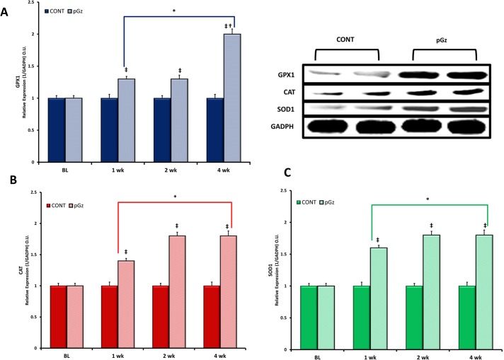
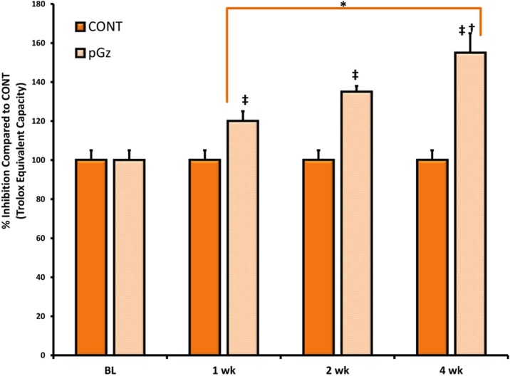
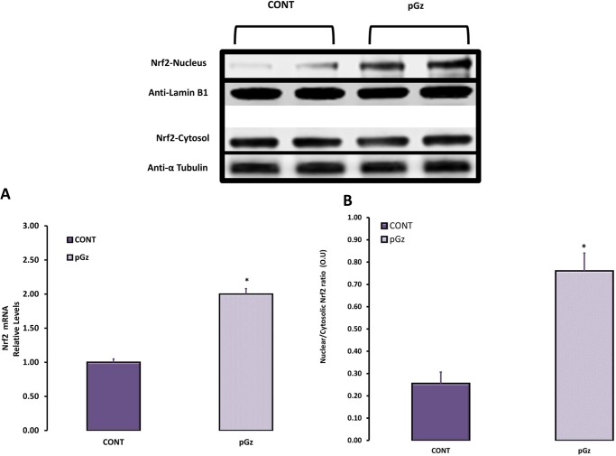
Induction of antioxidant enzymes by pGz was measured after 1, 2 and 4 wk. of daily pGz. Peak effect of enzymatic expression and total antioxidant capacity was seen after 4 wk. of pGz for GPX1, CAT, SOD1 (Figs 1 and 2). After 4 wk. of pGz, protein expression and upregulation of the antioxidant response element, transcription factor Nrf2, was significantly increased. Furthermore, pGz induced Nrf2 translocation from cytosol to nucleus (Fig 3).
Fig 1. Antioxidant Protein Expression is Induced by pGz.
This figure depicts the effects of 1, 2, and 4 wk. of daily pGz in normal mice on the expressions of (A). Glutathioneperoxidase-1 (GPX1), (B). Catalase (CAT), and (C). Superoxide Dismutase 1 (SOD1). The relative protein expressions of GPX1, CAT and SOD1 over time compared to time of CONT values showed that pGz significantly increased GPX1, CAT, and SOD1 expression at 1, 2 and 4 wk compared to time CONT (‡p < 0.01 1, 2, and 4 wk. pGz vs. CONT). One, 2, and 4 wk. of pGz significantly increased expression of GPX1, CAT, and SOD1 compared to baseline (BL) values for both CONT and pGz groups (* p< 0.01 1,2, and 4 wk. vs. BL values). Representative Immunoblots of protein expression of GPX1, CAT, SOD1 and protein quantity loading Glyceraldehyde 3-phosphate dehydrogenase (GADPH) after 4 wk. of pGz, for CONT and pGz groups showing significant expression of these compared to time CONT.
Fig 2. pGz Increases Total Antioxidant Capacity.
Total antioxidant capacity (Trolox Equivalent Capacity) was expressed as % Inhibition of control values, measured at baseline (BL) for CONT and pGz groups, and after 1, 2 and 4 wk. of pGz or time CONT. One, 2 and 4 wk. of daily pGz significantly increased antioxidant capacity compared to time CONT (‡p < 0.01 1, 2 and 4 wk. vs. time CONT). One, 2, and 4 weeks of pGz also significantly increased total antioxidant capacity compared to baseline (BL)values for both CONT and pGz (* p< 0.01 1,2 and 4 wk. vs. BL values for CONT and pGz groups). Optical Units = O.U.
Fig 3. The Expression and Translocation of Nrf2 Transcription Factor after pGz Treatment.
This figure depicts the expression and translocation of Nrf2 transcription factor after 4 wk of daily pGz or no pGz (CONT) (A). pGz significantly increased upregulation of the Antioxidant Response Element transcription factor Nrf2 compared to CONT (*p< 0.01) (B). Translocation of Nrf2 is expressed as the ratio of Nrf2 in nucleus and cytosol. pGz also significantly increased Nrf2 translocation to nucleus compared to CONT (*p< 0.01). Representative Immunoblots of Nrf2 in nucleus and cytosol with respective protein loading controls Anti-Laminin B1 (nucleus) and Anti-α Tubulin (cytosol) in CONT and pGz groups showed increased protein expression of Nrf2 in nucleus of pGz treated mice. Optical Units = O.U.
3.4 Effects of pGz in Models of Oxidative Stress
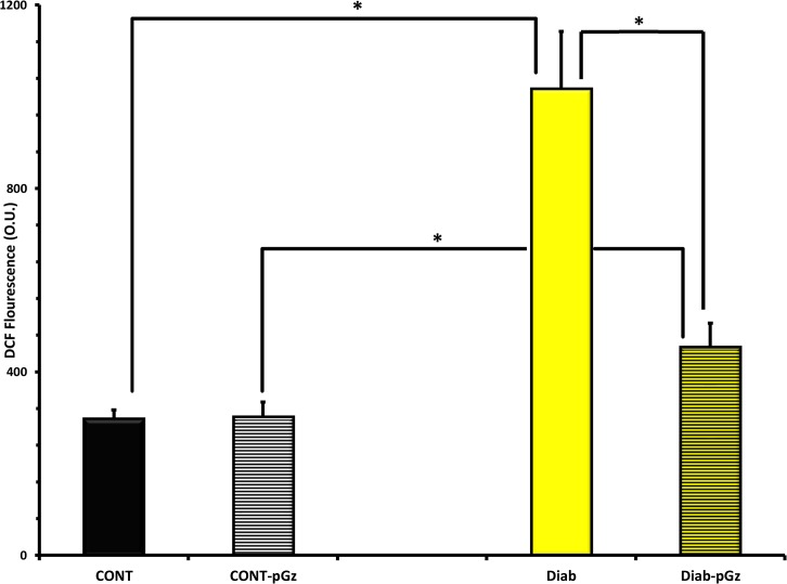
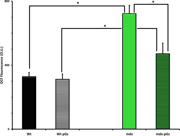
In order to study whether or not pGz reduces oxidative stress, we studied the effects of pGz on ROS production in two models of cardiovascular disease associated with elevated oxidative stress, type 1 diabetes and mdx. Type 1 Diabetes induces elevated cardiac oxidative stress as demonstrated in cardiomyocytes, ROS was 3.4 fold higher in diabetic mice than Wt (1017±28.0 to 298±4.3 O.U. (p< 0.001). Fourteen days of daily pGz application significantly reduced ROS from 1017±28.0 to 454 ±11.7 O.U, (p < 0.001) (Fig 4). The 12 mos age mdx mouse model of Duchene Muscular Dystrophy, is also characterized by significant amount of oxidative stress in heart with levels of ROS of 722±11.7 compared to 328±7.1 O.U in Wt, (p < 0.001), pGz also significantly attenuated oxidative stress in the mdx model from 722±11.7 to 471±15.0 O.U., (p < 0.001) (Fig 5).
Fig 4. The Effects of pGz treatment on Diabetes Induced Oxidative Stress in Cardiomyocytes.
The effects of pGz treatment for 14 days on ROS in cardiomyocytes in Control (CONT-pGz) and Diabetic mice (Diab-pGz) and their respective controls without pGz treatment in non diabetic (CONT) and diabetic (Diab). ROS was measured using the method of DCF fluorescence. Diabetes significantly increased ROS in cardiomyocytes (*p < 0.001 CONT vs. Diab). Treatment with pGz significantly reduced the diabetes induced increase in ROS (*p < 0.001 Diab vs. Diab-pGz and CONT-pGz vs Diab-pGz). Optical Units = O.U.
Fig 5. The Effects of pGz treatment on Duchene Muscular Dystrophy Induced Oxidative Stress in Cardiomyocytes.
Twelve month of age mdx mice had a significant increase in oxidative stress in cardiomyocytes compared to age matched wild type (Wt) controls (*p < 0.001). Treatment with pGz (mdx-pGz) for 14 days significantly reduced the levels of ROS measured by DCF fluorescence (*p<0.001) Optical Units = O.U.
Discussion
The present study demonstrates that one hour daily treatments of pGz to normal mice increased expression and phosphorylation of eNOS in the heart as a function of the number of treatments over time. Application of pGz through increased pulsatile shear stress activates eNOS through phosphorylation and genomic upregulation in cells and animal models in part mediated via the Akt/PI3K pathway [21] [18]. In the mdx model where eNOS and p-eNOS are decreased compared Wt, pGz also restored both. Further, we have also shown in other animal models in which eNOS and p-eNOS are also decreased such as whole body ischemia reperfusion injury [22], and focal myocardial ischemia [23] that pGz also significantly restores these. The clinical importance of increasing and activating eNOS has been reviewed by others [24–27].
Endogenous antioxidant enzymatic expressions and activity were also increased by pGz. Various methods to increase shear stress have been used to augment antioxidant levels. In-vitro studies have shown that pulsatile shear stress on the vascular endothelium increases Cu/Zn SOD (SOD1) and GPX1 [9, 28–31]. Exercise is an intervention which also increases shear stress. Aging mice studies have shown that chronic moderate treadmill exercise produces a mild effect at increasing the activities of Mn-SOD, SOD1, and catalase in brain, heart, liver, and kidney of mice exercised for 24 to 50 wk. After 24 weeks of aerobic exercise, antioxidant enzyme increased by 15–20% [13]. Additionally, the beneficial effects of exercise in diseased animal models have in part been shown to be related to augmentation of antioxidant defenses [32, 33]. Exercise may or may not be beneficial in mdx mice (model of Duchene Muscular Dystrophy, DMD). In this model, mdx mice running for 4 or 10 weeks accelerates ventricular dilatation and fibrosis [34–36] whereas another study showed that voluntary wheel running for one yr. produced positive exercise–induced remodeling in the heart [37].
Diabetic cardiomyopathy is a well-known complication of diabetes. Diabetic cardiomyopathy is characterized by early diastolic dysfunction and adverse structural remodeling leading to heart failure (HF). Pre-clinical studies confirm a major causal role for elevated myocardial ROS generation in diabetic cardiomyopathy [38]. In a type 1 diabetic rat model, 9 weeks of low intensity exercise provided protection from cardiomyopathy in part via augmentation of the antioxidant capacity [39]. Additionally, augmentation of extracellular SOD using a transgenic mouse model with type 1 diabetes protects from cardiac hypertrophy, fibrosis and dysfunction [40]. Thus, reduction of oxidative stress and augmentation of antioxidants could prevent/ameliorate the development and progression of diabetic cardiomyopathy [41, 42]. In the current study pGz increased antioxidant capacity and reduced ROS generation in diabetic cardiomyocytes, thus making it an attractive therapeutic modality.
A possible mechanism whereby pGz increases antioxidant capacity may involve the antioxidant response element (ARE) transcription factor Nrf2 which serves as the key controller of the redox homeostatic gene regulatory network. This factor was upregulated after pGz and translocated to the nucleus site of activity. Nrf2 regulates expression of genes containing antioxidant response element in their promoters e.g. heme oxygenase-1(HO-1), NAD(P)H quinone oxidoreductase 1(NQO1) [12, 43–49]. The Nrf2/Keap1/ARE signaling pathway has been used as a pharmacological therapeutic target [50, 51]. Nrf2 activity is reduced in diabetic cardiomyopathy and its activation has been shown to protect from diabetic cardiomyopathy[52]. Similarly, Nrf2 induction via sulforaphane has also been shown to improve muscle function and pathology in mdx [53].
There are limitations to the present work. We did not explore exposure to pGz for longer than 4 wk. and beneficial effects beyond this time period can only be speculated upon. We also did not address eNOS uncoupling in this study, the latter is highly unlikely in the setting of endogenous physiological production of NO in non-diseased mice.
Oxidative stress and vascular inflammation play a pivotal role in cardiovascular health and particularly in the aging human population [54]. The therapeutic value of enhancing antioxidant capacity for cardiovascular protection has been reviewed by others [51, 54, 55]. pGz is a noninvasive simple intervention, which does not require subject cooperation, and can be performed in persons with physical and or cognitive impairment. Since pGz has been safely used in humans with various cardiovascular diseases, our findings in these animal models are very clinically relevant [56–60]. In addition to the well-known salutary effects produced by activation and upregulation of eNOS, pGz also increases endogenous antioxidant capacity and reduces indices of oxidative stress, which have profound therapeutic potential.
Supporting Information
The Materials and Methods include; a) method for the determination of optimum endothelial vasodilatation by pGz in mice, b) methods for the determination of the time course of protein expression and phosphorylation of eNOS and Akt, c) methods for the determination of ROS in cardiomyocytes, d) supplemental description for methods for protein expression and phosphorylation, e) method for animal euthanasia. The Results section includes; a) representative tracing of the aortic pulse waveform in mice and the effects of pGz of varying frequency on the position of the dicrotic notch, b) figures for the effect of pGz frequency on mean arterial blood pressure and the change in a/b ratio, c) figures on the effects of duration of pGz on protein expression of eNOS and the ratio of p-eNOS/eNOS, d) figures on the effects of pGz on genomic upregulation of eNOS, e) figures on the effects of duration of pGz on protein expression of Akt and p-Akt/Akt, f) figures on the effect of 4 weeks of pGz on eNOS and p-eNOS protein expression in control (wt) and Duchenne Muscular Dystrophy (mdx) mouse model.
(DOCX)
Acknowledgments
The authors gratefully acknowledge the scientific contributions and the critical review of the manuscript from Marvin Sackner, M.D, and the technical support of Jaqueline Arias. This work was funded through a grant from the Florida Heart Research Institute.
Data Availability
All relevant data are within the paper and its Supporting Information files.
Funding Statement
This study was funded by a Grant to JAA from the Florida Heart Research Institute (www.floridaheart.org).
References
- 1. Valko M, Leibfritz D, Moncol J, Cronin MT, Mazur M, Telser J. Free radicals and antioxidants in normal physiological functions and human disease. Int J Biochem Cell Biol. 2007;39(1):44–84. 10.1016/j.biocel.2006.07.001 . [DOI] [PubMed] [Google Scholar]
- 2. Cingolani HE, Perez NG, Cingolani OH, Ennis IL. The Anrep effect: 100 years later. American journal of physiology. 2013;304(2):H175–82. 10.1152/ajpheart.00508.2012 . [DOI] [PubMed] [Google Scholar]
- 3. Zhang M, Shah AM. ROS signalling between endothelial cells and cardiac cells. Cardiovascular research. 2014;102(2):249–57. 10.1093/cvr/cvu050 . [DOI] [PubMed] [Google Scholar]
- 4. Sag CM, Wagner S, Maier LS. Role of oxidants on calcium and sodium movement in healthy and diseased cardiac myocytes. Free radical biology & medicine. 2013;63:338–49. 10.1016/j.freeradbiomed.2013.05.035 . [DOI] [PubMed] [Google Scholar]
- 5. Burgoyne JR, Mongue-Din H, Eaton P, Shah AM. Redox signaling in cardiac physiology and pathology. Circulation research. 2012;111(8):1091–106. 10.1161/CIRCRESAHA.111.255216 . [DOI] [PubMed] [Google Scholar]
- 6. Bjelakovic G, Nikolova D, Gluud C. Antioxidant supplements and mortality. Curr Opin Clin Nutr Metab Care. 2014;17(1):40–4. 10.1097/MCO.0000000000000009 . [DOI] [PubMed] [Google Scholar]
- 7. Myung SK, Ju W, Cho B, Oh SW, Park SM, Koo BK, et al. Efficacy of vitamin and antioxidant supplements in prevention of cardiovascular disease: systematic review and meta-analysis of randomised controlled trials. BMJ (Clinical research ed. 2013;346:f10 10.1136/bmj.f10 [DOI] [PMC free article] [PubMed] [Google Scholar]
- 8. Ye Y, Li J, Yuan Z. Effect of antioxidant vitamin supplementation on cardiovascular outcomes: a meta-analysis of randomized controlled trials. PLoS One. 2013;8(2):e56803 10.1371/journal.pone.0056803 [DOI] [PMC free article] [PubMed] [Google Scholar]
- 9. Takeshita S, Inoue N, Ueyama T, Kawashima S, Yokoyama M. Shear stress enhances glutathione peroxidase expression in endothelial cells. Biochemical and biophysical research communications. 2000;273(1):66–71. 10.1006/bbrc.2000.2898 . [DOI] [PubMed] [Google Scholar]
- 10. Rahal A, Kumar A, Singh V, Yadav B, Tiwari R, Chakraborty S, et al. Oxidative Stress, Prooxidants, and Antioxidants: The Interplay. BioMed research international. 2014;2014:761264 10.1155/2014/761264 [DOI] [PMC free article] [PubMed] [Google Scholar]
- 11. Li H, Horke S, Forstermann U. Vascular oxidative stress, nitric oxide and atherosclerosis.Atherosclerosis.2014;237(1):208–19. 10.1016/j.atherosclerosis.2014.09.001 . [DOI] [PubMed] [Google Scholar]
- 12. Muthusamy VR, Kannan S, Sadhaasivam K, Gounder SS, Davidson CJ, Boeheme C, et al. Acute exercise stress activates Nrf2/ARE signaling and promotes antioxidant mechanisms in the myocardium. Free radical biology & medicine. 2012;52(2):366–76. 10.1016/j.freeradbiomed.2011.10.440 . [DOI] [PMC free article] [PubMed] [Google Scholar]
- 13. Navarro A, Gomez C, Lopez-Cepero JM, Boveris A. Beneficial effects of moderate exercise on mice aging: survival, behavior, oxidative stress, and mitochondrial electron transfer. Am J Physiol Regul Integr Comp Physiol. 2004;286(3):R505–11. 10.1152/ajpregu.00208.2003 . [DOI] [PubMed] [Google Scholar]
- 14. Forstermann U, Li H. Therapeutic effect of enhancing endothelial nitric oxide synthase (eNOS) expression and preventing eNOS uncoupling. Br J Pharmacol. 2011;164(2):213–23. Epub 2011/01/05. 10.1111/j.1476-5381.2010.01196.x [DOI] [PMC free article] [PubMed] [Google Scholar]
- 15. Elrod JW, Calvert JW, Gundewar S, Bryan NS, Lefer DJ. Nitric oxide promotes distant organ protection: evidence for an endocrine role of nitric oxide. Proceedings of the National Academy of Sciences of the United States of America. 2008;105(32):11430–5. Epub 2008/08/08. doi: 0800700105 [pii] 10.1073/pnas.0800700105 [DOI] [PMC free article] [PubMed] [Google Scholar]
- 16. Adams JA, Bassuk J, Wu D, Grana M, Kurlansky P, Sackner MA. Periodic acceleration: effects on vasoactive, fibrinolytic, and coagulation factors. J Appl Physiol (1985). 2005;98(3):1083–90. 10.1152/japplphysiol.00662.2004 . [DOI] [PubMed] [Google Scholar]
- 17. Sackner MA, Gummels E, Adams JA. Nitric oxide is released into circulation with whole-body, periodic acceleration. Chest. 2005;127(1):30–9. 10.1378/chest.127.1.30 . [DOI] [PubMed] [Google Scholar]
- 18. Uryash A, Wu H, Bassuk J, Kurlansky P, Sackner MA, Adams JA. Low-amplitude pulses to the circulation through periodic acceleration induces endothelial-dependent vasodilatation. J Appl Physiol (1985). 2009;106(6):1840–7. Epub 2009/03/28. 10.1152/japplphysiol.91612.2008 . [DOI] [PubMed] [Google Scholar]
- 19. Altamirano F, Perez CF, Liu M, Widrick J, Barton ER, Allen PD, et al. Whole body periodic acceleration is an effective therapy to ameliorate muscular dystrophy in mdx mice. PLoS One. 2014;9(9):e106590 10.1371/journal.pone.0106590 [DOI] [PMC free article] [PubMed] [Google Scholar]
- 20. Uryash A, Bassuk J, Kurlansky P, Altamirano F, Lopez JR, Adams JA. Non-Invasive Technology That Improves Cardiac Function after Experimental Myocardial Infarction: Whole Body Periodic Acceleration (pGz). PLoS One. 2015;10(3):e0121069 10.1371/journal.pone.0121069 . [DOI] [PMC free article] [PubMed] [Google Scholar]
- 21. Wu H, Uryash A, Bassuk J, Kurlansky P, Giridharan GA, Shakeri M, et al. Mechanisms of Periodic Acceleration Induced Endothelial Nitric Oxide Synthase (eNOS) Expression and Upregulation Using an In Vitro Human Aortic Endothelial Cell Model. Cardiovascular Engineering and Technology. 2012;3(3):292–301. 10.1007/s13239-012-0096-4 [DOI] [Google Scholar]
- 22. Adams JA, Wu H, Bassuk JA, Arias J, Uryash A, Jorapur V, et al. Periodic acceleration (pGz) prior to whole body ischemia reperfusion injury provides early cardioprotective preconditioning. Life Sci. 2010;86(19–20):707–15. Epub Epub 2010 Mar 6. 10.1016/j.lfs.2010.02.022 . [DOI] [PubMed] [Google Scholar]
- 23. Uryash A, Wu H, Bassuk J, Kurlansky P, Adams JA. Preconditioning with periodic acceleration (pGz) provides second window of cardioprotection. Life sciences. 2012;91(5–6):178–85. Epub 2012/07/14. 10.1016/j.lfs.2012.06.031 . [DOI] [PubMed] [Google Scholar]
- 24. Lei J, Vodovotz Y, Tzeng E, Billiar TR. Nitric oxide, a protective molecule in the cardiovascular system. Nitric Oxide. 2013;35:175–85. 10.1016/j.niox.2013.09.004 . [DOI] [PubMed] [Google Scholar]
- 25. Katusic ZS, Austin SA. Endothelial nitric oxide: protector of a healthy mind. Eur Heart J. 2013. 10.1093/eurheartj/eht544 . [DOI] [PMC free article] [PubMed] [Google Scholar]
- 26. Zhang Y, Janssens SP, Wingler K, Schmidt HH, Moens AL. Modulating endothelial nitric oxide synthase: a new cardiovascular therapeutic strategy. American journal of physiology. 2011;301(3):H634–46. 10.1152/ajpheart.01315.2010 . [DOI] [PubMed] [Google Scholar]
- 27. Carnicer R, Crabtree MJ, Sivakumaran V, Casadei B, Kass DA. Nitric oxide synthases in heart failure. Antioxid Redox Signal. 2013;18(9):1078–99. 10.1089/ars.2012.4824 [DOI] [PMC free article] [PubMed] [Google Scholar]
- 28. Jones CI 3rd, Zhu H, Martin SF, Han Z, Li Y, Alevriadou BR. Regulation of antioxidants and phase 2 enzymes by shear-induced reactive oxygen species in endothelial cells. Annals of biomedical engineering. 2007;35(5):683–93. 10.1007/s10439-007-9279-9 . [DOI] [PubMed] [Google Scholar]
- 29. Inoue N, Ramasamy S, Fukai T, Nerem RM, Harrison DG. Shear stress modulates expression of Cu/Zn superoxide dismutase in human aortic endothelial cells. Circulation research. 1996;79(1):32–7. . [DOI] [PubMed] [Google Scholar]
- 30. Li R, Beebe T, Cui J, Rouhanizadeh M, Ai L, Wang P, et al. Pulsatile shear stress increased mitochondrial membrane potential: Implication of Mn-SOD. Biochemical and biophysical research communications. 2009;388(2):406–12. 10.1016/j.bbrc.2009.08.022 [DOI] [PMC free article] [PubMed] [Google Scholar]
- 31. Ando J, Yamamoto K. Effects of shear stress and stretch on endothelial function. Antioxid Redox Signal. 2011;15(5):1389–403. 10.1089/ars.2010.3361 . [DOI] [PubMed] [Google Scholar]
- 32. Vassort G, Turan B. Protective role of antioxidants in diabetes-induced cardiac dysfunction. Cardiovascular toxicology. 2010;10(2):73–86. 10.1007/s12012-010-9064-0 . [DOI] [PubMed] [Google Scholar]
- 33.Huynh K, Bernardo BC, McMullen JR, Ritchie RH. Diabetic cardiomyopathy: Mechanisms and new treatment strategies targeting antioxidant signaling pathways. Pharmacology & therapeutics. 2014. 10.1016/j.pharmthera.2014.01.003 . [DOI] [PubMed]
- 34. Costas JM, Nye DJ, Henley JB, Plochocki JH. Voluntary exercise induces structural remodeling in the hearts of dystrophin-deficient mice. Muscle & nerve. 2010;42(6):881–5. 10.1002/mus.21783 . [DOI] [PubMed] [Google Scholar]
- 35. Hourde C, Joanne P, Medja F, Mougenot N, Jacquet A, Mouisel E, et al. Voluntary physical activity protects from susceptibility to skeletal muscle contraction-induced injury but worsens heart function in mdx mice. The American journal of pathology. 2013;182(5):1509–18. 10.1016/j.ajpath.2013.01.020 . [DOI] [PubMed] [Google Scholar]
- 36. Nakamura A, Yoshida K, Takeda S, Dohi N, Ikeda S. Progression of dystrophic features and activation of mitogen-activated protein kinases and calcineurin by physical exercise, in hearts of mdx mice. FEBS Lett. 2002;520(1–3):18–24. . [DOI] [PubMed] [Google Scholar]
- 37. Selsby JT, Acosta P, Sleeper MM, Barton ER, Sweeney HL. Long-term wheel running compromises diaphragm function but improves cardiac and plantarflexor function in the mdx mouse. J Appl Physiol (1985). 2013;115(5):660–6. 10.1152/japplphysiol.00252.2013 [DOI] [PMC free article] [PubMed] [Google Scholar]
- 38. Boudina S, Abel ED. Diabetic cardiomyopathy revisited. Circulation. 2007;115(25):3213–23. 10.1161/CIRCULATIONAHA.106.679597 . [DOI] [PubMed] [Google Scholar]
- 39.Gimenes C, Gimenes R, Rosa CM, Xavier NP, Campos DHS, Fernandes AA. Low Intensity Physical Exercise Attenuates Cardiac Remodeling and Myocardial Oxidative Stress and Dysfunction in Diabetic Rats. Journal of diabetes research. 2015:10. [DOI] [PMC free article] [PubMed]
- 40. Call JA, Chain KH, Martin KS, Lira VA, Okutsu M, Zhang M, et al. Enhanced skeletal muscle expression of extracellular superoxide dismutase mitigates streptozotocin-induced diabetic cardiomyopathy by reducing oxidative stress and aberrant cell signaling. Circulation Heart failure. 2015;8(1):188–97. 10.1161/CIRCHEARTFAILURE.114.001540 . [DOI] [PMC free article] [PubMed] [Google Scholar]
- 41. Johansen JS, Harris AK, Rychly DJ, Ergul A. Oxidative stress and the use of antioxidants in diabetes: linking basic science to clinical practice. Cardiovasc Diabetol. 2005;4:5 10.1186/1475-2840-4-5 [DOI] [PMC free article] [PubMed] [Google Scholar]
- 42. Maritim AC, Sanders RA, Watkins JB 3rd. Diabetes, oxidative stress, and antioxidants: a review. Journal of biochemical and molecular toxicology. 2003;17(1):24–38. 10.1002/jbt.10058 . [DOI] [PubMed] [Google Scholar]
- 43. Bocci V, Valacchi G. Nrf2 activation as target to implement therapeutic treatments. Frontiers in chemistry. 2015;3:4 10.3389/fchem.2015.00004 . [DOI] [PMC free article] [PubMed] [Google Scholar]
- 44. Fledderus JO, Boon RA, Volger OL, Hurttila H, Yla-Herttuala S, Pannekoek H, et al. KLF2 primes the antioxidant transcription factor Nrf2 for activation in endothelial cells. Arteriosclerosis, thrombosis, and vascular biology. 2008;28(7):1339–46. 10.1161/ATVBAHA.108.165811 . [DOI] [PubMed] [Google Scholar]
- 45. Gounder SS, Kannan S, Devadoss D, Miller CJ, Whitehead KJ, Odelburg SJ, et al. Impaired Transcriptional Activity of Nrf2 in Age-Related Myocardial Oxidative Stress Is Reversible by Moderate Exercise Training. PLoS ONE. 2012;7(9):12 10.1371/journal.pone.0045697.g001 [DOI] [PMC free article] [PubMed] [Google Scholar]
- 46. Vriend J, Reiter RJ. The Keap1-Nrf2-antioxidant response element pathway: A review of its regulation by melatonin and the proteasome. Molecular and cellular endocrinology. 2015;401C:213–20. 10.1016/j.mce.2014.12.013 . [DOI] [PubMed] [Google Scholar]
- 47. Tkachev VO, Menshchikova EB, Zenkov NK. Mechanism of the Nrf2/Keap1/ARE signaling system. Biochemistry Biokhimiia. 2011;76(4):407–22. . [DOI] [PubMed] [Google Scholar]
- 48. Lehoux S. Redox signalling in vascular responses to shear and stretch. Cardiovascular research. 2006;71(2):269–79. 10.1016/j.cardiores.2006.05.008 [DOI] [PubMed] [Google Scholar]
- 49. Hosoya T, Maruyama A, Kang MI, Kawatani Y, Shibata T, Uchida K, et al. Differential responses of the Nrf2-Keap1 system to laminar and oscillatory shear stresses in endothelial cells. The Journal of biological chemistry. 2005;280(29):27244–50. 10.1074/jbc.M502551200 . [DOI] [PubMed] [Google Scholar]
- 50. Copple IM. The Keap1-Nrf2 cell defense pathway—a promising therapeutic target? Adv Pharmacol. 2012;63:43–79. 10.1016/B978-0-12-398339-8.00002-1 . [DOI] [PubMed] [Google Scholar]
- 51. Suzuki T, Motohashi H, Yamamoto M. Toward clinical application of the Keap1-Nrf2 pathway. Trends in pharmacological sciences. 2013;34(6):340–6. 10.1016/j.tips.2013.04.005 . [DOI] [PubMed] [Google Scholar]
- 52. Chen J, Zhang Z, Cai L. Diabetic cardiomyopathy and its prevention by nrf2: current status. Diabetes & metabolism journal. 2014;38(5):337–45. 10.4093/dmj.2014.38.5.337 [DOI] [PMC free article] [PubMed] [Google Scholar]
- 53. Sun C, Yang C, Xue R, Li S, Zhang T, Pan L, et al. Sulforaphane alleviates muscular dystrophy in mdx mice by activation of Nrf2. J Appl Physiol (1985). 2015;118(2):224–37. 10.1152/japplphysiol.00744.2014 . [DOI] [PubMed] [Google Scholar]
- 54. Golbidi S, Laher I. Exercise and the aging endothelium. Journal of diabetes research. 2013;2013:789607 10.1155/2013/789607 [DOI] [PMC free article] [PubMed] [Google Scholar]
- 55. Cardozo LF, Pedruzzi LM, Stenvinkel P, Stockler-Pinto MB, Daleprane JB, Leite M Jr., et al. Nutritional strategies to modulate inflammation and oxidative stress pathways via activation of the master antioxidant switch Nrf2. Biochimie. 2013;95(8):1525–33. 10.1016/j.biochi.2013.04.012 . [DOI] [PubMed] [Google Scholar]
- 56. Fukuda S, Shimada K, Kawasaki T, Kono Y, Jissho S, Taguchi H, et al. "Passive exercise" using whole body periodic acceleration: effects on coronary microcirculation. Am Heart J. 2010;159(4):620–6. Epub 2010/04/07. 10.1016/j.ahj.2009.12.034 . [DOI] [PubMed] [Google Scholar]
- 57. Miyamoto S, Fujita M, Inoko M, Oba M, Hosokawa R, Haruna T, et al. Effect on treadmill exercise capacity, myocardial ischemia, and left ventricular function as a result of repeated whole-body periodic acceleration with heparin pretreatment in patients with angina pectoris and mild left ventricular dysfunction. The American journal of cardiology. 2011;107(2):168–74. Epub 2010/12/07. 10.1016/j.amjcard.2010.09.007 . [DOI] [PubMed] [Google Scholar]
- 58. Rokutanda T, Izumiya Y, Miura M, Fukuda S, Shimada K, Izumi Y, et al. Passive exercise using whole-body periodic acceleration enhances blood supply to ischemic hindlimb. Arteriosclerosis, thrombosis, and vascular biology. 2011;31(12):2872–80. Epub 2011/09/24. 10.1161/ATVBAHA.111.229773 . [DOI] [PubMed] [Google Scholar]
- 59. Sackner MA. Whole Body Periodic Acceleration: “Passive Exercise” for Parkinson’s disease. Journal of Parkinsonism & Restless Legs Syndrome. 2012;2(1):1–5. 10.7157/jprls.2012.v2n1pp1-5 [DOI] [Google Scholar]
- 60. Sakaguchi M, Fukuda S, Shimada K, Izumi Y, Izumiya Y, Nakamura Y, et al. Preliminary observations of passive exercise using whole body periodic acceleration on coronary microcirculation and glucose tolerance in patients with type 2 diabetes. Journal of cardiology. 2012;60(4):283–7. 10.1016/j.jjcc.2012.05.006 . [DOI] [PubMed] [Google Scholar]
Associated Data
This section collects any data citations, data availability statements, or supplementary materials included in this article.
Supplementary Materials
The Materials and Methods include; a) method for the determination of optimum endothelial vasodilatation by pGz in mice, b) methods for the determination of the time course of protein expression and phosphorylation of eNOS and Akt, c) methods for the determination of ROS in cardiomyocytes, d) supplemental description for methods for protein expression and phosphorylation, e) method for animal euthanasia. The Results section includes; a) representative tracing of the aortic pulse waveform in mice and the effects of pGz of varying frequency on the position of the dicrotic notch, b) figures for the effect of pGz frequency on mean arterial blood pressure and the change in a/b ratio, c) figures on the effects of duration of pGz on protein expression of eNOS and the ratio of p-eNOS/eNOS, d) figures on the effects of pGz on genomic upregulation of eNOS, e) figures on the effects of duration of pGz on protein expression of Akt and p-Akt/Akt, f) figures on the effect of 4 weeks of pGz on eNOS and p-eNOS protein expression in control (wt) and Duchenne Muscular Dystrophy (mdx) mouse model.
(DOCX)
Data Availability Statement
All relevant data are within the paper and its Supporting Information files.


