Abstract
Biodegradable stents are increasingly being used for benign oesophageal conditions that include refractory strictures and perforations. Acute oesophageal necrosis has been reported with various other conditions but none due to the insertion of biodegradable stents. A 58-year-old male presented as an acute emergency in severe haemodynamic shock. Investigations confirmed an oesophageal perforation. He underwent an emergency surgical intervention that identified extensive necrosis of the oesophagus requiring thoracic oesophagectomy, cervical oesophagostomy and a feeding jejunostomy as a damage control procedure. This was followed a month later, by successful reconstruction using a gastric conduit. This is the first reported case of a necrosis of the oesophagus following insertion of two biodegradable stents for a benign oesophageal stricture and highlights this rare but very serious life-threatening complication.
INTRODUCTION
Biodegradable stents are increasingly being used for benign oesophageal conditions that include refractory strictures and perforations. Acute oesophageal necrosis has been reported with various other conditions but none due to the insertion of biodegradable stents.
We report a case of a necrosis of the oesophagus following insertion of two biodegradable stents for a benign oesophageal stricture.
CASE REPORT
A 58-year-old man was admitted to a District General Hospital (DGH) in severe haemodynamic shock. His past medical history included alcohol abuse, epilepsy and gastro-oesophageal reflux disease leading to refractory gastro-oesophageal junctional stricture that required two biodegradable stent insertions within 20 months. Medications included sodium valproate, thiamine and vitamin B compound.
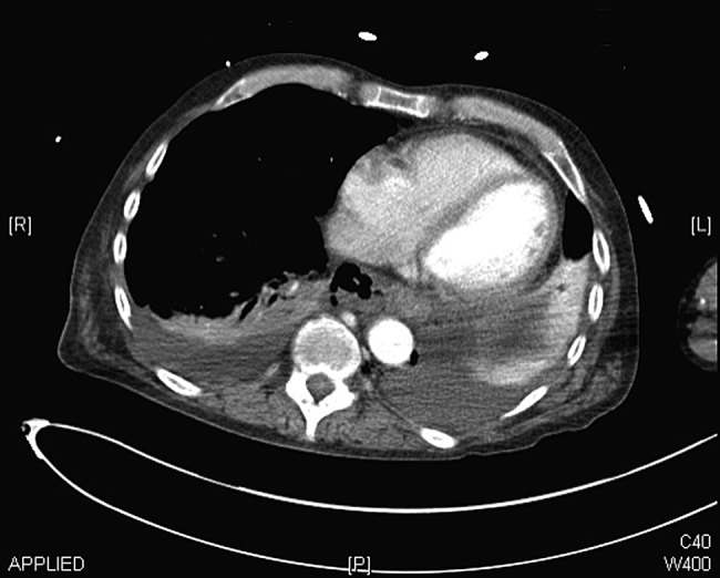
On admission, he had a Glasgow Coma Scale of 3 requiring aggressive resuscitation in the Intensive Care Unit (ICU). Contrast computed tomography (CT) chest showed mediastinitis, bilateral pleural effusions and free air around the distal posterior oesophagus (Fig. 1). A gastrograffin swallow confirmed an oesophageal perforation. He was transferred to the local specialist Upper Gastro-Intestinal Unit for further management.
Figure 1:

CT chest showing mediastinitis, bilateral pleural effusions and free air around the distal oesophagus.
The patient underwent emergency surgical intervention. A right postero-lateral thoracotomy was performed that showed extensive oesophageal necrosis (∼10 cm of sub-carinal oesophagus). Damage control surgery was performed due to the progressive intraoperative haemodynamic instability. The necrotic segment of the oesophagus was resected, and proximal and distal ends of the oesophagus were stapled followed by a cervical oesophagostomy and a feeding jejunostomy. Histology showed microscopic features of acute severe transmural inflammation with necrosis of the mucosa and patchy full thickness necrosis of the muscle wall. Vessels exhibited acute inflammatory thrombus and vasculitis, features that were consistent with acute oesophageal necrosis.
He was managed in ICU with maximal support where he made a successful recovery. A month later, the patient underwent successful reconstruction of the upper gastrointestinal tract using a gastric conduit. The proximal oesophagus was viable, however, 6 cm of the distal oesophagus looked unhealthy requiring further resection. A gastric conduit based on the right gastroepiploic arcade and the right gastric artery was created and was passed into the neck via a nasogastric (NG) tube guide. A hand-sewn oesophagogastric anastomosis was completed using interrupted 3-0 vicryl followed by the passage of a NG tube under vision. The patient made a steady progress and was finally discharged home following a 2-month stay in hospital.
His past history included long-standing severe reflux oesophagitis leading to worsening dysphagia in the last 2 years. He underwent six sessions of endoscopic balloon dilatations for an inflammatory oesophageal stricture at 29 cm with limited success. Histological specimen had confirmed Barrett's oesophagus.
He underwent a biodegradable stent insertion in view of the increased frequency of dilatations and the lack of long-term clinical success. Following balloon dilatation to a diameter of 10 mm under conscious sedation, a non-covered self-expandable biodegradable stent (SX-ELLA 31/25/31 mm × 60 mm) was placed endoscopically with its proximal end at 25 cm. There was good symptomatic relief for 8 months followed by the recurrence of obstructive symptoms. Endoscopy confirmed stent degradation. A second biodegradable stent (SX-ELLA 31/25/31 mm × 60 mm) was inserted due to the lack of success of the balloon dilatations. Insertions of both the stents were uneventful, with the acute life-threatening presentation 8 months following the second stent insertion.
DISCUSSION
Benign oesophageal strictures causing pain, dysphagia and malnutrition can have a significant impact on a patient's quality of life. Standard treatment, including serial endoscopic dilation using bougies or balloons can provide only short-term clinical improvement for some patients.
Oesophageal stents are useful tools for such patients with resistant dysphagias. Biodegradable self-expanding stents that are a recent inclusion to the treatment of refractory dysphagias for patients with benign oesophageal strictures have shown promising results [1]. They are easy to introduce, and their flexibility permits easier adjustment to the oesophageal stenosis. Their ability to provide patency without the need for stent removal and cases of stent migration leading to no harm has justified their usefulness in benign conditions of the oesophagus [2].
Our patient had a SX-ELLA biodegradable (BD) stent inserted for his refractory dysphagia. These stents are made of a biodegradable synthetic material (polydioxanone) and a semi-crystalline polymer monofilament that degrades by the random hydrolysis of its molecule ester bonds [3]. Following insertion, stent integrity and radial force are usually maintained for 6–8 weeks, and disintegration occurs between 11 and 12 weeks. Dual flared ends decrease the risk of migration, and radio-opaque markers in the middle and at both ends assist in their fluoroscopic deployment [4]. The length of the ELLA BD stent ranges between 60 and 135 mm, and the diameter of the flared ends and shaft are 30 and 25 mm, respectively [3]. Following their deployment, the stent progressively expands to its preformed diameter.
Reported complications of biodegradable stents include retrosternal pain and stent migration. However, there are some recent reports of biodegradable oesophageal stents eroding into the tracheobronchial tree causing airway compromise [5], severe tissue reaction with hyperplastic in- and overgrowth causing stent obstruction [3] and even oesophageal obstruction due to their collapse [3, 6].
Acute oesophageal necrosis, a very rare condition of unknown aetiology, is defined as ‘a dark, pigmented state of the oesophagus (“black oesophagus”), with mucosal and submucosal necrosis at histology’ [7]. Rejchrt et al. have reported three cases of acute oesophageal necrosis found on endoscopy, of which two patients died due to their underlying disease. Other predisposing factors identified for this life-threatening condition include anticardiolipin antibody syndrome, herpes oesophagitis, severe diabetic ketoacidosis, gastric volvulus and ruptured thoracic aortic aneurysm.
To our knowledge, this is possibly the first reported case of a biodegradable stent as a predisposing factor for acute oesophageal necrosis. We feel that as these stents are becoming increasingly popular especially for these difficult cases of benign refractory dysphagias, it is important to be aware of this serious life-threatening complication. This case highlights need for further long-term follow-up studies and the reporting of complications for these biodegradable stents.
CONFLICT OF INTEREST STATEMENT
None declared.
REFERENCES
- 1.Evrard S, Le Moine O, Lazaraki G, Dormann A, El Nakadi I, Devière J. Self-expanding plastic stents for benign oesophageal lesions. Gastrointest Endosc 2004;60:894–900. [DOI] [PubMed] [Google Scholar]
- 2.Holm AN, de la Mora Levy JG, Gostout CJ, Topazian MD, Baron TH. Self-expanding plastic stents in treatment of benign esophageal conditions. Gastrointest Endosc 2008;67:20–5. [DOI] [PubMed] [Google Scholar]
- 3.Repici A, Vleggaar FP, Hassan C. Efficacy and safety of biodegradable stents for refractory benign oesophageal strictures: the BEST (biodegradable esophageal stent) study. Gastrointest Endosc 2010;72:927–34. [DOI] [PubMed] [Google Scholar]
- 4.Irani S, Kozarek R. Esophageal stents: past, present and future. Techniques in Gastrointest Endosc 2010;12:178–90. [Google Scholar]
- 5.Katsanos K, Sabharwal T, Koletsis E, Fotiadis N, Roy-Choudhury S, Dougenis D et al. Direct erosion and prolapsed of oesophageal stents into the tracheobronchial tree leading to life-threatening airway compromise. J Vasc Interv Radiol 2009;20:1491–5. [DOI] [PubMed] [Google Scholar]
- 6.Rincon O, Madrigal A, Rodriguez B. Esophageal obstruction due to a collapsed biodegradable esophageal stent. Endoscopy 2011;43:E189–90. [DOI] [PubMed] [Google Scholar]
- 7.Rejchrt S, Douda T, Kopacova M, Siroky M, Repak R, Nozicka J et al. Acute oesophageal necrosis (black oesophagus)—report of three cases. Folia Gastroenterol Hepatol 2004;2:87–91. [Google Scholar]


