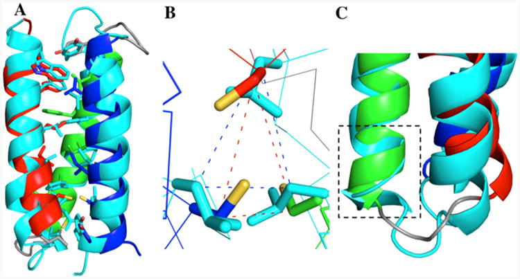
Figure 5.

(A) An overlay of α3DIV (helix 1: red, helix 2: green, and helix 3: blue) and α3D (cyan). The backbone (N, Cα, C,O) rms was determined on PYMOL to be 1.75 Å. (B). Top-down view of the mutation site (18, 28, and 67), displaying superimposed Cys/Leu residues. (C) Gain of helical content in helix 2 for residues 26–28.
