
Figure 3.
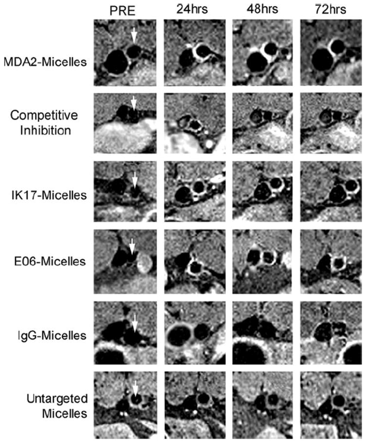
Representative abdominal aorta (arrows) enhancement in apoE−/− mice as a function of time after injection of 0.075–mmol Gd/kg micelles. The vessel to the left of the aorta is the inferior vena cava (IVC). Although arterial flow was saturated to allow delineation of the arterial wall, some enhancement in the IVC may be observed because of flow artifacts.