

Abstract
Morphological variation of normal oral structures such as double frenum and fusion together in a patient is rare. Sometimes such scenarios may mislead the diagnosis, affecting treatment planning and prognosis. Hence a thorough evaluation of patients with such morphological defects is necessary. This case report describes a case of double frenum and fusion, and the multiple challenges the clinician faces.
Background
The median maxillary labial frenum is a post-eruptive remnant of the tectolabial bands.1 2 Syndromes associated with different frenal attachments include: Ehlers-Danlos syndrome, infantile hypertrophic pyloric stenosis, holoprosencephaly, Ellis-van Creveld syndrome and orofacial-digital syndrome. In addition to abnormal oral frenum observed in syndromic conditions, anomalous frenum is encountered without other associated phenotypic features of genetic or chromosomal states.
Fused teeth arise through union of two normally separated tooth germs. The process of fusion involves epithelial and mesenchymal germ layers resulting in irregular tooth morphology, and fusion may be total or partial and leads to reduced number of teeth in the arch.3 Fusion can occur between adjacent teeth of the normal complement4 5 or between a tooth and an adjacent supernumerary.6 7 Fusion of the lateral incisor and cuspid is the most common manifestations in the primary dentition, and symmetrical occurrence is often found.8 Grahnen and Granath9 reported that fused teeth are more common in deciduous than in permanent dentition.
Presence of both morphological variations of fusion and double frenum in these patients presents a challenge in diagnosis and hence a through history and clinical examination are necessary to rule out possible syndromes.
Case presentation
A 4-year-old boy with a large front tooth was brought to our department by his mother. The patient was healthy and the family history and a through examination did not suggest any possible syndromes. However, the mother did mention nursing difficulty during the boy's infancy.
Oral examination revealed fused teeth involving 61 and 62 (figure 1). Fifty-two was missing. In addition, the patient also had an abnormal double median maxillary frenum. The alveolar gingiva surrounding the fused teeth was healthy and free of plaque accumulation. No carious lesion was found.
Figure 1.
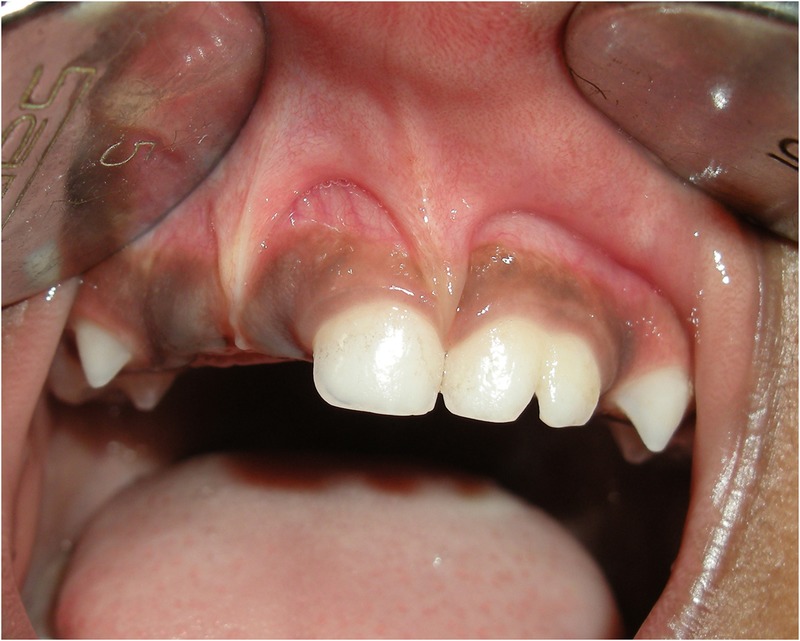
Intraoral view.
Investigations
Periapical radiographs of the region revealed that the roots of 61 and 62 had two evident pulp chambers. Tooth buds of permanent maxillary central and lateral incisors were present.
Differential diagnosis
Clinical features suggestive of syndromes associated with abnormal frenum and fused teeth were not present in this patient. Hence this case can only be an abnormal presentation without associated phenotypic features of genetic or chromosomal states.
Treatment
Frenal attachments that encroach on the marginal gingiva distend the gingival sulcus, fostering plaque accumulation, increasing the rate of progression of periodontal recession and thereby leading to recurrence after treatment. In this patient, since hygiene was good, treatment was not rendered for the double frenum for the time being.
Since exfoliation times are usually different for each tooth involved in the fusion, consideration should be given to the variations in root resorption. In the present case, the exfoliation time for the central and lateral incisors was similar and no significant problems were expected. Management of fused primary teeth in this patient only included observation and allowance of normal exfoliation.
Outcome and follow-up
Follow-up at regular intervals of 6 months until exfoliation of primary teeth and eruption of permanent teeth has been advised to this patient as there is a probability of loss of arch length.
Discussion
In newborns, abnormal maxillary frenum and ankyloglossia, separate or together, may compromise breastfeeding.10 These abnormal attachments also contribute to poor latching onto the mother's nipple, difficult or painful nursing, and impaired labial movements for a full and engaging smile.11 In this patient, history confirmed nursing difficulties.
Absence of the inferior labial and lingual frenum has been described in Ehlers-Danlos syndrome.12 The absence or hypoplasia of mandibular frenum represents an important diagnostic tool in detection of infantile hypertrophic pyloric stenosis.13 Absence of labial maxillary frenum is a characteristic feature of holoprosencephaly.14 Continuous or broad maxillary labial frenula and multiple small mandibular labial frenula are seen in Ellis-van Creveld syndrome.15 Abnormal supernumerary frenula and hypertrophied lingual frenum are features of orofacial-digital syndrome.16 However, in this case, there were no findings suggestive of any syndrome.
Mader17 discussed about the possible clinical problems related to appearance, spacing and periodontal conditions brought about by fused teeth. The clinical implications of fused teeth include the probability of loss of arch length, caries susceptibility of the fissure, and missing corresponding permanent incisors. Fusion of the primary lateral incisor and cuspid might result in early loss of the cuspid with potential loss of arch length or midline shift. To prevent this complication, preservation of arch space and form should be considered.18 In this case, the fusion was between the central and lateral incisor, and only observation and allowance of normal exfoliation was required.
Learning points.
Occurrence of morphological abnormalities such as double frenum and fusion should be viewed with caution because of the possibility of diagnosing a syndrome.
Problems of nursing in infants with frenal abnormalities must not be overlooked.
Potential loss of arch length or midline shift must be kept in mind when fused and/or missing teeth are present.
Footnotes
Competing interests: None declared.
Patient consent: Obtained.
Provenance and peer review: Not commissioned; externally peer reviewed.
References
- 1.Dewel BF. The normal and the abnormal labial frenum: clinical differentiation. J Am Dent Assoc 1946;33:318–29. 10.14219/jada.archive.1946.0261 [DOI] [PubMed] [Google Scholar]
- 2.Henry SW, Levin MP, Tsaknis PJ. Histologic features of superior labial frenum. J Periodontol 1976;47:25–8. 10.1902/jop.1976.47.1.25 [DOI] [PubMed] [Google Scholar]
- 3.Tannenbaum KA, Alling EE. Anomalous tooth development. Case reports of gemination and twinning. Oral Surg Oral Med Oral Pathol 1963;16:883–7. 10.1016/0030-4220(63)90326-8 [DOI] [PubMed] [Google Scholar]
- 4.Surmont PA, Martens LC, De Craene LG. A complete fusion in the primary human dentition: a histological approach. ASDCJ Dent Child 1988;55:362–7. [PubMed] [Google Scholar]
- 5.Nik-Hussein NN. Bilateral symmetrical fusion of primary and permanent mandibular lateral incisors and canines. J Pedod 1989;13:378–3. [PubMed] [Google Scholar]
- 6.Himelhoch DA. Separation of fused primary incisors: report of case. ASDCJ Dent Child 1988;5:294–7. [PubMed] [Google Scholar]
- 7.Camm JH, Wood AJ, Gemination, fusion and supernumerary tooth in the primary dentition: report of case. ASDCJ Dent Child 1989;56:60–1. [PubMed] [Google Scholar]
- 8.Pindborg JJ. Pathology of the dental hard tissues. Copenhagan: Munksgaard, 1970:47–54. [Google Scholar]
- 9.Grahnen H, Granath LE. Numerical variations in primary dentition and their correlation with the permanent dentition. Odontol Rec 1961;12:348. [Google Scholar]
- 10.Weissinger D, Miller M. Breastfeeding difficulties as the result of tight lingual and labial frena. J Hum Lact 1995;11:313–16. 10.1177/089033449501100419 [DOI] [PubMed] [Google Scholar]
- 11.Kotlow LA. The influence of the maxillary frenum on the development and pattern of dental caries on anterior teeth in breastfeeding infants: prevention, diagnosis, and treatment. J Hum Lact 2010;26:304–8. 10.1177/0890334410362520 [DOI] [PubMed] [Google Scholar]
- 12.De Felice C, Toti P, Di Maggio G et al. Absence of the inferior labial and lingual frenula in Ehlers-Danlos syndrome. Lancet 2001;357:1500–2. 10.1016/S0140-6736(00)04661-4 [DOI] [PubMed] [Google Scholar]
- 13.Jenista JA. Mandibular frenulum as a sign of infantile hypertrophic pyloric stenosis. J Pediatr 2001;138:447 10.1067/mpd.2001.109192 [DOI] [PubMed] [Google Scholar]
- 14.Martin RA, Jones KL. Absence of the superior labial frenulum in holoprosencephaly: a new diagnostic sign. J Pediatr 1998;133:151–3. 10.1016/S0022-3476(98)70198-2 [DOI] [PubMed] [Google Scholar]
- 15.Hunter ML, Roberts GJ. Oral and dental anomalies in Ellis-van Creveld syndrome (chondroectodermal dysplasia): report of a case. Int J Paediatr Dent 1998;8:153–7. 10.1046/j.1365-263X.1998.00069.x [DOI] [PubMed] [Google Scholar]
- 16.Dodge JA, Kernohan DC. Oral facial digital syndrome. Arch Dis Child 1967;42:214–19. 10.1136/adc.42.222.214 [DOI] [PMC free article] [PubMed] [Google Scholar]
- 17.Mader CL. Fusion of teeth. J Am Dent Assoc 1979;98:62–4. 10.14219/jada.archive.1979.0037 [DOI] [PubMed] [Google Scholar]
- 18.Eidelman E. Fusion of maxillary primary central and lateral incisors bilaterally. Pediatr Dent 1981;3:346–7. [PubMed] [Google Scholar]