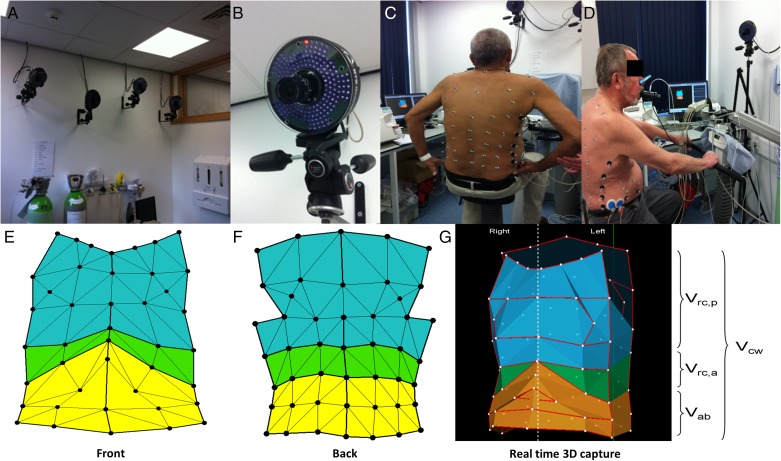
Figure 1 –
Optoelectronic plethysmography. A, B, Infrared cameras. C, D, Marker positioning. E-G, Geometric model. 3D = three-dimensional; Vab = abdominal compartment volume; Vcw = total chest wall volume; Vrc,a = abdominal ribcage volume; Vrc,p = pulmonary ribcage volume. (The patients provided written consent for the use of the photographs.)

