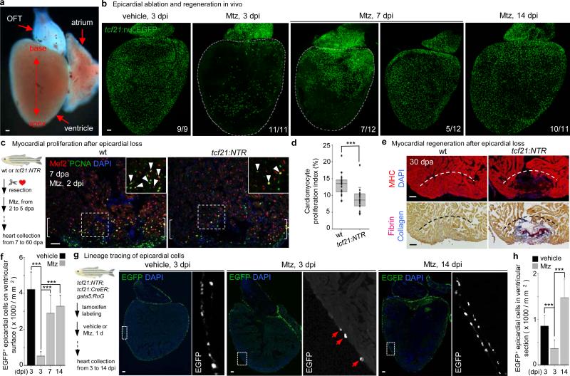
Figure 1. Epicardial ablation and regeneration.
a, Adult zebrafish heart. OFT, outflow tract. b, tcf21:NTR; tcf21:nucEGFP adults were incubated with Mtz or vehicle, and hearts collected by random sampling at 3, 7, or 14 days post-incubation (dpi). Proportion of total animals with indicated phenotype is in lower right corner. All 3 dpi ventricles showed major ablation, averaging ~90% loss. c, (Left) Schematic for tests of epicardial ablation on muscle regeneration. (Right) Ventricular cardiomyocyte proliferation at 7 dpa. Brackets, injury site. Arrowheads, proliferating cardiomyocytes. d, Quantified PCNA+ cardiomyocyte indices in injury sites in experiments from (c). ***P < 0.001; Mann-Whitney Rank Sum test; n = 18 (wt) and 19 (tcf21:NTR) animals from two experiments. e, Section images of ventricles at 30 dpa, assessed for muscle recovery (MHC) and scar indicators (fibrin, collagen). One of 11 tcf21:nucEGFP and 8 of 12 tcf21:NTR; tcf21:nucEGFP ventricles showed myocardial gaps. Dashed line, approximate resection plane. **P < 0.01; Fisher Irwin exact test. f, Quantified EGFP+ nuclei from experiments in (b). ***P < 0.001; Student's two-tailed t-test. g, (Left) CreER-based strategy for permanent labeling of tcf21+ progeny. (Right) Section images of lineage-labeled EGFP+ epicardial progeny through 14 dpi, indicating derivation from pre-existing epicardium. Arrows at 3 dpi, EGFP+ cells spared by epicardial ablation. h, Quantified EGFP+ cells from experiments in (g). ***P < 0.001; Student's two-tailed t-test; n = 10 (vehicle, 3 dpi), 13 (Mtz, 3 dpi), and 15 (Mtz, 14 dpi). White dashed lines in (b), ventricle. Insets in (c, g), high magnifications of boxed areas. Scale bars, 50 μm. Error bars, s.d.

