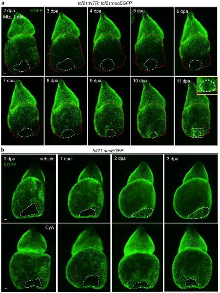
Extended Data Figure 4. Epicardial regeneration after ventricular resection.
a, Hearts were removed from tcf21:NTR; tcf21:nucEGFP fish immediately after ventricular resection injuries, followed by 24 hours of Mtz and a 24-hour washout ex vivo. A base-to-apex pattern of epicardial regeneration was observed, in this example covering the apical wound by 11 dpa (n = 18; behavior seen in all samples). Epicardial coverage of resection injuries in these ablation experiments is delayed compared to ventricles recovering with an intact epicardium (b, Top). Yellow boxed area, magnified view of the apical wound. b, Hearts were removed from tcf21:nucEGFP clutchmates immediately after apical resection injury and cultured ex vivo, before random separation two treatment groups. Epicardial cells covered the wound area by 3 dpa (n = 11; behavior seen in all samples), unless treated with CyA (n = 26; failed coverage in 20 of 26 ventricles). Red dashed lines in (a), ventricle. White dashed lines in (a, b), apical wounds. Scale bars, 50 μm.

