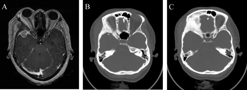
Figure 6. Preoperative MRI and CT imaging of right middle fossa meningioma.
(A) Axial T1-weight MRI with contrast revealing a right middle fossa meningioma with associated hyperostosis of surrounding bone and invasion of the temporalis muscle. (B and C) Extensive bony hyperostosis of the right lateral orbit and pterion.

