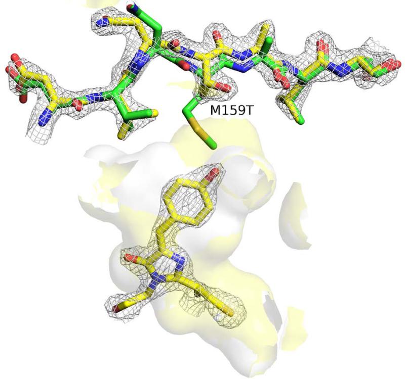
Figure 2.
The cavity available to the cis anionic chromophore in the M159T structure (yellow surface) is enlarged relative to Dronpa (white surface) as a result of the reduced side chain as well as a 1.4 Å ‘outward retraction’ of the side chain (Dronpa-M159T: yellow scheme / Dronpa: green scheme 2Z1O 10). The cavity volume shown does not include crystal waters. 2Fo-Fc electron density is contoured at the 2.0 sigma level for chromophore and residues 156-171 for the Dronpa-M159T structure.

