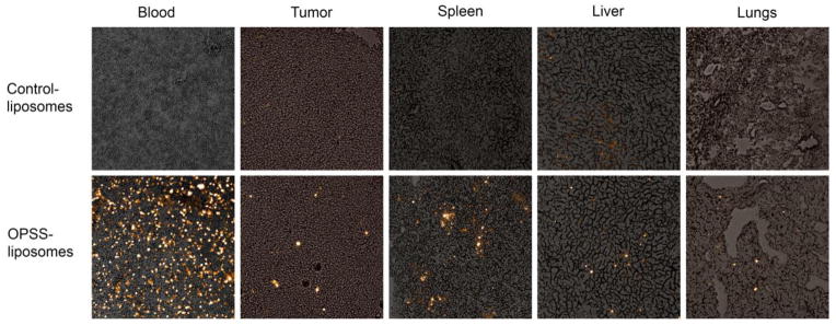
Figure 4.

Histology images of OPSS- and control-liposomes in the blood and various tissues. Images show rhodamine labeled liposomes taken up by cells in the blood, tumor, spleen, liver and lungs. The top panel are tissues from mice injected with control-liposomes and the bottom panel are tissues from mice injected with OPSS-liposomes.
