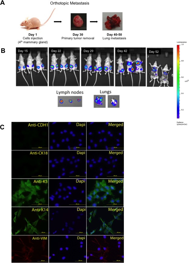
Figure 1. Orthotopic metastasis of JygMC(A) cells and epithelial mesenchymal characterization in vitro.

Lung metastasis by orthotopic cell injection in Balb/C nude mice. B. Representation of bioluminescent imaging of animals. Animals imaged at day 15, 22, 29, 42 and 52 post JygMC(A)-GFP/Luc cell injection. Ex-vivo imaging of lymph nodes and lung metastases. C. Immunofluorescence of luminal epithelial and basal mesenchymal markers in JygMC(A) parental cells. Epithelial marker: CDH1 (Alexa 488), luminal marker: CK18 (Alexa 488), basal marker: K5 and K14 (Alexa 488) and mesenchymal marker: Vimentin (Alexa 594). Nuclear staining in blue (DAPI). Scale bars: 200 μm.
