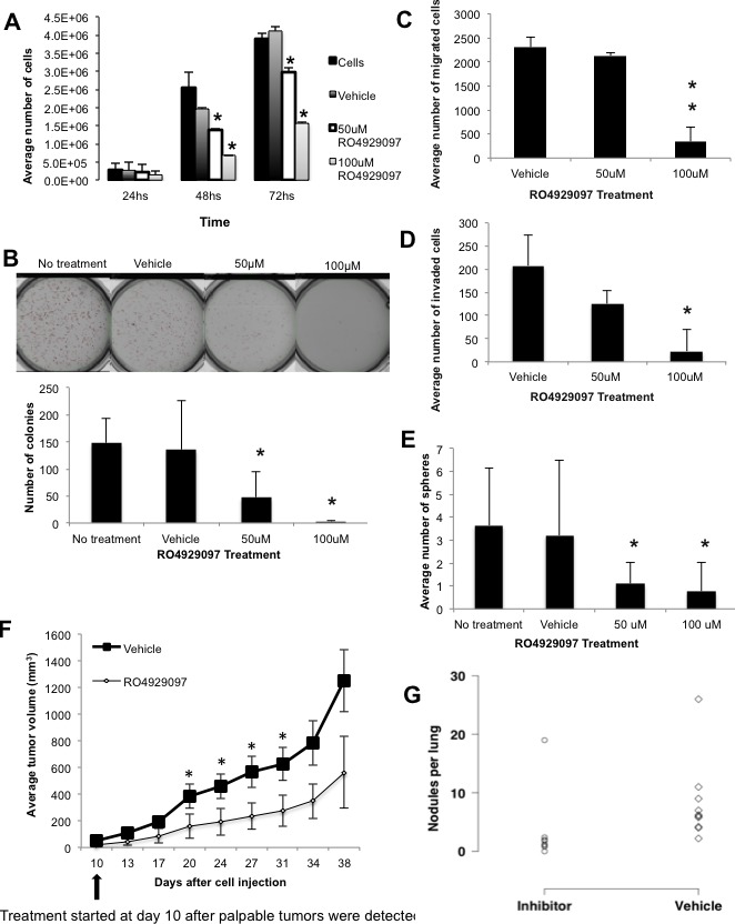
Figure 6. In vitro and In vivo effects of the gamma-secretase inhibitor RO4929097 in the JygMC(A) cell line and mouse model.

Proliferation assay. JygMC(A)-GFP/Luc cells were seeded in 12-well dishes in triplicate at 5×10³cells/well and cultured for 24, 48 and 72 hrs. Cells were then harvested and counted. Data are representative of two independent experiments in triplicate ±SD, *P < 0.0001, as compared to control vehicle-treated cells. B. Colony formation assay in soft agar. A total of 1.5×10⁴cells were cultured for 10 days. Colonies were stained with NitroBlue Tetrazolium and quantified using Gelcount. Data are representative of two independent experiments in triplicate ±SD, *P < 0.005, as compared to control vehicle-treated cells. C. Boyden chamber migration and D. invasion assays. A total of 4×10⁴cells in serum-free medium were seeded on the top chambers, and 2% or 5% FBS-containing medium was placed in the bottom wells as a chemoattractant. Cells were incubated for 24hrs. Cells that migrated/invaded through the membrane were stained and counted using a light microscope. Data are expressed as the average of cells in ten fields from each membrane (20X). Data are representative of two independent experiments in duplicate ±SD, *P < 0.01 and **P < 0.001, as compared to control vehicle-treated cells. E. Sphere formation assay. A total of 10,000 cells were cultured for 10 days on non-adherent plates and treated with different concentrations of RO4929097. Data are representative of two independent experiments in sixplicate ±SD, *P < 0.001, as compared to control vehicle-treated cells. F. Nude mice bearing mammary tumors (n = 10/group) were dosed orally following the schedule of 60mg/Kg every other week (7days on/7days off) or vehicle for 4 weeks and primary tumor volumes are represented in the graph, ±SD, *P < 0.05, as compared to control vehicle-treated animals. G. Number of pulmonary nodules per animal in control vehicle-treated and RO4929097-treated animals (*P < 0.05, one-sided values; Wilcoxon rank-sum test).
