Abstract
We have developed and tested a novel, ionizing-radiation Quantum Imaging Detector (iQID). This scintillation-based detector was originally developed as a high-resolution gamma-ray imager, called BazookaSPECT, for use in single-photon emission computed tomography (SPECT). Recently, we have investigated the detector’s response and imaging potential with other forms of ionizing radiation including alpha, neutron, beta, and fission fragment particles. The confirmed response to this broad range of ionizing radiation has prompted its new title. The principle operation of the iQID camera involves coupling a scintillator to an image intensifier. The scintillation light generated by particle interactions is optically amplified by the intensifier and then re-imaged onto a CCD/CMOS camera sensor. The intensifier provides sufficient optical gain that practically any CCD/CMOS camera can be used to image ionizing radiation. The spatial location and energy of individual particles are estimated on an event-by-event basis in real time using image analysis algorithms on high-performance graphics processing hardware. Distinguishing features of the iQID camera include portability, large active areas, excellent detection efficiency for charged particles, and high spatial resolution (tens of microns). Although modest, iQID has energy resolution that is sufficient to discriminate between particles. Additionally, spatial features of individual events can be used for particle discrimination. An important iQID imaging application that has recently been developed is real-time, single-particle digital autoradiography. We present the latest results and discuss potential applications.
Keywords: Charged particle imaging detectors, Ionizing radiation, BazookaSPECT, Digital autoradiography
1. Introduction
Single-event imaging detectors that are sensitive to photons (gamma/X rays) and particles (alphas, betas, neutrons, fission fragments, auger electrons, etc.) are important in a number of applications. Examples of medical imaging applications include single-photon emission computed tomography (SPECT), gamma-ray scintigraphy, digital radiography, SPECT/CT, and autoradiography. Both scintillation and semiconductor technologies exist, each with trade-offs in terms of cost, counting-rate capability, spatial resolution, energy resolution, and active area.
One such detector developed at the Center for Gamma-Ray Imaging is BazookaSPECT [1-3]. It is a scintillation detector that combines image intensifiers and CCD/CMOS cameras for high-resolution gamma-ray imaging applications. Our latest objective has been to explore the detector’s response and imaging potential with other forms of ionizing radiation including alpha, neutron, beta, and fission fragment particles. We present an overview of the technology and discuss recent results demonstrating the camera’s sensitivity to a broad range of ionizing radiation, which has prompted its new title: iQID (ionizing-radiation Quantum Imaging Detector).
2. Materials and methods
2.1. iQID imaging system
iQID operates on the principle of using electro-optical gain to amplify scintillation light from an event before imaging onto a CCD or CMOS camera sensor. This optical gain is provided by an image intensifier, which amplifies scintillation light while also preserving spatial information. iQID typically uses microchannel plate (MCP) image intensifiers, which have luminous gains ranging from 104 to 106 or higher, depending on the number of MCPs used. With intensifiers having two or more MCPs, even single optical photons can be detected at an appropriate MCP voltage. In contrast to photo-multiplier tube (PMT) type imaging detectors, where scintillation light from a single event is spread out across an array of PMTs, iQID directly forms an image of the light distribution generated from the particle or photon interaction. We typically use image intensifiers with fiber-optic (FO) input windows. The FO window allows scintillators to be placed in close proximity with the intensifier for high light-collection efficiency without the need of lenses to image scintillation light onto the photocathode. The output window of the intensifier is then imaged onto the CCD/CMOS camera typically using conventional CCTV camera lenses. A single interaction appears as a flash of light confined to a small region of pixels, which we call an event cluster. Although it is possible to image individual events using monolithic crystals [4], high-resolution imaging is typically done using structured scintillators, e.g., microcolumnar CsI(Tl) and LaBr3 or thin phosphor screens [5,6]. These scintillators restrict the lateral spread of scintillation light, resulting in less uncertainty when using pixel data to estimate the 2D or 3D interaction position.
Depending on the frame rate of the camera and source activity, multiple events can occur within an image frame. To keep events that interact within the same temporal window spatially separated, the camera frame rate is adjusted to the proper number of frames per second (FPS). CCDs that have a global (or frame) shutter, in contrast to CCDs that use a rolling shutter readout are preferred. This ensures that light collection starts and ends at the same time for all pixels. Neglecting any minimal frame overhead time, e.g., image transfer time (45 μs per frame for the iQID camera discussed below), the maximum integration time for each pixel is then 1/FPS. An image frame with multiple gamma-ray events is shown in Fig. 1. Note that each flash is a single gamma-ray interaction. If event clusters do overlap, spatial features and maximum-likelihood position estimation techniques can be used to resolve and separate events [7,8]. Using high-speed cameras, counting rates of the order 450k counts per second are possible (assuming uniform illumination) using relatively inexpensive CCD/CMOS cameras [7,9].
Fig. 1.
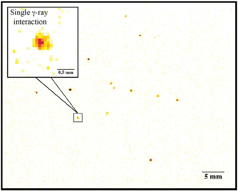
57Co gamma-ray interactions in a gadolinium oxysulfide (Gadox) X-ray scintillation screen.
To keep pace with the continual development of faster cameras having an ever increasing number of pixels, we utilize advances in high-performance graphics processing technology and multi-core CPUs to achieve real-time event detection and position estimation. We refer to this image analysis technique as frame parsing [9]. When performing long image acquisitions using fast cameras, potentially operating at hundreds of frames per second, storage of individual frames and post-processing is unattractive and impractical. Using NVIDIA graphics processing units (GPUs) and the CUDA programming platform, events are identified in real time and only relevant data are computed and stored as a listmode event entry. These include a 2D position estimate, time stamp, summed pixel values within an event cluster, and all associated event pixels. Additionally, a separate image of the 2D position estimates is generated in real time, where pixel values correspond to the number of events detected at a given pixel location. A visual example of the frame processing algorithm is shown in Fig. 2.
Fig. 2.
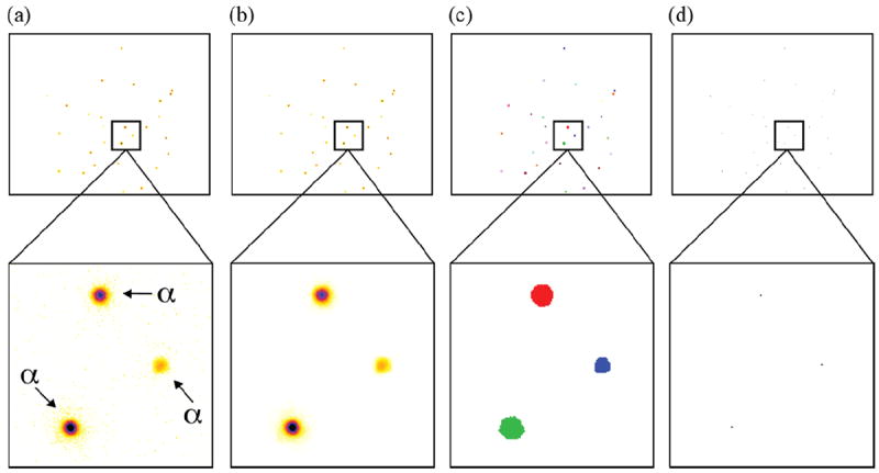
iQID frame parsing algorithm: (a) raw CCD camera frame where the magnified region shows three alpha particles, (b) median-filtered image with noisy pixels removed, (c) filtered image with individual particles identified using a connected components labeling algorithm [10,11], and (d) the estimated 2D interaction position. The position estimate, time stamp, summed pixel values, and all associated event pixels are concatenated and written to disk as a listmode event entry.
A notable feature of the iQID camera is the ability to readily increase detector area using a fiber-optic (FO) taper. For example, the active area of a typical 25 mm diameter image intensifier can be increased 16 × using a 4:1 (100 mm:25 mm) fiber-optic taper [3]. Even with light loss incurred by imaging with the taper, individual events are still detectable.
2.2. PNNL iQID
An image of the iQID camera system at Pacific Northwest National Laboratory (PNNL) is shown in Fig. 3. The PNNL iQID camera uses a 40 mm diameter ProxiVision MCP–PROXIFIER® with 2 MCPs in a V-stack assembly. It has fiber-optic input/output widows with a bialkali photocathode and P43 phosphor. The detector area is increased to 125 mm diameter using an Incom Inc. FO taper. The camera is a Point Grey Research, Inc. Flea® 3 that uses an ON Semiconductor® VITA1300 CMOS sensor. The camera uses a USB 3.0 interface and acquires images at rates up to 150 fps (1280 × 1024 pixels) and 400 fps (640 × 512 pixels).
Fig. 3.
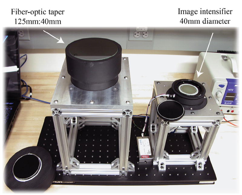
PNNL iQID camera. The detector is composed of a 40 mm diameter image intensifier and the detector area can be expanded to 125 mm using a fiber-optic taper. The system operates using a laptop computer and has been developed to be highly portable to accommodate a variety of imaging experiments and is flexible for testing various imaging configurations and components.
2.3. Scintillators and source fabrication
For initial testing of iQID’s response to alpha particles and fission fragments, we used silver doped zinc sulfide (ZnS:Ag) scintillation screens. ZnS:Ag is a conventional material for detecting alpha particles and one of the brightest scintillators ~95,000 photons/MeV [12].
As a historical side note, ZnS was used by Rutherford in his experiments that led to discovery of the atomic nucleus. Additionally, in 1903 William Crookes invented a device for observing individual nuclear disintegrations, called the spinthariscope (from the Greek word scintillation), using radium salt suspended above a zinc sulfide screen [13,14]. A magnifying lens and the human eye serves as the imaging detector for the spinthariscope. The device requires dark adaptation and individual alpha decays are seen as flashes of light, even with the naked eye. The spinthariscope became a popular amusement in the early 1900s and later a popular children’s educational toy during the 1940–1950s. In fact, in 1947 you could get a Lone Ranger spinthariscope ring for 15 cents and a Kix cereal box top.
The isotope we used for alpha and fission fragment experiments is 252Cf. Approximately 3% of its decays are via spontaneous fission (SF) and 97% alpha decay. The energies of the primary alpha emissions are 6.075 MeV (15.7% emission intensity) and 6.118 MeV (84.2% emission intensity), and the mean kinetic energies of daughter fission fragments are 80.1 MeV and 105.71 MeV, respectively [15]. 252Cf spontaneous fission decay also produces neutrons and betas particles. As will be discussed later, the light output from alpha and fission fragment interactions in ZnS:Ag screens is high enough that iQID can be operated essentially beta and gamma/X-ray blind when detecting alpha particles and alpha-particle blind when detecting fission fragments.
To reduce energy loss and ensure high detection efficiency, we fabricated a 252Cf source by stippling ~1/3 microliter drops of 252Cf in 0.1 M nitric acid solution directly onto a ZnS:Ag screen. The activity of each drop contained approximately 1 becquerel (Bq). After the solution dried, a second ZnS:Ag disk was placed in contact with the first to provide ~100% (4π) detection efficiency for both alpha particles and fission products. The source disk was placed between layers of transparent tefion film that was then heat sealed around the edges. Fig. 4 shows an image of the 252Cf source and the fabrication technique.
Fig. 4.
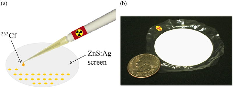
Fabrication of a 252Cf ZnS:Ag source. (a) Drops of a 252Cf in 0.1 M nitric acids were stippled on a ZnS:Ag phosphor disk and dried before imaging. ~1 Bq per drop. (b) Sealed ZnS:Ag 252Cf source that is placed in contact with the iQID detector for imaging.
For neutron detection we use 6LiF/ZnS powder screens. Lithium-6 has a relatively high cross-section (520 barns at 1 Å [16]) for thermal neutrons with the reaction 6Li +1n → 3H (2.75 MeV) + 4He (2.05 MeV). 6LiF/ZnS scintillators yield ~160k optical photons per neutron capture [16]. Thermal neutrons can also be detected at very high efficiency after neutron capture in Gadolinium-157 and -155 from the production of internal conversion electrons with energies ranging from 29 to 246 keV [17]. Examples of gadolinium-based neutron scintillators include CsI(Tl) sandwiched between thin layers of GdF3 [18] and phosphor powders made of gadolinium oxysulfide (Gd2O2S), also routinely used for X-ray imaging.
For beta particle detection, a number of scintillator options are available including monolithic, structured, and plastic scintillators as well as phosphor screens. To demonstrate iQID beta detection we use Gadox and BaFBr phosphor screens.
3. Results
3.1. Particle detection
Images of the iQID camera’s response to alpha particles and fragments from spontaneous fission are shown in Fig. 5, with magnified regions of a selected alpha particle and fission fragment. The pixel colorbar scale of the top magnified images shows the amplitude difference while the bottom shows the spatial extent of the light distribution between particles. The light generated from fission fragments and alphas particles is high enough that the intensifier is operated at the minimum gain settings.
Fig. 5.
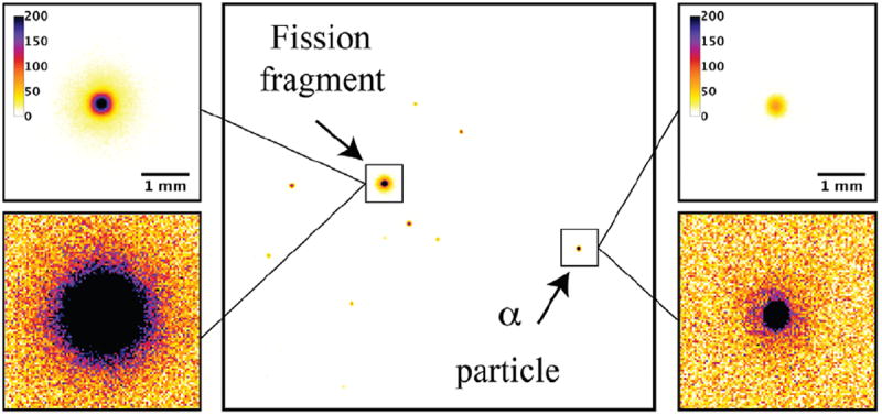
Fission fragments and alphas particle interactions in ZnS:Ag from a 252Cf source. (For interpretation of the references to color in this figure caption, the reader is referred to the web version of this paper.)
Fig. 6 shows thermalized 252Cf neutrons detected via the two example pathways previously described: 4He (2.05 MeV) and 3H (2.75 MeV) particles after neutron capture in 6Li (Fig. 6(a)) using a 6LiF/ZnS:Ag screen and a Gd2O2S:Tb (Gadox) screen for conversion electrons (29–246 keV) after neutron capture in 155,157Gd (Fig. 6(b)). The intensifier gain is significantly higher when detecting conversion electrons thereby making iQID more susceptible to detection of background events. To validate that we were indeed seeing conversion electrons produced by neutron capture in gadolinium, we imaged in conjunction with Gadox another common X-ray scintillator screen, CaWO4 (L–Plus®), which is of comparable X-ray sensitivity but not sensitive to thermal neutrons. The location of the two screens is superimposed on events in Fig. 6(b).
Fig. 6.
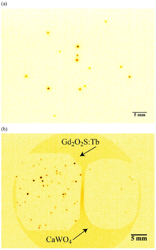
iQID thermal neutron detection. Thermalized neutrons from a 252Cf source are detected (a) with a 6LiF/ZnS:Ag screen via the production of 3H (2.05 MeV) and 4He (2.75 MeV) particles and (b) via conversion electrons (29–246 keV) in a Gd2O2S:Tb (Gadox). Note that the intensifier gain for conversion electron detection is significantly higher compared to that needed for alpha-particle detection. A non-neutron-sensitive X-ray screen, CaWO4 (L–Plus®), was imaged in conjunction with the Gadox screen for validating neutron detection above background. Also, a thick lead disk was placed on the screens to suppress background events.
iQID’s sensitivity to beta particles is demonstrated in Fig. 7. A series of selected beta-particle interactions were taken using a 90Sr/90Y source, where 90Y emits betas with an endpoint energy of 2.28 MeV. The images show interesting tracks produced by betas particles interacting in a BaFBr intensifying screen at highly oblique angles.
Fig. 7.
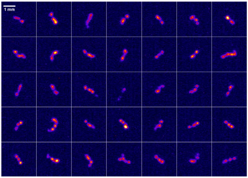
Selected beta tracks from a 90Sr/90Y source detected using a BaFBr phosphor screen.
3.2. Position estimation
Pixel signals from an event cluster can be used to estimate the 2D interaction location of the ionizing-radiation particle or photon. If interactions are not identified on a quantum or event-by-event basis and scintillation light is merely summed across image frames, the resultant image will have significant blur caused by both the lateral spread of scintillation light and additional noise sources such as electronic noise acquired during pixel readout. This effect is demonstrated in Fig. 8(a) using the 252Cf source. In contrast, if the interaction location of individual events is estimated to a single pixel, the spatial resolution significantly improves, as shown in Fig. 8(b, c). Taking this approach further, we can compute a sub-pixel estimate of the interaction location that leads to finer sampling of the activity distribution. This is demonstrated in Fig. 8(d), where the location of each event is estimated to the nearest pixel quadrant. The spatial resolution improvement for sub-pixel position estimation with alpha particles will be quantified in a subsequent publication relating to the detector’s use in digital autoradiography with alpha emitters.
Fig. 8.
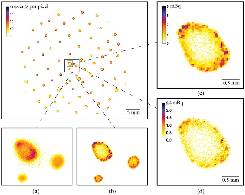
Spatial distribution of alpha particles and fission fragments from stippled drops of 252Cf, ~1 Bq per drop. (a) Magnified sub-region of the source that is generated by summing light from each event cluster. Note the induced image blur caused by this method in contrast to identifying and estimating the position of each particle to the nearest pixel (b). (c, d) Activity distribution of a source drop, where the colorbar scale is in units of millibecquerels, with event positions estimated to the nearest pixel (40 × 40 μm2 pixels) (c) and sub-pixel quadrant (d) (20 × 20 μm2 pixels). (For interpretation of the references to color in this figure caption, the reader is referred to the web version of this paper.)
3.3. Activity estimation
The short range of alpha particles and fission fragments allows for very high detection efficiency, even using only a few tens of microns of scintillation material. The iQID camera has effectively a 100% detection and geometric efficiency (4π) with the 252Cf source, as the californium-252 atoms are sandwiched between the two ZnS:Ag scintillation screens. The combination of high efficiency (geometric + detection), timing information, and position estimation to ≤ 100 μm for each alpha and fission fragment particle allows for activity quantification at the mBq per pixel level, with effective pixel areas of the order 40 × 40 μm2, for example. Fig. 8(c, d) shows sub-region images of the 252Cf source where the pixel colorbar scale is in units of millibecquerels. The effective pixel size in these images is 40 × 40 μm2 (estimated to nearest pixel) and 20 × 20 μm2, respectively.
3.4. Particle discrimination
Although the energy resolution of iQID is relatively poor compared to other detector technologies, an energy estimate can be obtained simply by summing the pixel values of an event cluster and then selecting an energy threshold to discriminate between particles. Additional methods for particle discrimination include the use of spatial features such as spatial variance, kurtosis, and eccentricity [7,19]. To test the detector’s capability for discriminating between gamma rays/betas and alphas/fission fragments we fabricated a multi-isotope source. Similar to the method described above for fabricating the 252Cf source, we stippled drops of 241Am, 90Sr/90Y, and 252Cf on to a ZnS:Ag disk at an activity of 20 Bq total per isotope dispersed over 10–15 drops. Fig. 9(a) shows the source fabrication method. High-intensity emissions of interest from 241Am include alphas ranging in energy 5.442–5.485 MeV and 59.5 keV gamma rays. Emissions from 90Sr/90Y include beta particles with endpoint energies of 546 keV and 2.28 MeV. In addition to the previously discussed fission fragments and alpha particle decays from 252Cf, high-energy beta particles are emitted with a yield of 5.8 per fission event [20].
Fig. 9.
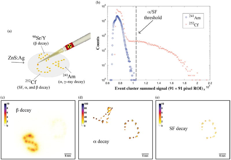
(a) Gamma-ray, alpha, beta, and spontaneous fission (SF) fragment source. Drops of solution (252Cf “C”, 241Am “A”, 90Sr/90Y “S”) were stippled on a ZnS:Ag phosphor disk and dried before imaging at ~20 Bq total per isotope. (b) Histograms of 241Am alphas and 252Cf alphas and SF fragments with a threshold selected to distinguish between alphas and fission fragments. (c) Beta-particle distribution generated using a Gadox phosphor screen. Note the relatively weak activity distribution in “C” from betas emitted during SF in 252Cf compared to betas in “S” from 90Sr/90Y. (d, e) Alpha and SF fragment distributions. Pixel colorbar scale is in units of the number of particles detected during the imaging acquisition. (For interpretation of the references to color in this figure caption, the reader is referred to the web version of this paper.)
When using iQID with ZnS:Ag to detect alphas and fission fragments, the detector operates essentially blind to other particles due to the high amount of light that is generated. However, other particles from the same source can be detected using an additional scintillator and an opaque screen between the ZnS:Ag. To detect betas and gammas with our multi-isotopic source, we used a Gadox screen with an emulsion layer thick enough to block scintillation light produced by the alphas and fission fragments interacting in the ZnS.
Particle discrimination is demonstrated in Fig. 9. Alpha particles are separated from spontaneous fission fragments by applying an energy threshold, where the energy is estimated by summing a 91 × 91 region of pixels around each event. To determine an approximate alpha particle energy cutoff, we extracted alpha-particle events from 241Am and generated an energy histogram of the events (Fig. 9(b)). We then used the energy of the alpha particle with the highest energy as the alpha/spontaneous fission threshold. This is an approximate threshold as 252Cf alphas have higher energy compared to 241Am, 6.118 MeV (84.2% emission intensity) vs 5.485 MeV (84.5% emission intensity). Approximate alpha-only and SF-only images of the sources are shown in Fig. 9(d, e). After separating alphas from fission fragment decays using this technique, we estimate the decay rates for 252Cf at 3.01% for spontaneous fission and 96.99% for alphas, a very good estimate where other published values place the rate 3.092% and 96.908% [21].
An iQID image of the beta source distribution is shown in Fig. 9(c). The faint “C” represents the distribution of betas emitted from 252Cf during spontaneous fission and the intense “S” distribution represents betas emitted by 90Sr/90Y. Note the decreased spatial resolution due to the long travel range of the beta particles compared to alphas and fission fragments. Another reason for the reduced resolution is that the current position estimation algorithm is not optimized for betas, as it does not make use of track information. Since a beta particle will become more ionizing toward the end of its trajectory, an image of its track at this point will show an increased signal density per unit length. For betas that sufficiently interact in the scintillator to capture this effect in their track, we anticipate position estimation algorithms that fully utilize track information will provide improved spatial resolution. Another feature in the images to note is the lack of any gamma-ray events from 241Am (59.5 keV), which are filtered using a pixel threshold when identifying events during frame parsing.
4. Discussion and conclusion
We have successfully developed a high-resolution imaging detector that is sensitive to a broad range of ionizing radiation including gamma rays, alpha, beta, neutron, and fission fragment particles. An important feature of the iQID camera is a real-time dynamic imaging capability. It is highly portable and does not require cooling and can be operated using a laptop computer, potentially even with a smartphone in the near future. iQID provides fine sampling of scintillation-event light distributions, which are then processed for high-resolution position estimation, even to sub-pixel levels. Images generated by the iQID camera do not contain noise sources such as electronic or CCD noise as they are removed during the event-detection process. Relatively large-area iQIDs can be produced using fiber-optic tapers, which we have currently tested in areas up to 12.5 cm in diameter.
The detector has very high sensitivity, particulary for alpha, beta, and fission fragment particles. We have successfully imaged source distributions at the mBq per pixel level. Its high sensitivity, spatial resolution, and temporal information for each event make iQID an attractive technology for digital autoradiography. We have demonstrated that iQID can be used to characterize the spatial profile of low-activity (~0.1 Bq), calibration sources such as disks that are electroplated with alpha-emitting isotopes [22]. Also, we are currently investigating its application for imaging tissue sections in research areas such as radioimmunotherapy, where alpha- and beta-particle emitters are being investigated as potential therapy agents in cancer treatment. Microdosimetry is important in these applications and iQID’s features of high spatial resolution and superb activity quantification make it a very promising technology.
Acknowledgments
This work is supported by the Laboratory Directed Research and Development Program at Pacific Northwest National Laboratory, a multiprogram national laboratory operated by Battelle for the U.S. Department of Energy. B.W. Miller is grateful for the support of a Linus Pauling Distinguished Postdoctoral Fellowship at PNNL. iQID camera development is in collaboration with the Center for Gamma-Ray Imaging, NIH Grant P41EB002035.
References
- 1.Miller B, Barber H, Barrett H, Wilson D, Chen L. IEEE Nuclear Science Symposium Conference Record; 2006. p. 3540. [Google Scholar]
- 2.Miller B, Barrett H, Furenlid L, Barber H, Hunter R. Nuclear Instruments and Methods in Physics Research, Section A. 2008;591:272. doi: 10.1016/j.nima.2008.03.072. [DOI] [PMC free article] [PubMed] [Google Scholar]
- 3.Miller BW, Barber HB, Barrett HH, Liu Z, Nagarkar VV, Furenlid LR. Progress in BazookaSPECT: high-resolution dynamic scintigraphy with large-area imagers. International Society for Optics and Photonics on SPIE Optical Engineering+ Applications. :85080F–85080F. doi: 10.1117/12.966810. [DOI] [PMC free article] [PubMed] [Google Scholar]
- 4.Park R, Miller B, Jha A, Furenlid L, Hunter W, Barrett H. A prototype detector for a novel high-resolution PET system: BazookaPET; IEEE Nuclear Science Symposium and Medical Imaging Conference (NSS/MIC); pp. 2123–2127. [DOI] [PMC free article] [PubMed] [Google Scholar]
- 5.Nagarkar V, Gupta T, Miller S, Klugerman Y, Squillante M, Entine G, Inc R, Watertown M. IEEE Transactions on Nuclear Science. 1998;NS-45:492. [Google Scholar]
- 6.Bhandari H, Gelfandbein V, Miller S, Agarwal A, Miller B, Barber H, Nagarkar V. IEEE Transactions on Nuclear Science. 2013;NS-60:3. [Google Scholar]
- 7.Miller B. High-resolution gamma-ray imaging with columnar scintillators and CCD/CMOS sensors, and FastSPECT III: a third-generation stationary SPECT imager (Ph D thesis) University of Arizona, Department of Optical Sciences; Tucson, Arizona: 2011. [Google Scholar]
- 8.Barrett H, Hunter W, Miller B, Moore S, Chen Y, Furenlid L. IEEE Transactions on Nuclear Science. 2009;NS-56:725. doi: 10.1109/tns.2009.2015308. [DOI] [PMC free article] [PubMed] [Google Scholar]
- 9.Miller B, Barber H, Furenlid L, Moore S, Barrett H. Penetrating Radiation Systems and Applications X. 2009;7450:74500C. [Google Scholar]
- 10.Suzuki K, Horiba I, Sugie N. Fast connected-component labeling based on sequential local operations in the course of forward raster scan followed by backward raster scan. Proceedings of 15th International Conference on Pattern Recognition; 2000. pp. 434–437. [Google Scholar]
- 11.Kalentev O, Rai A, Kemnitz S, Schneider R. Journal of Parallel and Distributed Computing. 2011;71:615. [Google Scholar]
- 12.Dorenbos P. Nuclear Instruments and Methods in Physics Research Section A: Accelerators, Spectrometers, Detectors and Associated Equipment. 2002;486:208. doi: 10.1016/s0168-9002(01)01386-9. [DOI] [PubMed] [Google Scholar]
- 13.Crookes W. Proceedings of the Royal Society of London. 1902;71:405. [Google Scholar]
- 14.Kolar Z, den Hollander W. Applied Radiation and Isotopes. 2004;61:261. doi: 10.1016/j.apradiso.2004.03.056. [DOI] [PubMed] [Google Scholar]
- 15.Whetstone SL. Physical Review. 1963;131:1232. [Google Scholar]
- 16.van Eijk CW. Radiation Measurements. 2004;38:337. [Google Scholar]
- 17.Harms A, McCormack G. Nuclear Instruments and Methods. 1974;118:583. [Google Scholar]
- 18.Shestakova I, Tipnis S, Gaysinskiy V, Antal J, Bobek L, Nagarkar V. IEEE Transactions on Nuclear Science. 2005;NS-52:1109. [Google Scholar]
- 19.Miller B, Barber H, Barrett H, Chen L, Taylor S. Proceedings of SPIE. 2007;6510:65100N. doi: 10.1117/12.710109. [DOI] [PMC free article] [PubMed] [Google Scholar]
- 20.Wark DL. The β spectrum following fission from 252Cf (Ph D thesis) Vol. 1987 California Institute of Technology; [Google Scholar]
- 21.Miller A. Neutron Capture Therapy. Vol. 2012. Springer; Berlin Heidelberg: Californium-252 as a neutron source for BNCT; pp. 69–74. http://dx.doi.org/10.1007/978-3-642-31334-9_5. [Google Scholar]
- 22.Dion MP, Liezers M, Farmer OT, Miller BW, Morley SM, Barinaga CJ, Eiden GC. Journal of Radioanalytical and Nuclear Chemistry. 2014 submitted for publication. [Google Scholar]


