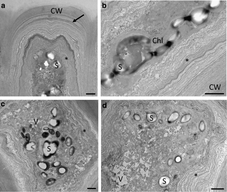
Fig. 5.
Transmission electron micrographs of desiccated (73 % RWC) and rehydrated (2 h in ASW) samples of U. compressa. a Desiccation leads to undulations of the inner cell wall layers and the cytoplasm appears dense due to shrinkage of the protoplast followed by the innermost pectic cell wall layers (asterisk). The outermost fibrillar cell wall layers surrounding a periclinal pectic layer (arrow) do not change in shape. b Detail of the cell wall after desiccation. The innermost pectic cell wall layers (asterisk) are attached to the shrunken protoplast. c, d Numerous stark grains and vacuoles are visible upon rehydration. Undulation of the inner pectic cell wall layers (asterisk) is still visible. Scale bars 1 µm

