Description
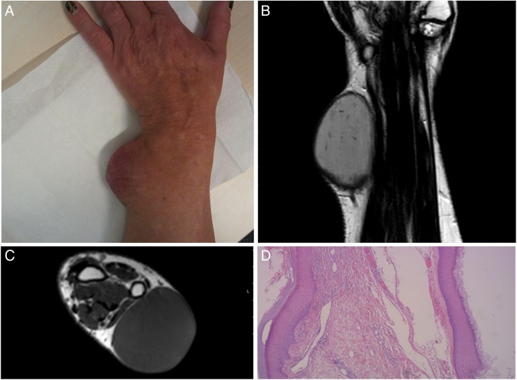
A 69-year-old woman presented with a painful swelling on her left wrist (figure 1A). The tumour had been present for years, but was neglected by the patient until development of local pain during compression. Medical history included only hypertension. Physical examination revealed a subcutaneous tumour (7 cm ×4 cm× 4 cm) fixed to the skin on the lateral side of the left wrist. There was no impairment of function and sensibility of the left wrist, hand and digits. Considering the large diameter and planning surgical resection, a MRI was performed to evaluate the type and extent of the tumour.1
Figure 1.
(A) A subcutaneous tumour at the ulnar–volar side of the left wrist. (B) Coronal MRI and (C) transversal MRI of the left upper limb showing a large epidermoid cyst. (D) Histology showing the epidermoid cyst wall.
The radiological diagnosis pointed to a giant epidermoid cyst of the wrist (figure 1B, C), which was confirmed by histopathology after excision (figure 1D). Sebaceous, keratin, epidermal or epithelial cysts are eponyms. Cysts can develop when epidermis is trapped subcutaneously, for example, after trauma. Epidermal cysts are often encountered in the clinical practice and normally appear around the third and fourth decade on the trunk, neck, face and behind the ears, and more often in male patients. Diagnosis is based on the clinical appearance and palpation of a freely movable subcutaneous mass, often with a central punctum. In total, 95% of subcutaneous tumours on the wrist are benign. In approximately 60%, the ganglion is the most common. Giant cell tumours of the tendon sheath, epidermoid cysts, trichilemmal cysts, lipomas, hibernomas and liposarcomas, are found less frequently.2 In contrast to benign tumours, malignant tumours are often fixed to the surrounding tissues and may feel non-homogeneous on palpation.2 Ultrasound or MRI can make malignant tumours less likely, but histopathology is needed for a definitive diagnosis. Whenever there is doubt on the seriousness, an incisional biopsy or complete removal should be performed after imaging.
Learning points.
The differential diagnosis of a large subcutaneous tumour at the wrist should include an epidermoid cyst.
Although 95% of tumours of the wrist are benign, oncological surgical principles should always be followed.
Footnotes
Competing interests: None declared.
Patient consent: Obtained.
Provenance and peer review: Not commissioned; externally peer reviewed.
References
- 1.Peh WC, Truong NP, Totty WG et al. Pictorial review: magnetic resonance imaging of benign soft tissue masses of the hand and wrist. Clin Radiol 1995;50:519–25. doi:10.1016/S0009-9260(05)83185-X [DOI] [PubMed] [Google Scholar]
- 2.Nahra ME, Bucchieri JS. Ganglion cysts and other tumor related conditions of the hand and wrist. Hand Clin 2004;20:249–60, v doi:10.1016/j.hcl.2004.03.015 [DOI] [PubMed] [Google Scholar]


