Abstract
A 4-month-old male child presented with right undescended testis and left inguinal hernia with funiculitis. Ultrasonography showed funiculitis on the left side testis along with presence of 1.5 × 1 cm testis like structure just above left testis and empty right scrotal sac without any evidence of mullerian structures. On diagnostic laparoscopy, right testicular vessels were crossing from right to left and had uterus with both testes in left hernia sac. Mobilization of vessels, division of uterus, and hernia repair was done laparoscopically. On the review of literature, there is only one case report of total laparoscopic repair of transversetesticular ectopia (TTE) with hernia without persistent mullerian duct (PMDS). The uniqueness of our case is that it had TTE with hernia and PMDS, which were totally managed by laparoscopy. On 6 months of follow-up, both the testes are palpable in scrotum.
Keywords: Laparoscopy, persistent mullerian duct syndrome, transversetesticular ectopia
INTRODUCTION
Transversetesticular ectopia (TTE) is a rare form of testicular ectopia in which one of the testes crosses midline and occupies contralateral side of hemiscrotum. Thirty percent of TTE patients have persistent mullerian duct (PMDS) structures. More than hundred such cases were reported in the literature. TTE with persistent mullerian duct syndrome usually presents as undescended testis with contralateral inguinal hernia. Diagnostic laparoscopy with open exploration an dorchiopexy is a routine practice. Laparoscopy is being increasingly used for diagnostic as well as therapeutic management of undescended testis. Evans et al. were first to report total laparoscopic correction of TTE.[1] Here we describe total laparoscopic correction of TTE with PMDS.
CASE REPORT
A 4-month-old male child was brought to uswith complaint of pain in the left inguinal region for 3 days. He was also had swelling in the same region since 20 days. On clinical examination, he had left-sided irreducible inguinal hernia with tenderness. Right scrotum was empty with testis not palpable. Left testis was normal and was present in left scrotum. Ultrasonography showed funiculitis of the left cord structures and 1.5 × 1 cm testis-like structure present just above the left testes.
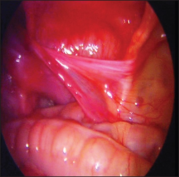
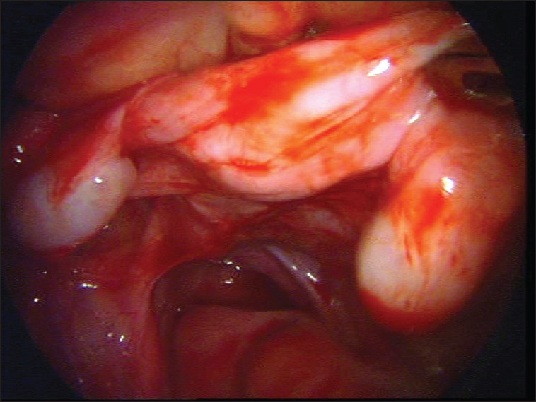
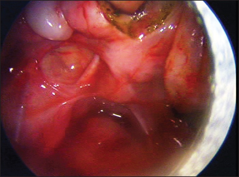
After recovery of funiculitis, we posted the patient for diagnostic laparoscopy followed by definitive repair. During the procedure, operating surgeon was at the head end. A 5 mm port was inserted at umbilicus forthe camera and two working ports were inserted 5 cm away on either side of the umbilical. Atlaparoscopy, right-sided internal ring was open with gubernaculum passing through it. Right-sided testicular vessels crossed from right to left, entering in left internal ring [Figure 1]. Contents of left inguinal canal were reduced. There was a uterus in between two testes [Figure 2]. Both vas deferences were running along the sides of uterus. Small hernia sac was also present. We divided the uterus in midline with help of bipolar cautery [Figure 3]. After mobilization of right testicular vessels we found adequate length to bring it out through right-sided internal ring. Small incision taken on both side of median raphe of scrotum. Laparoscopic-assisted tunnel was created on the right side of inguinal canal. Both testes brought out and fixed in the subdartous pouch. Left-sided hernial sac closed with intracaropreal suturing.
Figure 1.

Photograph showing right-sided testicular vessels crossing midline to left and entering into internal ring
Figure 2.

Photograph showing uterus in between two testis
Figure 3.

Photograph showing division of uterus in midline
We successfully managed transverse testicular ectopia with persistent mullerian duct syndrome with hernia laparoscopicaly. On 6 months of follow-up, both the testes are palpable in scrotum.
DISCUSSION
Transverses testicular ectopia is a rare form of testicular ectopia in which testis is present in contralateral side of hemiscrotum. More than 100 such cases were reported in the literature. The first case is reported by Lenhossek in 1886, he found it during an autopsy of adult man.[2]
Several theories have been postulated to explain the etiology. Berg suggested that both testes arise from same germinal ridge.[3] Gupta suggested that adherence of developing wolffian duct with mullerian duct causes descend of both testis on the same side.[4] Freys proposed possibility of formation of defective gubernaculums.[5]
Three types of TTE are described: (1) TTE with hernia (40–50%), (2) TTE with PMDS (30%), (3) TTE with hypospadias, pseudohermaphrodite, or some complexity (20%).[6] Testicular duplication is also described in the literature. TTE without hernia is also described. Hutson et al.[7] classified PMDS into three groups: Group A - the testes are in the position of the normal ovaries (female type); Group B - one testis is found in a hernia sac orscrotum along with the uterus and tubes (male type); Group C - both testes are found in the same hernia sac with associated tubes and the uterus (male type). Our patient belongs to Group C.
Most common presentation is the absence of testis on the one side with inguinal hernia on opposite side. Ultrasonography and magnetic resonant imaging helps to diagnose this condition but most of them are diagnosed at the time of surgery. Laparoscopy is best for localization of ectopic testis. Hormonal assessment is not done routinely.
Various methods to manage this condition have been described, like herniotomy with trans-septal fixation of testis, that is, Ombredanne technique, fixation on opposite side through suprapubic subcutaneous tunnel, and staged procedure. Persistent mullerian structures needed to be managed separately.
Laparoscopy is being increasingly used for diagnostic and correction of undescended testes and hernia. Gohari et al.[8] in 2003 described various laparoscopic procedures for management of PMDS. Dean and shah reported laparoscopically assisted correction of TTE.[9] In 2007, Evans reported total laparoscopic correction of TTE.[1] The dividedmeso-orchium and right testis was brought through right inguinal canal. Deshpande[10] described the role of laparoscopy in TTE.
Uniqueness of our case is that our case was having TTE with hernia and PMDS, which was totally managed by laparoscopy. Division of uterus in midline and mobilization of cord and vessels carried out by laparoscopy. Laparoscopic route is feasible and convenient for management of such cases. Exploration of groin could be very well avoided by using laparoscopy. We are first to describe total laparoscopic repair for TTE with PMDS.
Footnotes
Source of Support: Nil
Conflict of Interest: None declared.
REFERENCES
- 1.Evans K, Desai A. Total laparoscopic correction of transverse testicular ectopia. J Pediatr Urol. 2008;4:245–6. doi: 10.1016/j.jpurol.2007.07.008. [DOI] [PubMed] [Google Scholar]
- 2.Von Lenhossek M. Ectopia testis transversa. Anat Anz (Jena) 1886;1:376. [Google Scholar]
- 3.Berg AA., 3rd Transverse Ectopy of the Testis. Ann Surg. 1904;40:223–4. [PMC free article] [PubMed] [Google Scholar]
- 4.Gupta RL, Das P. Ectopia testis transversa. J Indian Med Assoc. 1960;35:547–9. [PubMed] [Google Scholar]
- 5.Frey HL, Rajfer J. Role of the gubernaculum and intraabdominal pressure in the process of testicular descent. J Urol. 1984;131:574–9. doi: 10.1016/s0022-5347(17)50507-8. [DOI] [PubMed] [Google Scholar]
- 6.Gauderer MW, Grisoni ER, Stellato TA, Ponsky JL, Izant RJ., Jr Transverse testicular ectopia. J Pediatr Surg. 1982;17:43–7. doi: 10.1016/s0022-3468(82)80323-0. [DOI] [PubMed] [Google Scholar]
- 7.Hutson JM, Chow CW, Ng WT. Persistent mullerian duct syndrome with transverse testicular ectopia. Pediatr Surg Int. 1987;2:191–4. [Google Scholar]
- 8.El-Gohary MA. Laparoscopic management of persistent mullerian duct syndrome. Pediatr Surg Int. 2003;19:533–6. doi: 10.1007/s00383-003-0984-7. [DOI] [PubMed] [Google Scholar]
- 9.Dean GE, Shah SK. Laparoscopically assisted correction of transverse testicular ectopia. J Urol. 2002;167:1817. [PubMed] [Google Scholar]
- 10.Deshpande AV, La Hei ER. Impact of laparoscopy on the management of transverse testicular ectopia. J Laparoendosc Adv Surg Tech A. 2009;19:443–6. doi: 10.1089/lap.2008.0106. [DOI] [PubMed] [Google Scholar]


