Abstract
[Purpose] The purpose of this study was to determine the relationship between navicular drop and plantar flexion torque of the first and second-fifth metatarsophalangeal joints. [Subjects] Ten healthy young men participated in this study. [Methods] The Pearson product-moment correlation coefficient was calculated to determine the relationship between navicular drop and plantar flexion torque of the first and second-fifth metatarsophalangeal joints. [Results] Significant negative correlations were observed between navicular drop and plantar flexion torques in the lengthened position of the intrinsic toe plantar flexion muscles, but no correlations were found between navicular drop and plantar flexion torques in the neutral position of the ankle and metatarsophalangeal joints. Moreover, the intrinsic toe plantar flexion muscles were found to contribute to the formation of the medial longitudinal arch. [Conclusion] Navicular drop correlates with metatarsophalangeal joint muscle strength in plantar flexion where the intrinsic toe muscles are capable of exerting force.
Key words: Toe flexor strength, Intrinsic foot muscle, Medial longitudinal arch
INTRODUCTION
The human foot is composed of three arch structures, namely the medial longitudinal arch (MLA), lateral longitudinal arch, and transverse arch. These arch structures reduce pressure on the soft tissue by dispersing the loading stress and increasing the efficiency of force transmission by functioning as a spring during walking1). The MLA has been measured to evaluate the role of arch structure in a physical therapy setting, with the navicular bone being regarded as a landmark of the arch apex, as it is palpable and deviates minimally. Some methods of MLA measurement have been previously described, including navicular drop (ND), in which the difference in navicular height between the standing and sitting positions is measured2). The height and general shape of the MLA are maintained by the plantar fascia, ligaments, and intrinsic and extrinsic muscles of the foot3). Previous studies have reported that ND is related to lower limb disability4, 5) and that it can be decreased after toe exercise6); taken together, these findings suggest that toe muscle strength may relate to arch structure and lower limb disability.
There are two types of toe flexor muscles, namely extrinsic and intrinsic muscles. The extrinsic muscles running across the ankle joint include the flexor hallucis longus (FHL) and flexor digitorum longus. On the other hand, the intrinsic muscles do not run across the ankle joint and do not contribute to ankle movement. Both the intrinsic and extrinsic muscles are stretched by dorsiflexion of the metatarsophalangeal (MTP) joints, whereas only the extrinsic muscles are relaxed by plantar flexion of the ankle joint7). The exhibited maximal torque is affected by the joint angle8), and a relationship between joint angle and plantar flexion torque of the MTP joints has been reported previously9).
Thus, these previous studies suggest that excessive ND can cause lower limb disability and that this can be controlled by toe exercise. However, the dynamometer used in the previous studies measured only toe grip strength, i.e., the plantar flexion force of all toes at the plantar flexion position of the MTP joint10). Moreover, the relationship between ND and the measurement position of plantar flexion torque of the MTP joints has not been studied. Therefore, the aims of this study were to clarify the relationship between ND and plantar flexion torque of the first and second-fifth MTP joints and to determine the differences between neutral positions of the MTP and ankle joints and stretching position of the intrinsic muscles. In a previous study, the MLA was found to be raised by contraction of the intrinsic foot muscles11). Accordingly, we hypothesized that the relationship between ND and plantar flexion torque of the MTP joints would change with different measuring positions, and we believe that the results of this study will provide valuable information for exercise and risk management to prevent lower limb disabilities.
SUBJECTS AND METHODS
Ten healthy men participated in this study. The mean values ± standard deviations (SDs) for their age, height, and body weight were 23.8 ± 1.5 years, 1.69 ± 0.04 m, and 58.4 ± 8.1 kg, respectively. The exclusion criterion was a lower limb orthopedic disorder. This study was approved by the Ethics Committee on Human Research of Waseda University (approval number: 2013-195). The authors provided all subjects with an explanation of the purpose and methods of this study and details of the study protocol, and written informed consent was obtained from all subjects.
Navicular height was defined as the distance from the floor to the tubercle of the navicular bone, as measured with a caliper. Palpation of the tubercle of the navicular bone was performed by a physical therapist who had more than 3 years of experience in orthopedic physical therapy. ND was calculated by subtracting the navicular height in the standing position from that in the sitting position.
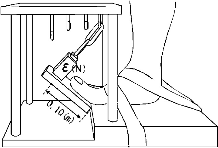
A custom-made MTP joint plantar flexion torque meter device was used to measure MTP joint plantar flexion torque. The subjects were seated on a chair, with their trunk, thighs, lower thighs, and dominant foot fastened to the chair and the torque meter device. After warming up, each subject performed maximal voluntary isometric contraction of plantar flexion of the first MTP joint, followed by that of the second-fifth MTP joint for 3 s to determine the maximal voluntary contraction (MVC) torque. The plantar flexion torque was calculated as the tensile force of a strain gauge (TU-BR 500N, TEAC, Japan) multiplied by the 0.10-m lever arm of the force plate (Fig. 1). We converted the tensile force data from analog to digital (Power Lab, AD instruments, Australia) via an amplifier (DPM-711B, Kyowa Electronics, Japan) using a personal computer. MVC torque was measured at two positions: at a neutral position of the ankle and MTP joints and at 45° dorsiflexion of the MTP joints with 20° plantar flexion of the ankle joint. The measured MVC torque values were normalized by body weight. MVC torque measurements were performed after ND measurements.
Fig. 1.
Structure of the torque meter used to measure isometric plantar flexion torque at the metatarsophalangeal (MTP) joints. MTP joint plantar flexion torque was calculated using the following formula: plantar flexion torque (Nm) = strain ε (N) × moment arm 0.10 (m).
Further, we measured MVC torque again on a different day to determine the test-retest repeatability of these measurements, which was accomplished by calculating the mean coefficient of variance (mean/SD) and intraclass correlation coefficient (ICC [1,1]). To assess the ICCs, we used the criteria advocated by Fleiss12), in which an ICC > 0.75 is defined as excellent reliability. A Pearson correlation coefficient was used to test the correlation between the ND and MVC torques. For all tests, statistical significance was set at p < 0.05.
RESULTS
The mean coefficients of variance of the MVC torques of the first MTP and second-fifth MTP joints measures were 7.6% and 7.4%, respectively, and the corresponding ICCs (1.1) were 0.81 and 0.87, respectively.
Significant negative correlations were seen between the ND and the MVC torques at 20° plantar flexion of the ankle joint with 45° dorsiflexion of the MTP joints of the first MTP joint (r = −0.78, p < 0.01) and second-fifth MTP joints (r = −0.82, p < 0.01) (Table 1). However, no significant correlations were found between the ND and MVC torques at the neutral position of the ankle and first MTP joint (r = −0.52, p > 0.05) and second-fifth MTP joints (r = −0.38, p > 0.05) (Table 1).
Table 1. Relationship between navicular drop ratio and maximal voluntary contraction torque ratio of the plantar flexion of the metatarsophalangeal joints.
| Position of the MTP and ankle joints | Measured joints | MVC torque (N/kg) |
NDs (mm) |
Correlation Coefficients |
|---|---|---|---|---|
| Neutral | First MTP | 0.13 ± 0.03 | 7.4 ± 2.5 | −0.52 |
| Second-fifth MTP | 0.09 ± 0.03 | −0.38 | ||
| 45° dorsiflexion of the MTP joints, 20° plantar flexion of the ankle joint | First MTP | 0.17 ± 0.03 | −0.78* | |
| Second-fifth MTP | 0.12 ± 0.02 | −0.82* |
Mean ± SDs; MTP joint: metatarsophalangeal joint; MVC torque: maximal voluntary contraction torque; ND: navicular drop; *p < 0.01
DISCUSSION
The ICCs (1.1) for both the first and second-fifth MTP joints were more than 0.75 in this study and showed excellent reliability12), and the test-retest repeatability of the measurements of MVC torque were confirmed.
Significant negative correlations were observed between ND and MVC torque at 45° dorsiflexion of the MTP joints with 20° plantar flexion of the ankle joint. On the other hand, there was no significant correlation between ND and MVC torque at the neutral position, indicating that ND relates to intrinsic muscle strength.
MVC torque is affected by muscle length, and muscle length in turn is known to be affected by the joint angle8,13). In a previous study, the muscle length of the FHL was found to vary an average of 0.22 mm by 1° plantar flexion or dorsiflexion of the first MTP joint and 0.50 mm by 1° plantar flexion or dorsiflexion of the ankle joint7). Furthermore, according to the same study, at 45° dorsiflexion of the first MTP joint, the muscle length of the FHL increased approximately 10 mm from that in neutral position, whereas at 20° plantar flexion of the ankle joint, the length of the FHL decreased approximately 10 mm from that in neutral position. In the present study, the length variation of the extrinsic muscle was countered and the intrinsic muscles were lengthened by 20° plantar flexion of the ankle joint and 45° dorsiflexion of the first MTP joint. At this position, a higher torque can be exerted even though the length of the extrinsic muscle remains constant8). In addition, it has been reported that toe flexion exercises performed during plantar flexion of the ankle joint are an effective method of intrinsic foot flexor strength training14). According to these findings, it appears as if the intrinsic muscles are capable of exerting force in this position. Furthermore, it has been reported that the MLA was raised upon inducing contraction of the intrinsic muscles via electrical stimulation11).
At the neutral position of the ankle and MTP joints, both the intrinsic and extrinsic muscles can produce similar force. However, the produced force ratio of the extrinsic muscle to the intrinsic muscles is relatively large at the neutral position of the ankle and MTP joints as compared to at 20° plantar flexion of the ankle joint with 45° dorsiflexion of the MTP joints, and this is likely the reason for the lack of a significant correlation between MVC torque and ND in this condition. Taken together, these results suggest that the intrinsic foot muscles contribute greatly to the construction of the MLA and that ND relates to maximal plantar flexion torque of MTP joints at 20° plantar flexion of the ankle joint with 45° dorsiflexion of the first MTP joint. These results support a previous finding that ND is correlated with electromyographic activity of the abductor hallucis muscle15).
Moreover, these results indicate that the strength of the intrinsic toe plantar flexion muscles is related to ND, suggesting that exercises targeting the intrinsic muscles may not only help improve excessive ND but also prevent sport-related disorders caused by excessive ND.
The main limitation of this study was that the MLA was affected not only by the toe flexor muscle but also by the muscle that did not contribute to MTP joint plantar flexion (e.g., the tibialis posterior and peroneus longus muscles). Future studies need to examine these effects as well. Furthermore, we did not investigate extrinsic muscle length according to variations of the MTP joint angle. Therefore, we instead discussed the results based on the length variation of the FHL. Future studies aimed at assessing the flexor digitorum longus length according to variations of the second to fifth MTP joints are needed.
In conclusion, we found that ND relates to MTP joint muscle strength in plantar flexion where the intrinsic toe plantar flexion muscles are capable of exerting force. This result suggests that exercises targeting the intrinsic muscles may help improve excessive ND.
REFERENCES
- 1.Kanai S: Kinesiology of foot arch. In: Evaluation method of foot and shoe for physical therapist. Tokyo: Bunkoudou, 2013, p 25 (in Japanese). [Google Scholar]
- 2.Brody DM: Techniques in the evaluation and treatment of the injured runner. Orthop Clin North Am, 1982, 13: 541–558. [PubMed] [Google Scholar]
- 3.Neumann DA: Kinesiology of the Musculoskeletal System: Foundations for Rehabilitation. St. Louis: Mosby, 2002, pp 477–521. [Google Scholar]
- 4.Bennett JE, Reinking MF, Pluemer B, et al. : Factors contributing to the development of medial tibial stress syndrome in high school runners. J Orthop Sports Phys Ther, 2001, 31: 504–510. [DOI] [PubMed] [Google Scholar]
- 5.Beckett ME, Massie DL, Bowers KD, et al. : Incidence of hyperpronation in the ACL injured knee: a clinical perspective. J Athl Train, 1992, 27: 58–62. [PMC free article] [PubMed] [Google Scholar]
- 6.Mulligan EP, Cook PG: Effect of plantar intrinsic muscle training on medial longitudinal arch morphology and dynamic function. Man Ther, 2013, 18: 425–430. [DOI] [PubMed] [Google Scholar]
- 7.Refshauge KM, Chan R, Taylor JL, et al. : Detection of movements imposed on human hip, knee, ankle and toe joints. J Physiol, 1995, 488: 231–241. [DOI] [PMC free article] [PubMed] [Google Scholar]
- 8.Williams M, Stutzman L: Strength variation through the range of joint motion. Phys Ther Rev, 1959, 39: 145–152. [DOI] [PubMed] [Google Scholar]
- 9.Goldmann JP, Brüggemann GP: The potential of human toe flexor muscles to produce force. J Anat, 2012, 221: 187–194. [DOI] [PMC free article] [PubMed] [Google Scholar]
- 10.Kurihara T, Yamauchi J, Otsuka M, et al. : Maximum toe flexor muscle strength and quantitative analysis of human plantar intrinsic and extrinsic muscles by a magnetic resonance imaging technique. J Foot Ankle Res, 2014, 7: 26. [DOI] [PMC free article] [PubMed] [Google Scholar]
- 11.Kelly LA, Cresswell AG, Racinais S, et al. : Intrinsic foot muscles have the capacity to control deformation of the longitudinal arch. J R Soc Interface, 2014, 11: 20131188. [DOI] [PMC free article] [PubMed] [Google Scholar]
- 12.Fleiss JL: Statistical Methods for Rates and Proportions, 2nd ed. New York: John Wiley and Sons, 1981, p 218. [Google Scholar]
- 13.Gordon AM, Huxley AF, Julian FJ: The variation in isometric tension with sarcomere length in vertebrate muscle fibres. J Physiol, 1966, 184: 170–192. [DOI] [PMC free article] [PubMed] [Google Scholar]
- 14.Hashimoto T, Sakuraba K: Assessment of effective ankle joint positioning in strength training for intrinsic foot flexor muscles: a comparison of intrinsic foot flexor muscle activity in a position intermediate to plantar and dorsiflexion with that in maximum plantar flexion using needle electromyography. J Phys Ther Sci, 2014, 26: 451–454. [DOI] [PMC free article] [PubMed] [Google Scholar]
- 15.Nam KS, Kwon JW, Kwon OY: The relationship between activity of abductor hallucis and navicular drop in the one-leg standing position. J Phys Ther Sci, 2012, 24: 1103–1106. [Google Scholar]


