SUMMARY
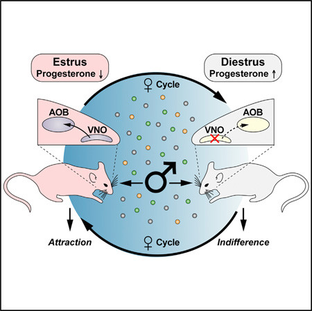
Females may display dramatically different behavior depending on their state of ovulation. This is thought to occur through sex-specific hormones acting on behavioral centers in the brain. Whether incoming sensory activity also differs across the ovulation cycle to alter behavior has not been investigated. Here, we show that female mouse vomeronasal sensory neurons (VSNs) are temporarily and specifically rendered “blind” to a subset of male-emitted pheromone ligands during diestrus yet fully detect and respond to the same ligands during estrus. VSN silencing occurs through the action of the female sex-steroid progesterone. Not all VSNs are targeted for silencing; those detecting cat ligands remain continuously active irrespective of the estrous state. We identify the signaling components that account for the capacity of progesterone to target specific subsets of male-pheromone responsive neurons for inactivation. These findings indicate that internal physiology can selectively and directly modulate sensory input to produce state-specific behavior.
Graphical Abstract
INTRODUCTION
Across evolution, males and females differ both in physical features as well as their behavioral responses (Yang and Shah, 2014). How is it that females may respond to the world differently than males? Moreover, a female’s behavior can change dramatically based on her reproductive state, yet little is known about the neural targets on which sex hormones act. An individual’s behavior is influenced by sensory information gathered from the external environment as well as one’s immediate internal physiological state. The nocturnal mouse relies on its olfactory system to survey the rich chemical environment in order to inform behavior (Liberies, 2014; Touhara and Vosshall, 2009). A subset of detected chemosignals is thought to be specialized to signal social behavior between individuals (pheromones) and warn of potential predators (kairomones) (Karlson and Luscher, 1959; Wyatt, 2010). While pheromones and kairomones promote stereotyped behavior, this reliable response is only true when the receiving animals are controlled for age, gender, dominance, and anxiety. It is commonly understood that male-emitted chemosignals elicit aggression from dominant males while juveniles and subordinate males respond to the same cues with indifference. Similarly, a female’s attraction and receptivity behavior toward male sensory cues is most robust when she is in the state of estrus yet the same sensory cues generate indifference or even aggression from a female in diestrus. How neurons that respond to the same chemosensory environment generate such different behavior responses based on the internal state of the receiver is largely unknown.
Olfactory circuits directly project to regions of the limbic system that express sex-hormone receptors, including the amygdala and hypothalamus (Morris et al., 2004; Yang and Shah, 2014). Manipulation of cells from these nuclei has been shown to alter certain sex-specific behaviors (Juntti et al., 2010; Lee et al., 2014; Yang et al., 2013). However, these brain regions are molecularly and functionally heterogeneous and mechanistic study of how sex-steroid receptor expressing neurons contribute to behavior has been difficult (Yang and Shah, 2014). In contrast, the olfactory system is highly ordered (Touhara and Vosshall, 2009). Recently, ligands that stimulate the vomeronasal sensory neurons (VSNs, which comprise an olfactory subsystem) have been purified from complex secretions. Ligands from male mouse urine, including major urinary proteins (MUPs), have been shown to promote attraction (preference) behavior while FELD4 from cat saliva promotes defensive (fear-like) behavior (Papes et al., 2010; Roberts et al., 2010). Isolation of purified ligands with known bioactivity allows systematic identification of the subset of sensory neurons that initiate sex-specific behavior. These ordered neurons can be used to subsequently activate and identify the responsive neural subsets that generate behavior in the limbic system. At least two basic coding hypotheses could underlie sex-specific behavior. The first assumes that the function of a sensory system is to reliably monitor the environment, therefore it is expected that chemosensory detection neurons will display stable activity responses to available ligands irrespective of internal state. Subsequent “downstream” neurons in the activity circuit would then be charged with monitoring internal state, integrating available information, and commanding the appropriate behavioral output. This is a likely scenario based on the complicated task and growing evidence supports this as a mechanism of coding (Yang and Shah, 2014). The second hypothesis is based on the observation that our sensory “perception” changes with internal state. When we are hungry, food smells more delicious. Although it has not previously been described, it is a formal possibility that this could occur through a modification of the response properties of the sensory detectors themselves. In this model, changes in behavior would be a direct result of increase, or decrease, in sensory neural activity. The isolation of pure ligands that elicit state-dependent activity now enables evaluation of this second model of behavioral control.
Here, we identify an olfactory behavior that changes with the female’s internal estrous state. She displays attraction toward major urinary proteins, MUPs, during estrus and indifference during diestrus. This change in behavior provides a platform to test whether sensory detection is modified by internal state. We use calcium imaging to monitor neural activity and find that female hormones act directly on a subset of mouse VSNs to prevent MUPs from initiating sex-specific behavior. We discover that the hormone progesterone is the key signal that silences MUP-responsive VSNs. Importantly; progesterone does not impair all VSNs from detecting ligands. We find that VSNs that detect FELD4, which is a potential predator signal emitted by cats (Papes et al., 2010), escape progesterone silencing and are ligand responsive throughout the entire estrous cycle. Biochemistry and genetic ablation reveals that MUP-responsive VSNs express unique primary signal transduction elements that are targets of phosphorylation-mediated inactivation initiated by the progesterone receptor membrane component1 protein (PGRMC1) (Bashour and Wray, 2012; Meyer et al., 1996; Peluso et al., 2008; Petersen et al., 2013). These results provide evidence for the second coding hypothesis (sensory modulation) to also underlie sex-specific behavior. Through sensory modulation males and females do not sense the environment equally. Instead, olfaction is dynamic; with females regularly “blind” to subsets of cues each estrous cycle in order to regulate sex-specific behavior.
RESULTS
Female Attraction to MUPs Changes with Her State of Estrus
Because female mouse behavior is normally assayed in a complex social environment the subset(s) of neurons that are differentially active in estrus or diestrus have been difficult to identify and have not been mechanistically studied. In order to investigate mechanisms to explain how female behavior dramatically differs across the ovulation cycle, we sought to control the stimulus assay using purified ligands. Male-emitted pheromones that promote attraction behavior have recently been described (major urinary proteins; MUPs) (Roberts et al., 2010, 2012). Recombinant MUP20 produced in bacteria and purified, rMUP, is sufficient to elicit attraction in a simple robust assay (Roberts et al., 2010), but the extent to which this behavior varies across the female estrous cycle has not been investigated. Even though only rMUP20 has been demonstrated to underlie attraction behavior, we elected to assay all five of the MUPs naturally emitted by our test strain males as a pool because they are detected as a blend in physiological contexts (Kaur et al., 2014). To determine if the estrous state alters a female’s response to this pool of rMUPs, we tested the behavioral effects of in a two-choice assay with females staged either in estrus or diestrus (Figures S1A–S1C). We found that the behavioral response to these stimuli did vary across the female’s estrous cycle. Only estrous staged females displayed a preference for rMUPs (or MUP-enriched native urine fractions), while diestrous-staged females were behaviorally indifferent to rMUP ligands (Figure 1A–B, S1D). The identification of a simple odor-mediated assay that elicits either attraction during estrus or indifference during diestrus provides an experimental platform to mechanistically uncover how the female generates this behavioral difference.
Figure 1. Female Sensory Response Varies with Estrous State.
(A) Schematic representation of estrous cycle, indicating proestrus (light gray), estrus (pink), metestrus (light gray), and diestrus (dark gray).
(B) Preference index from two choice behavior assay conducted on estrous- and diestrous-staged females with rMUP stimuli versus biologically non-relevant control odor (p = 0.002; estrus n = 12, diestrus n = 10).
(C) Percentage of VSNs from estrous and diestrous females showing calcium influx to rMUPs (p = 2.57 × 10⁻⁵; 2,811 and 2,726 cells imaged, respectively).
(D) Overlaid representative calcium influx traces of individual VSNs from estrous and diestrous females in response to stimulation with rMUPs and male urine.
(B and C) Two-tailed t test. All values in mean ± SEM. p < 0.05, **p < 0.01, ***p < 0.001, ****p < 0.0001; ns, not significant. Pink bars, estrus; dark gray bars, diestrus.
See also Figure S1.
MUPs Fail to Activate VSNs during Diestrus
The vomeronasal organ is a specialized olfactory subsystem known to detect MUP pheromones (Chamero et al., 2012; Dulac and Torello, 2003; Kaur et al., 2014; Stowers and Logan, 2010). Pheromone-responsive circuits in the brain include subsets of sexually dimorphic nuclei expressing sex-steroid receptors thought to be primarily responsible for changes in sex-specific behavior (Manoli et al., 2013; Morris et al., 2004; Yang and Shah, 2014). However, the formal possibility that behavior regulation also occurs by altering signaling in the pheromone-detecting sensory neurons has not been previously evaluated. To test this possibility, we acutely dissociated VSNs from estrous and diestrous females and assayed their response to native and rMUPs by calcium imaging (Chamero et al., 2007; Kaur et al., 2013, 2014). Upon perfusion of rMUPs (or MUP-enriched native urine fractions) ~5% of the total cells from estrous staged VSNs showed a calcium influx (Figures 1C, 1D, S1E, and S1F). This amount of activity is similar to response levels previously described for male VSNs (Chamero et al., 2007; Kaur et al., 2014). Surprisingly, when we performed the same analysis of VSNs from diestrous-staged females, only 1 % of the neurons were activated by rMUPs (Figures 1C, 1D, S1E, and S1F). This indicates that a female’s VSN sensory capability to detect rMUPs is not static; instead it is differentially responsive depending on the female’s internal state. Approximately 80% of the rMUP-detecting VSNs change their sensitivity across the female’s estrous cycle, with the highest sensory detection occurring during estrus and the lowest during diestrus. This finding is consistent with the ability of sensory neural activity to underlie behavioral differences to rMUPs across the estrous cycle.
Progesterone Signals the Female’s Estrous State to VSNs
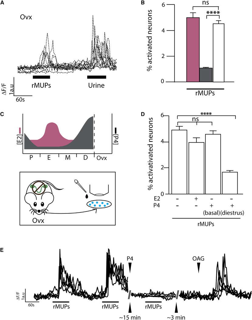
If the sensory detectors are indeed changing their response properties across the estrous cycle, then there must be internal signals that report the female’s estrous state to the VSNs. The ovaries are the significant production source for the female sex-steroid hormones progesterone (P4) and estradiol (E2). Females lacking ovaries fail to synthesize significant circulating E2 or P4. To determine if circulating female hormones alter sensory neural activity, we manipulated hormone production by surgical removal of the ovaries (ovx) and analyzed VSN activity by calcium imaging in response to rMUPs. We found that VSNs from ovx females robustly respond to rMUPs similar to estrous-staged intact females (Figures 2A and 2B). This indicates that rMUP-elicited activity from female VSNs is silenced by key signals from intact ovaries.
Figure 2. Progesterone Silences Sensory Activity.
(A) Overlaid representative calcium influx traces of individual VSNs from ovariectomized (ovx) females in response to stimulation with rMUPs and male urine.
(B) Percentage of VSNs from ovx females showing calcium influx to rMUPs compared to estrous- and diestrous-staged females (1,939; 2,811; and 2,726 cells imaged, respectively).
(C) Upper panel: representation of estrogen (pink, E2) and progesterone (black, P4) surges in cycling and ovariectomized females (Joshi et al., 2010). Lower panel: experimental design of calcium imaging; acute culture of VSNs from ovx females with the addition of hormones/drugs prior to perfusion of ligand stimuli.
(D) Percentage of VSNs from ovx females showing calcium influx to rMUPs alone or with addition of E2 (200 pM) or P4 at basal (5 nM) or diestrus (40 nM) concentrations (3,292; 2,545; 3,286; 2,712 cells imaged, respectively).
(E) Overlaid calcium influx traces of VSNs responding to consecutive pulses of rMUPs, followed by P4 incubation and third pulse of rMUPs, followed by a pulse of DAG analog, 1-Oleoyl-2-acetyl-sn-glycerol (OAG).
(B and D) One-way ANOVA followed by Bonferroni correction. All values in mean ± SEM. *p < 0.05, **p < 0.01, ***p < 0.001, ****p < 0.0001; ns, not significant. White bars, ovx; pink bars, estrus; dark gray bars, diestrus.
See also Figure S2.
In a normally cycling female, both hormones are present at basal levels throughout the cycle but E2 transiently surges prior to estrus while P4 synthesis increases during diestrus (Figure 2C, upper) (Fata et al., 2001; Joshi et al., 2010; Wood et al., 2007). To determine which hormone silences the response to rMUPs (or MUP-enriched native urine fractions) in diestrus we added either background (basal) or cycle-specific surging levels of E2 or P4 (Joshi et al., 2010; Wood et al., 2007) directly into culture media with acutely dissociated VSNs from ovx females and analyzed their activity by calcium imaging (Figure 2C, lower). While the addition of E2 or basal levels of P4 did not significantly alter rMUP signaling, we found that the addition of the diestrous level of progesterone was sufficient to attenuate VSN response to rMUPs (or MUP-enriched native urine fractions) similar to VSNs from intact diestrous females (Figures 2D and S2A). MUP sensory responses require the TRPC2 channel (Chamero et al., 2007). Therefore, we ensured that the subset of rMUP-responsive neurons silenced by P4 remained healthy by perfusing with a known TRPC2 channel activator 1-oleoyl-2-acetyl-sn-glycerol (OAG) (Lucas et al., 2003) (Figure S2B). This treatment generated robust calcium influx indicating that P4 does indeed silence, not damage, rMUP-responsive neurons (Figures 2E and S2B). These experiments further indicate that the action of P4 can occur ex vivo following the removal of the VSNs from the nose; therefore, the sensory silencing of the VNO during diestrus is not likely to be simply a consequence of circuit feedback from hormone detecting central nuclei in the brain. Rather, it indicates that female VSNs are themselves direct targets of the action of P4. Together these results reveal that P4 acts directly on female VSNs to inhibit their ability to sense rMUPs.
VSNs Express Progesterone Responsive Proteins
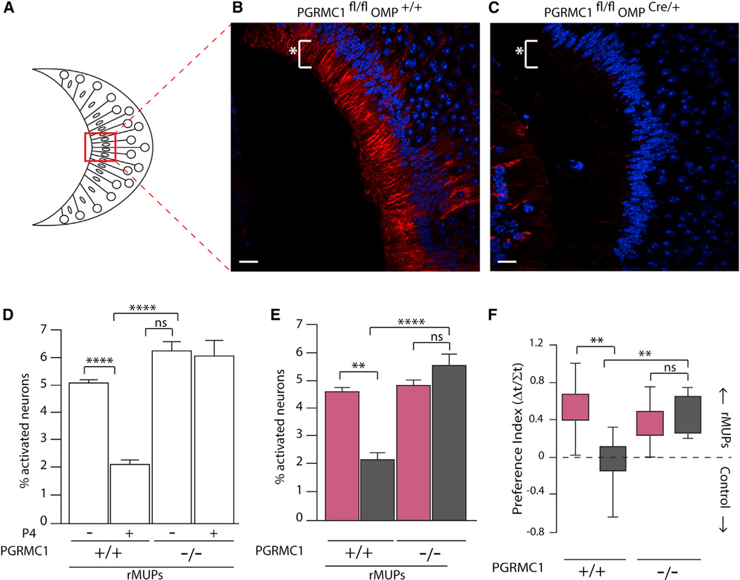
For the sensory neurons to modulate rMUP-promoted attraction behavior, they must have a mechanism to directly monitor the presence of P4. However, VSNs have not been described to express progesterone responsive proteins. We performed RNA sequencing (RNA-seq) analysis on total vomeronasal organ (VNO) cDNA (Ibarra-Soria et al., 2014) and identified several candidates including progesterone receptor membrane-component protein PGRMC1 (Bashour and Wray, 2012; Meyer et al., 1996; Peluso et al., 2008; Petersen et al., 2013; Kaluka et al., 2015). We confirmed its relevance by pre-incubating ovx VSNs with the PGRMC1 antagonist, A205 (Bashour and Wray, 2012), followed by incubation with P4 and performed calcium imaging with rMUP stimulation. Blocking the ability of PGRMC1 to respond to P4 did indeed enable rMUPs to efficiently activate VSNs (Figure S3A). Immunohistochemistry with anti-PGRMC1 confirmed PGRMC1 protein expression in VSNs (Figures 3A, 3B, and S3B–S3D). If PGRMC1 is indeed necessary to mediate P4’s direct effects on rMUP-detecting VSNs, than it should provide the molecular means to further investigate VSN sensory silencing. Therefore, we created an olfactory sensory neuron-specific deletion of Pgrmc1 by crossing olfactory marker protein-Cre, Omp-Cre (Eggan et al., 2004), to a floxed allele of Pgrmc1 (Figures 3B, 3C, and S3E–S3I).
Figure 3. Silencing of VSN Activity by Progesterone Requires PGRMC1.
(A–C) Schematic of coronal VNO epithelium to orient (B) and (C). Immunohistochemical staining for PGRMC1 in (B) Pgrmc1fl/flOmp+/+ and (C) Pgrmc1fl/flOmpCre/+ mice. Scale bar, 20 µm, white asterisk indicates VSN dendrites.
(D)Percentage of VSNs showing calcium influx to rMUPs from ovx Pgrmc1fl/flOmp+/+ and Pgrmc1fl/flOmpCre/+ females treated with or without 40 nM P4 (2,112; 2,041; 2,077; and 2,085 cells imaged, respectively).
(E) Percentage of VSNs showing calcium influx to rMUPs from estrous and diestrous Pgrmc1fl/flOmp+/+ and Pgrmc1fl/fl OmpCre/+ females (2,543; 2,039; 2,549; and 2,223 cells imaged, respectively).
(F) Preference index from two choice behavior assay conducted on estrous and diestrous Pgrmc1fl/flOmp+/+ and Pgrmc1mlOmpCre/+ females (n = 8, 9, 8, and 8, respectively).
(D–F) One-way ANOVA followed by Bonferroni correction. All values in mean ±SEM. *p <0.05, **p< 0.01, ***p< 0.001, ****p< 0.0001; ns, not significant. White bars, ovx; pink bars, estrous; dark gray bars, diestrus.
See also Figure S3.
We evaluated the ability of VSNs to directly respond to P4 signals by three important criteria. First, we determined the ability of VSNs lacking PGRMC1 to respond to the silencing effects of P4 ex vivo. We used calcium imaging to evaluate VSNs from Pgrmc1−/− and Pgrmc1+/+ ovx females and found mutant neurons to be fully responsive to rMUPs; even in the presence of diestrous levels of P4 (Figure 3D). This confirms that wild-type VSNs express specific signaling elements that function to inhibit rMUP-elicited neural activity in the presence of P4. Second, we investigated the ability of rMUP-detecting VSNs to undergo silencing during a natural estrous cycle from gonad-intact animals under the native production of P4. This evaluation was feasible because we found the olfactory-specific deletion of Pgrmc1 had no effect on mutant females to display a regular estrous cycle (Figure S3J). Using calcium imaging we assayed the ability of VSNs from Pgrmc1−/− diestrous-staged females to respond to rMUPs and observed that the mutant VSNs remained consistently responsive to rMUPs across both estrus and diestrus (Figure 3E). This reveals a molecular mechanism through PGRMC1 for P4 to silence rMUP signaling during natural diestrus. Third, we determined if the different female behavior responses to rMUPs across the estrous cycle was dependent upon P4 detection by sensory neurons. We assayed the behavior of mutant females in and found them to be continuously attracted to rMUPs in both estrus and diestrus (Figures 3F, S3K, and S3L). The variation from attraction (in estrus) to indifferent behavior (in diestrus) observed in wild-type females was replaced with constant preference in animals lacking functional PGRMC1. Since these animals only lacked PGRMC1 in olfactory tissues and displayed normal estrous cycles, the lack of cycle-specific variation in behavior can be primarily attributed to defects in sensory signaling. Together, these data reveal that VSNs express specific signaling elements, such as PGRMC1, that enable them to detect and respond to naturally circulating female hormones. The ability of VSNs to monitor circulating female hormones results in differing sensory responses to rMUPs, and correspondingly different behavioral responses, depending on her stage of the estrous cycle.
Not All VSNs Are Silenced by Progesterone
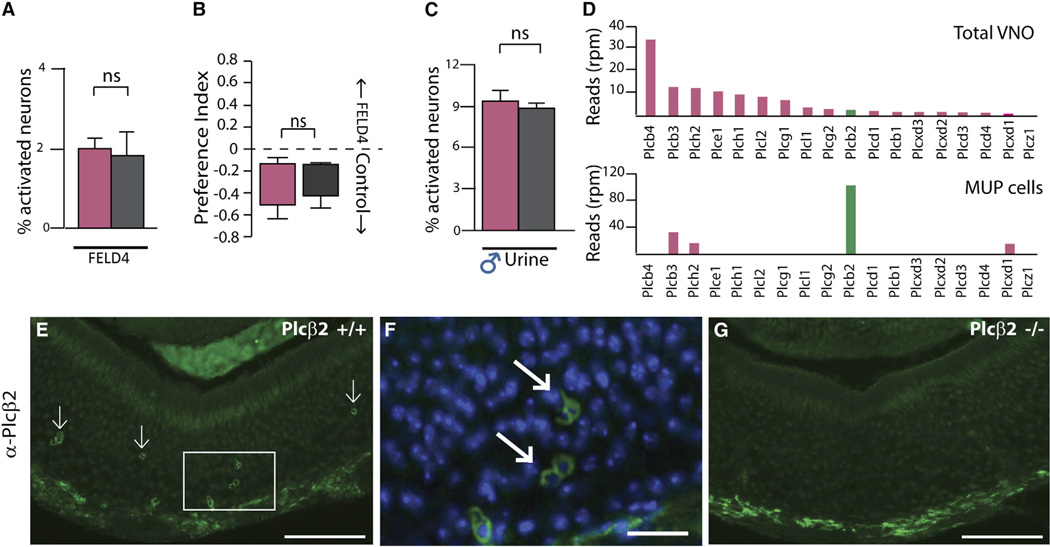
In addition to male odors, the VNO also functions to detect predator odors (Isogai et al., 2011; Papes et al., 2010). P4 silencing of VSNs that warn of danger would potentially threaten the survival of the female during diestrus. To determine if P4 silences all female VSNs during diestrus we evaluated their ability to respond to the cat-emitted kairomone, FELD4, both directly by calcium imaging and functionally by monitoring aversion behavior (Figures 4A and 4B) (Papes et al., 2010). Through both methods, we found no evidence for estrous cycle-specific sensory modulation. Irrespective of the female’s estrous state, FELD4 consistently activated VSNs and resulted in constant behavioral aversion (Figures 4A and 4B). Moreover, we found that overall VSN activity is not silenced during diestrus in response to total male urine (Figures 4C and S4), which in addition to MUPs contains many uncharacterized VNO ligands of unknown function. Therefore, the impairment of sensory response during diestrus is due to P4’s selective modulation on the subset of rMUP detecting VSNs.
Figure 4. VSNs that Detect the Cat Ligand FELD4 Are Not Silenced in Diestrus.
(A) Percentage of VSNs from estrous and diestrous females showing calcium influx to FELD4 (p = 0.615, 2,506 and 4,231 cells imaged).
(B) Preference index from two choice behavior assay conducted on estrous and diestrous females comparing FELD4 against biologically non-relevant control odor(p = 0.225, estrus n = 9, diestrus n = 10).
(C) Percentage of VSNs from estrous and diestrous females showing calcium influx to male urine (p = 0.371, 3,396 and 3,188 cells imaged, respectively).
(D and E) RNA deep sequencing reads (reads per million [rpm]) for PLC family in total unstimulated VNO (top panel) and MUP responsive neurons (bottom panel). Anti-PLCβ2 staining in VNO epithelium from (E) Plcβ2+/+. Scale bar, 70 µm.
(F and G) Inset from (E) with nuclear staining (scale bar, 10 µm) and (G) Plcβ2−/−(scale bar, 70 µm).
(A–C) Two-tailed t test. All values in mean ± SEM. p < 0.05, **p < 0.01, ***p < 0.001, ****p < 0.0001; ns, not significant. White bars, ovx; pink bars, estrus; dark gray bars, diestrus.
See also Figure S4.
Primary Signal Transduction Elements Are Specific to MUP-Responsive VSNs
PGRMC1 is expressed by many VSNs (Figures 3B and S3B–S3D), yet we find that the rMUP-responsive neurons are the only detectable subset that is silenced during diestrus. This indicates that there must be an additional molecular component that targets the silencing action of P4 specifically to the rMUP-responsive VSN subset. VNO sensory neurons are known to molecularly specialize by their expression of one out of ~350 sensory receptors that couple either to Gαi2 or Gαo, however, other primary signaling components, including phospholipase C (PLC), diacylglyceride, and the primary sensory transduction channel TRPC2 are thought to be similarly expressed by all VSNs (Chamero et al., 2012; Dulac and Torello, 2003; Lucas et al., 2003). To identify essential signaling components that are enriched in MUP-responsive neurons, we performed RNA-seq to analyze libraries prepared from either total (unstimulated) VNO tissue (Figure 4D, upper) or pooled individual single VSNs that were activated by a native fraction of male urine that is primarily composed of native MUPs (Figure 4D, lower) (Keydar et al., 2013). We found one member of the PLC family (Kadamur and Ross, 2013), PLCβ2, expressed at low levels in the total VNO library and high levels in MUP-responsive neurons (Figure 4D). Correspondingly, immunohistochemistry of the VNO epithelium revealed that PLCβ2 is not expressed in all sensory neurons; instead, we find its expression limited to a small number of VSNs, consistent with the production of a specialized sensory response (Figures 4E–4G).
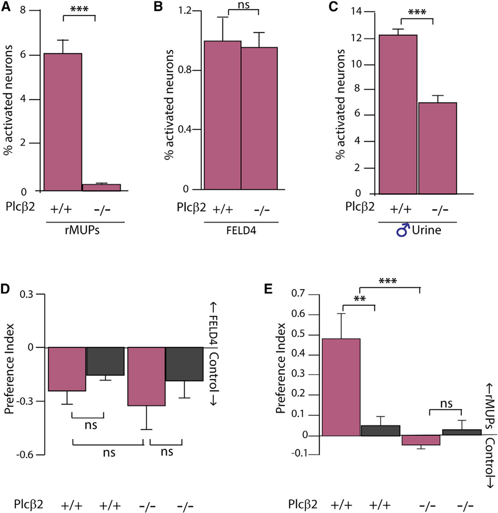
To serve as an effective target of P4 silencing, PLCβ2 must be primary signal transduction element in rMUP responsive neurons but not primary to sensory transduction in FELD4 or total male urine responsive VSNs. We first assayed the functional role of PLCβ2 in rMUP, FELD4, and total male urine signaling by comparing the ability of VSNs from Plcβ2−/− and Plcβ+/+ litter-mates to detect rMUPs by calcium imaging (Figures 5A–5C). VSNs from estrous PLCβ2−/− females failed to respond to rMUP stimulation (Figure 5A), but were activated robustly by non-rMUP stimuli (Figures 5B and 5C). This indicates that PLCβ2 is indeed a primary signal transduction element in rMUP-responsive neurons, but is not required for activity in VSNs that detect cat or other male mouse urine ligands. To evaluate whether PLCβ2 generates rMUP signals that are meaningful to the female, we assayed estrous- and diestrous-staged Plcβ2−/− and Plcβ+/+ littermates for behavioral responses. We found PLCβ2 mutant animals both fully able to detect and avoid FELD4 yet failed to behaviorally respond with attraction to rMUPs during estrus (Figures 5D and 5E). Since the PLCβ2 mutation is not limited to the olfactory sensory neurons, the results from this analysis must be interpreted with caution; however they are consistent with the calcium imaging data. Overall, our analysis indicates that PLCβ2 expression is functionally necessary for VSNs to detect and respond to rMUPs, but is dispensable for primary signaling in subsets of FELD4 detecting VSNs.
Figure 5. PLCβ2 Is a Primary Signaling Component in rMUP Detecting VSNs but Not in FELD4 Detecting VSNs.
(A–E) Percentage of VSNs from Plcβ2+/+ (1,842 cells imaged) and Plcβ2−/− (2,023 cells imaged) estrous staged females showing calcium influx to (A) rMUPs (p = 0.00014), (B) FELD4 (p = 0.9305), and (C) male urine (p = 0.0004). Preference index from two choice behavior assay conducted on Plcβ2+/+ (estrus n = 8; diestrus n = 4) and Plcβ2−/− (estrus n = 7, diestrus n = 3) female mice with (D) FELD4 and (E) rMUPs. (A–C) Two-tailed t test. (D and E) One-way ANOVA followed by Bonferroni correction. All values in mean ± SEM. *p < 0.05, **p < 0.01, ***p < 0.001, ****p < 0.0001; ns, not significant. Pink bars, estrus; dark gray bars, diestrus.
Primary Signaling in MUP-Responsive VSNs Is a Target of Progesterone Silencing
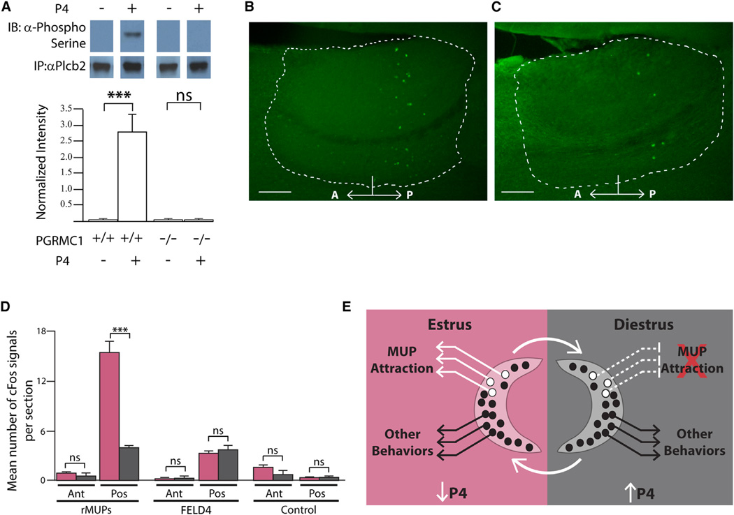
What is special about PLCβ2 that enables it to prevent rMUP activation in the presence of P4? PGRMC1 is known to influence intracellular signaling through the initiation of a phosphorylation cascade (Bashour and Wray, 2012; Su et al., 2012). To determine if phosphorylation could serve as a means to alter rMUP signaling we performed calcium imaging on ovx VSNs and found that the silencing of rMUP signaling by P4 was abolished following kinase inhibition (Figures S5A and S5B). This supports a role for modulation of PLCβ2 activity by phosphorylation. PLC phosphorylation is known to activate some PLC isoforms while it can conversely inhibit the function of other PLC isoforms (Gresset et al., 2012; Litosch, 2002). Phosphorylation of PLCβ2 on serine residues has been shown to silence its signaling activity (Liu and Simon, 1996). In order to determine if the effects of P4-mediated phosphorylation occur on serine residues of PLCβ2, we performed an immunoprecipitation of PLCβ2 from ovx Pgrmc1−/− and Pgrmc1+/+ littermate VSNs following treatment with P4 (Figure 6A). We found that in the absence of P4, PLCβ2 is not subjected to serine phosphorylation. Further, in the presence of P4 serine phosphorylation of PLCβ2 is robust in Pgrmc1+/+ females yet undetectable in Pgrmc1−/− littermates (Figure 6A). This indicates that the inactivating serines of PLCβ2 are phosphorylated by PGRMC1-dependant signaling in the presence of P4. VSNs that do not require PLCβ2 for primary sensory activity (such as those that detect FELD4 or components of total male urine) escape this mechanism of silencing and remain active irrespective of the female’s estrous state.
Figure 6. VSN Sensory Silencing Changes Neural Circuit Activity in Accessory Olfactory Bulb.
(A–C) Immunoprecipitation followed by anti-phosphoserine immunoblot for PLCβ2 from VNOs of Pgrmc1fl/flOmpCre/+ and Pgrmc1fl/fl Omp+/+ treated with or without 40 nM P4 (top panel); phosphoserine density normalized to total PLCβ2 density (bottom panel). Sagittal section of accessory olfactory bulb (AOB, dashed white outline) from (B) estrous and (C) diestrous mice showing cFOS expression after two-choice test with rMUPs (A, anterior; P, posterior; scale bars, 100 µm).
(D) Average number of cFOS positive cells in glomerular and mitral layers per section from three animals in each state, following two-choice assay against either rMUPs, FELD4, or no odor control (Ant, anterior; Pos, posterior).
(E) Schematic representation of differences in MUP responsive VSNs in the presence of high P4 during diestrus compared to low P4 during estrus, which results in specific changes in sensory attraction behavior.
(A) Two-tailed t test. (D) One-way ANOVA followed by Bonferroni correction. All values in mean ± SEM. *p < 0.05, **p < 0.01, ***p < 0.001, ****p < 0.0001; ns, not significant. Pink bars, estrus; dark gray bars, diestrus; white bars, ovx.
See also Figure S5.
Neural Circuit Activity Changes with Sensory Silencing
In order to regulate differences in behavior effectively, VNO sensory silencing would need to alter the activity of the downstream circuit from estrus to diestrus. rMUPs are known to be detected by VSNs that express V2Rs and Gαo that project axons to the posterior accessory olfactory bulb (AOB) (Chamero et al., 2007; Dulac and Torello, 2003; Papes et al., 2010). We used the two-choice test to stimulate VSNs in freely behaving females and then performed immunohistochemistry against the immediate early gene, cFOS, as a proxy to detect recent neural activity across the AOB. During estrus, when the VSNs robustly detect rMUPs, we found cFOS expression restricted to a zone extending across the glomerular, mitral, and granule cell layers of the posterior AOB, consistent with activation of a limited number of glomeruli (Figures 6B, 6D, S5C, S5D, and S5F). However, corresponding with sensory silencing, the cFOS expression in the downstream neurons was significantly attenuated in diestrous females (Figures 6C, 6D, S5D, and S5F). Importantly, the number of cells showing cFOS expression following exposure to the cat kairomone, FELD4, during the two-choice assay was similar between estrous- and diestrous-staged females (Figures 6D and S5E). This is consistent with VSN signaling and the female’s behavior response to FELD4 (Figures 4A and 4B). These findings indicate that VSN sensory silencing alters the response of the downstream neural circuit and provides an effective mechanism to underlie the changes in olfactory-mediated behavior.
DISCUSSION
Cycling Female Hormones Block a Subset of VNO Sensory Detection
Among many species, female behavior varies with the reproductive cycle. Diverse sensory information such as a partner’s appearance, vocalizations, or emitted odors promote female mating behavior during the ovulatory phase yet lead to indifference, or even aggression, during other stages of the reproductive cycle. The primary purpose of a sensory system is to reliably monitor the environment. Therefore, it was thought that irrespective of her state of ovulation a female receives and transmits all available sensory information to the brain where decision-making centers determine the appropriate behavioral response. Here, we show that the estrous cycle-specific hormone, progesterone, directly acts on a subset of vomeronasal sensory neurons to block the transmission of sensory detection normally elicited by a subset of male-emitted pheromones; MUPs. As the estrous cycle repeats, females are regularly rendered “blind” to this subset of ligands during diestrus (Figure 6E). While food “appears” less appetizing following a large meal, imagine that upon reaching an internal state of satiety food was no longer visible; the photoreceptors were specifically blocked from sending light reflections of food to the brain. This inconceivable scenario is analogous to our discovery of a female’s specific inhibition of MUP sensory activity in the diestrous state. Such state-specific modulation of sensory signaling has not been described in mammals.
Primary Signal Transduction Components Specify Sensory Neuron Activity
Whether or not an olfactory sensory neuron responds to a particular ligand is thought to be defined by its specific expression of an odorant receptor (Touhara and Vosshall, 2009). Here, we find that the primary signal transduction molecule PLCβ2 and PGRMC1 also specify the extent to which small subsets of VSNs are activated by ligands. Our data indicate that PLCβ2 function is necessary for primary signaling in response to rMUPs but dispensable for the detection of the cat odor, FELD4, and many unknown ligands contained in male urine. This indicates that other VSNs rely on different PLC isoforms for primary signaling. It will be of interest to determine if there is systematic variation in ligand detection, state responsive detectors and primary signal transduction components across other VSN subsets. Such heterogeneity of response capabilities would render olfactory sensory neurons poised as a peripheral center that integrates internal state with external chemosignals.
Internal Self-Censoring of Available Sensory Cues
In the course of a normal day, a female detects many sensory cues that the brain “decides” not to act upon. The evolution of a molecular mechanism preventing small subsets of sensory signals from being detected during a particular internal state is extraordinary and we do not know the reason for this precaution. It may be that the attraction of male-emitted MUPs provides such a distraction to a female in diestrus that the species has evolved a mechanism to temporarily eliminate their sensation. Alternatively, the silencing of MUP detection may be related to a more fundamental, currently unknown, physiological or behavioral response of diestrous females to male-emitted MUPs perhaps decreasing the female’s fitness. Our data indicate that olfactory attraction to MUPs is not sufficient to generate sexual receptivity behavior in diestrus (Figures S3K and S3L). Female receptivity is known to be activated by the production of estrogen that is largely absent during diestrus (Yang and Shah, 2014). While it is difficult to outline a rationale for an individual to self-censor their sensory detection, the ability to silence incoming sensory signals does produce an absolute method to regulate one’s behavior to a particular subset of cues while still retaining the ability to detect and respond to others. Protein modification is rapid and reversible providing the ability to censure incoming sensory information on a timescale compatible with changing internal state and external environment. Deviation from baseline of other internal states such as hunger, anxiety, and pregnancy also alter an individual’s predicted behavior. Our study raises the possibility that state-variation of other olfactory-mediated behaviors may also arise from sensory modulation, which can be investigated once other behavior-generating ligands have been purified. Our finding of a mechanism by which small subsets of sensory neurons can be rapidly silenced abrogates the brain’s need to process, and its ability to act upon, particular subsets of information. Sensory silencing in this context can account for the state-dependent regulation of behavior.
EXPERIMENTAL PROCEDURES
Estrous Staging of Female Mice
Female C57BL/6J mice (8–10 weeks old) were analyzed. Twenty microliter aliquots of sterile 1 × PBS were used for a vaginal lavage. Five microliters of the resulting suspension was examined using phase contrast microscopy. Mice showing an abundance of cornified epithelial cells were scored as estrus and mice with ~100% blood cells were scored as diestrus (Figure S1B). Mice were staged for at least a full cycle to ensure only normally cycling mice were used for subsequent experiments.
VSN Stimuli
Urine was collected fresh from group-housed adult C57BL/6J male mice, from multiple cages and combined for each experiment. To fractionate, it was applied to Amicon Ultra Centrifugal Filters (10,000 MWCO regenerated cellulose, Millipore). The HMW fraction was washed by adding sterile 1 × PBS (equal to starting urine volume) and centrifugation at 7,000 rpm, room temperature, five times. The washed protein fraction was diluted to starting urine volume with 1× PBS. MUP7 (EMBL: EU882230), MUP10 (EMBL: EU882231), MUP19 (EMBL: EU882232), MUP20 (EMBL: EU882234), and MUP3 (EMBL: EU882235) rMUPs corresponding to the five C57BL/6J MUPs excreted in male urine and FELD4 (Genbank NM_001009233) were expressed and purified as previously described (Kaur et al., 2014; Papes et al., 2010). For calcium imaging: urine, HMW fraction, and rMUPs were diluted 1:300 from native concentrations.
Behavior
Two-choice assay: 8- to 10-week-old estrous-staged female C57BL/6J mice were used for two-choice assays. Mice were habituated for 1 hr in assay cages in the behavior room for 2 days. On the day of the assay, mice were staged for estrus by vaginal cytology. Either 20 µl HMW, rMUP mix (4 µl each, at 5 mg/ml MUP concentration) or FELD4 (4 µl, 15 mg/ml) as stimuli or 1× PBS-MBP (10 mg/ml) control were applied in random patterns to 1 × 1 inch squares of odorless blotting paper (Fisher Scientific, #05–714-4) and stuck to opposite walls of the arena at ~7.5 cm from the bedding using odorless transparent tape. Mice were allowed to investigate for 10 min and video recorded under red light. Video analysis software, The Observer (Noldus), was used to score the frequency and duration of visits. Investigation of the paper by contact with the snout was scored as a visit. All mice were naive to the stimulus and used only once, except some females used for Figures 5D and 5E were repeatedly used for a total of four behavioral assays (once in estrus versus rMUPs, once in estrus versus FELD4, once in diestrus versus rMUPs, and once in diestrus versus FELD4). All animal procedures were in compliance with institutional guidelines established and approved by the Institutional Animal Care and Use Committee.
Calcium Imaging
Calcium imaging was performed as described (Kaur et al., 2013). For all hormone and drug supplementation experiments, VNO from ovariectomized mice were used. Acute culture of VSNs was carried out in phenol-free DMEM (Cellgro, 17–205-CV), supplemented with Charcoal:Dextran stripped FBS (Gemini Bio-Products, 100–119) and L-glutamine. Hormones and drugs used in various assays were dissolved methanol or 0.001% DMSO, as recommended by manufacturer. These were added at indicated concentrations in culture media prior to incubation at 37°C.
See also the Supplemental Experimental Procedures.
Supplementary Material
Highlights.
Females display different behaviors to MUP pheromones depending on their estrous state
Diestrous female MUP-detecting sensory neurons are directly silenced by progesterone
Primary signal transduction heterogeneity allows selective sensory neuron silencing
Sensory silencing abrogates the brain’s need to process irrelevant information
ACKNOWLEDGMENTS
This work was supported by NIH-NIDCD grants DC009413 and DC006885 (L.S.), the Ellison Medical Foundation NR-SS-0107-12 (L.S.), R21 RR030264 (J.J.P. and J.K.P.), the Wellcome Trust (098051) (D.W.L. and X.l.-S.), and NIH DC010857 and DC012095 (H.M). S.D. is grateful to J. Lat, J. Orman, and D. Huff for animal care support.
Footnotes
SUPPLEMENTAL INFORMATION
Supplemental Information includes Supplemental Experimental Procedures and five figures and can be found with this article online at http://dx.doi.org/10.1016/j.cell.2015.04.052.
AUTHOR CONTRIBUTIONS
S.D, P.C., and L.S. initiated the study. S.D. performed all experiments. J.K.P. and J.J.P. generated the conditional PGRMC1 mouse. X.l.-S. and D.W.L. prepared and analyzed the total VNO RNA-seq libraries. K.R.S. isolated single rMUP-responsive VSNs for RNA-seq analysis. M.-S.C. and H.M. prepared and analyzed the pooled rMUP-responsive VSNs by RNA-seq. All authors contributed to the preparation of the manuscript.
REFERENCES
- Bashour NM, Wray S. Progesterone directly and rapidly inhibits GnRH neuronal activity via progesterone receptor membrane component 1. Endocrinology. 2012;153:4457–4469. doi: 10.1210/en.2012-1122. [DOI] [PMC free article] [PubMed] [Google Scholar]
- Chamero P, Marton TF, Logan DW, Flanagan K, Cruz JR, Saghatelian A, Cravatt BF, Stowers L. Identification of protein pheromones that promote aggressive behaviour. Nature. 2007;450:899–902. doi: 10.1038/nature05997. [DOI] [PubMed] [Google Scholar]
- Chamero P, Leinders-Zufall T, Zufall F. From genes to social communication: molecular sensing by the vomeronasal organ. Trends Neurosci. 2012;35:597–606. doi: 10.1016/j.tins.2012.04.011. [DOI] [PubMed] [Google Scholar]
- Dulac O, Torello AT. Molecular detection of pheromone signals in mammals: from genes to behaviour. Nat. Rev. Neurosci. 2003;4:551–562. doi: 10.1038/nrn1140. [DOI] [PubMed] [Google Scholar]
- Eggan K, Baldwin K, Tackett M, Osborne J, Gogos J, Chess A, Axel R, Jaenisch R. Mice cloned from olfactory sensory neurons. Nature. 2004;428:44–49. doi: 10.1038/nature02375. [DOI] [PubMed] [Google Scholar]
- Fata JE, Chaudhary V, Khokha R. Cellular turnover in the mammary gland is correlated with systemic levels of progesterone and not 17beta-estradiol during the estrous cycle. Biol. Reprod. 2001;65:680–688. doi: 10.1095/biolreprod65.3.680. [DOI] [PubMed] [Google Scholar]
- Gresset A, Sondek J, Harden TK. The phospholipase C isozymes and their regulation. Subcell. Biochem. 2012;58:61–94. doi: 10.1007/978-94-007-3012-0_3. [DOI] [PMC free article] [PubMed] [Google Scholar]
- Ibarra-Soria X, Levitin MO, Saraiva LR, Logan DW. The olfactory transcriptomes of mice. PLoS Genet. 2014;10:e1004593. doi: 10.1371/journal.pgen.1004593. [DOI] [PMC free article] [PubMed] [Google Scholar]
- Isogai Y, Si S, Pont-Lezica L, Tan T, Kapoor V, Murthy VN, Dulac C. Molecular organization of vomeronasal chemoreception. Nature. 2011;478:241–245. doi: 10.1038/nature10437. [DOI] [PMC free article] [PubMed] [Google Scholar]
- Joshi PA, Jackson HW, Beristain AG, Di Grappa MA, Mote PA, Clarke CL, Stingl J, Waterhouse PD, Khokha R. Progesterone induces adult mammary stem cell expansion. Nature. 2010;465:803–807. doi: 10.1038/nature09091. [DOI] [PubMed] [Google Scholar]
- Juntti SA, Tollkuhn J, Wu MV, Fraser EJ, Soderborg T, Tan S, Honda S, Harada N, Shah NM. The androgen receptor governs the execution, but not programming, of male sexual and territorial behaviors. Neuron. 2010;66:260–272. doi: 10.1016/j.neuron.2010.03.024. [DOI] [PMC free article] [PubMed] [Google Scholar]
- Kadamur G, Ross EM. Mammalian phospholipase C. Annu. Rev. Physiol. 2013;75:127–154. doi: 10.1146/annurev-physiol-030212-183750. [DOI] [PubMed] [Google Scholar]
- Kaluka D, Batabyal D, Chiang BY, Poulos TL, Yeh SR. Spectroscopic and mutagenesis studies of human PGRMC1. Biochemistry. 2015;54:1638–1647. doi: 10.1021/bi501177e. [DOI] [PMC free article] [PubMed] [Google Scholar]
- Karlson P, Luscher M. Pheromones’: a new term for a class of biologically active substances. Nature. 1959;183:55–56. doi: 10.1038/183055a0. [DOI] [PubMed] [Google Scholar]
- Kaur A, Dey S, Stowers L. Live cell calcium imaging of dissociated vomeronasal neurons. Methods Mol. Biol. 2013;1068:189–200. doi: 10.1007/978-1-62703-619-1_13. [DOI] [PubMed] [Google Scholar]
- Kaur AW, Ackels T, Kuo TH, Cichy A, Dey S, Hays C, Kateri M, Logan DW, Marton TF, Spehr M, Stowers L. Murine pheromone proteins constitute a context-dependent combinatorial code governing multiple social behaviors. Cell. 2014;157:676–688. doi: 10.1016/j.cell.2014.02.025. [DOI] [PMC free article] [PubMed] [Google Scholar]
- Keydar I, Ben-Asher E, Feldmesser E, Nativ N, Oshimoto A, Restrepo D, Matsunami H, Chien MS, Pinto JM, Gilad Y, et al. General olfactory sensitivity database (GOSdb): candidate genes and their genomic variations. Hum. Mutat. 2013;34:32–41. doi: 10.1002/humu.22212. [DOI] [PMC free article] [PubMed] [Google Scholar]
- Lee H, Kim DW, Remedios R, Anthony TE, Chang A, Madisen L, Zeng H, Anderson DJ. Scalable control of mounting and attack by Esr1+ neurons in the ventromedial hypothalamus. Nature. 2014;509:627–632. doi: 10.1038/nature13169. [DOI] [PMC free article] [PubMed] [Google Scholar]
- Liberies SD. Mammalian pheromones. Annu. Rev. Physiol. 2014;76:151–175. doi: 10.1146/annurev-physiol-021113-170334. [DOI] [PMC free article] [PubMed] [Google Scholar]
- Litosch I. Novel mechanisms for feedback regulation of phospholipase C-beta activity. IUBMB Life. 2002;54:253–260. doi: 10.1080/15216540215673. [DOI] [PubMed] [Google Scholar]
- Liu M, Simon MI. Regulation by cAMP-dependent protein kinease of a G-protein-mediated phospholipase C. Nature. 1996;382:83–87. doi: 10.1038/382083a0. [DOI] [PubMed] [Google Scholar]
- Lucas P, Ukhanov K, Leinders-Zufall T, Zufall F. A diacylglycerol-gated cation channel in vomeronasal neuron dendrites is impaired in TRPC2 mutant mice: mechanism of pheromone transduction. Neuron. 2003;40:551–561. doi: 10.1016/s0896-6273(03)00675-5. [DOI] [PubMed] [Google Scholar]
- Manoli DS, Fan P, Fraser EJ, Shah NM. Neural control of sexually dimorphic behaviors. Curr. Opin. Neurobiol. 2013;23:330–338. doi: 10.1016/j.conb.2013.04.005. [DOI] [PMC free article] [PubMed] [Google Scholar]
- Meyer O, Schmid R, Scriba PC, Wehling M. Purification and partial sequencing of high-affinity progesterone-binding site(s) from porcine liver membranes. FEBS. 1996;239:726–731. doi: 10.1111/j.1432-1033.1996.0726u.x. [DOI] [PubMed] [Google Scholar]
- Morris JA, Jordan CL, Breedlove SM. Sexual differentiation of the vertebrate nervous system. Nat. Neurosci. 2004;7:1034–1039. doi: 10.1038/nn1325. [DOI] [PubMed] [Google Scholar]
- Papes F, Logan DW, Stowers L. The vomeronasal organ mediates interspecies defensive behaviors through detection of protein pheromone homologs. Cell. 2010;141:692–703. doi: 10.1016/j.cell.2010.03.037. [DOI] [PMC free article] [PubMed] [Google Scholar]
- Peluso JJ, Romak J, Liu X. Progesterone receptor membrane component-1 (PGRMC1) is the mediator of progesterone’s antiapoptotic action in spontaneously immortalized granulosa cells as revealed by PGRMC1 small interfering ribonucleic acid treatment and functional analysis of PGRMC1 mutations. Endocrinology. 2008;149:534–543. doi: 10.1210/en.2007-1050. [DOI] [PMC free article] [PubMed] [Google Scholar]
- Petersen SL, Intlekofer KA, Moura-Conlon PJ, Brewer DN, Del Pino Sans J, Lopez JA. Novel progesterone receptors: neural localization and possible functions. Front. Neurosci. 2013;7:164. doi: 10.3389/fnins.2013.00164. [DOI] [PMC free article] [PubMed] [Google Scholar]
- Roberts SA, Simpson DM, Armstrong SD, Davidson AJ, Robertson DH, McLean L, Beynon RJ, Hurst JL. Darcin: a male pheromone that stimulates female memory and sexual attraction to an individual male’s odour. BMC Biol. 2010;8:75. doi: 10.1186/1741-7007-8-75. [DOI] [PMC free article] [PubMed] [Google Scholar]
- Roberts SA, Davidson AJ, McLean L, Beynon RJ, Hurst JL. Pheromonal induction of spatial learning in mice. Science. 2012;338:1462–1465. doi: 10.1126/science.1225638. [DOI] [PubMed] [Google Scholar]
- Stowers L, Logan DW. Sexual dimorphism in olfactory signaling. Curr. Opin. Neurobiol. 2010;20:770–775. doi: 10.1016/j.conb.2010.08.015. [DOI] [PMC free article] [PubMed] [Google Scholar]
- Su C, Cunningham RL, Rybalchenko N, Singh M. Progesterone increases the release of brain-derived neurotrophic factor from glia via progesterone receptor membrane component 1 (Pgrmc1)-dependent ERK5 signaling. Endocrinology. 2012;153:4389–4400. doi: 10.1210/en.2011-2177. [DOI] [PMC free article] [PubMed] [Google Scholar]
- Touhara K, Vosshall LB. Sensing odorants and pheromones with chemosensory receptors. Annu. Rev. Physiol. 2009;71:307–332. doi: 10.1146/annurev.physiol.010908.163209. [DOI] [PubMed] [Google Scholar]
- Wood GA, Fata JE, Watson KL, Khokha R. Circulating hormones and estrous stage predict cellular and stromal remodeling in murine uterus. Reproduction. 2007;133:1035–1044. doi: 10.1530/REP-06-0302. [DOI] [PubMed] [Google Scholar]
- Wyatt TD. Pheromones and signature mixtures: defining species-wide signals and variable cues for identity in both invertebrates and vertebrates. J. Comp. Physiol. A Neuroethol. Sens. Neural Behav. Physiol. 2010;196:685–700. doi: 10.1007/s00359-010-0564-y. [DOI] [PubMed] [Google Scholar]
- Yang CF, Shah NM. Representing sex in the brain, one module at a time. Neuron. 2014;82:261–278. doi: 10.1016/j.neuron.2014.03.029. [DOI] [PMC free article] [PubMed] [Google Scholar]
- Yang CF, Chiang MC, Gray DC, Prabhakaran M, Alvarado M, Juntti SA, Unger EK, Wells JA, Shah NM. Sexually dimorphic neurons in the ventromedial hypothalamus govern mating in both sexes and aggression in males. Cell. 2013;153:896–909. doi: 10.1016/j.cell.2013.04.017. [DOI] [PMC free article] [PubMed] [Google Scholar]
Associated Data
This section collects any data citations, data availability statements, or supplementary materials included in this article.


