

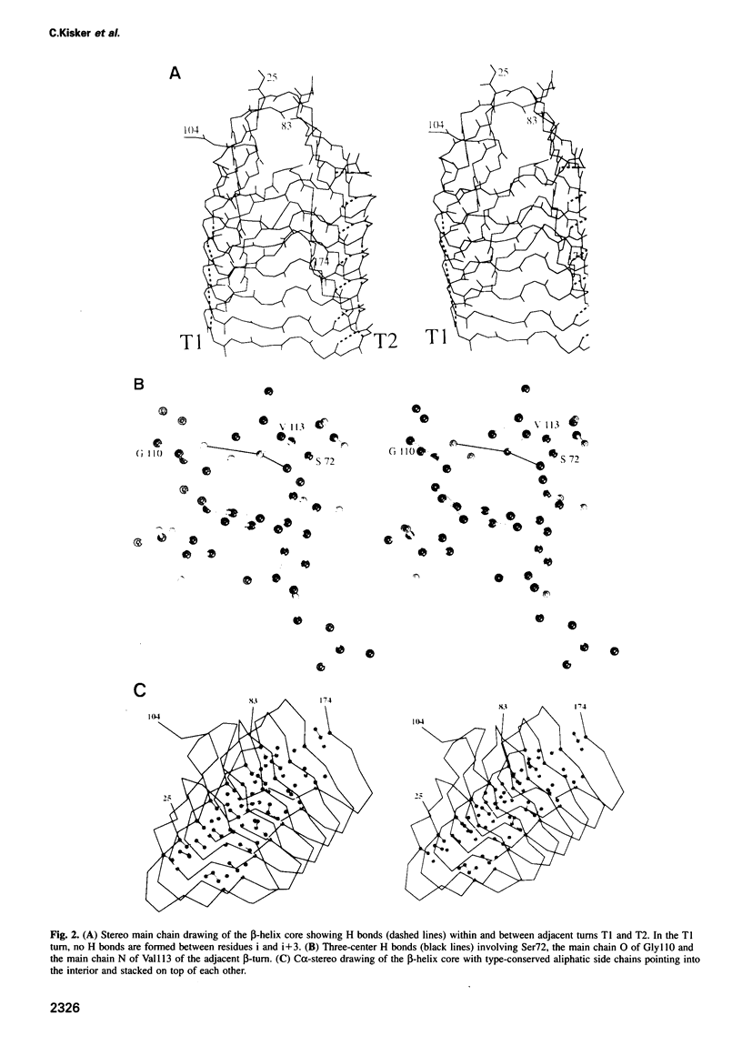
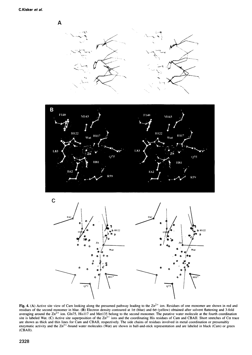
Abstract
A carbonic anhydrase from the thermophilic archaeon Methanosarcina thermophila that exhibits no significant sequence similarity to known carbonic anhydrases has recently been characterized. Here we present the structure of this enzyme, which adopts a left-handed parallel beta-helix fold. This fold is of particular interest since it contains only left-handed crossover connections between the parallel beta-strands, which so far have been observed very infrequently. The active form of the enzyme is a trimer with three zinc-containing active sites, each located at the interface between two monomers. While the arrangement of active site groups differs between this enzyme and the carbonic anhydrases from higher vertebrates, there are structural similarities in the zinc coordination environment, suggestive of convergent evolution dictated by the chemical requirements for catalysis of the same reaction. Based on sequence similarities, the structure of this enzyme is the prototype of a new class of carbonic anhydrases with representatives in all three phylogenetic domains of life.
Full text
PDF


Images in this article


Selected References
These references are in PubMed. This may not be the complete list of references from this article.
- AEvarsson A., Brazhnikov E., Garber M., Zheltonosova J., Chirgadze Y., al-Karadaghi S., Svensson L. A., Liljas A. Three-dimensional structure of the ribosomal translocase: elongation factor G from Thermus thermophilus. EMBO J. 1994 Aug 15;13(16):3669–3677. doi: 10.1002/j.1460-2075.1994.tb06676.x. [DOI] [PMC free article] [PubMed] [Google Scholar]
- Abrahams J. P., Leslie A. G. Methods used in the structure determination of bovine mitochondrial F1 ATPase. Acta Crystallogr D Biol Crystallogr. 1996 Jan 1;52(Pt 1):30–42. doi: 10.1107/S0907444995008754. [DOI] [PubMed] [Google Scholar]
- Alber B. E., Ferry J. G. A carbonic anhydrase from the archaeon Methanosarcina thermophila. Proc Natl Acad Sci U S A. 1994 Jul 19;91(15):6909–6913. doi: 10.1073/pnas.91.15.6909. [DOI] [PMC free article] [PubMed] [Google Scholar]
- Bjorkman P. J., Saper M. A., Samraoui B., Bennett W. S., Strominger J. L., Wiley D. C. Structure of the human class I histocompatibility antigen, HLA-A2. Nature. 1987 Oct 8;329(6139):506–512. doi: 10.1038/329506a0. [DOI] [PubMed] [Google Scholar]
- Fukuzawa H., Suzuki E., Komukai Y., Miyachi S. A gene homologous to chloroplast carbonic anhydrase (icfA) is essential to photosynthetic carbon dioxide fixation by Synechococcus PCC7942. Proc Natl Acad Sci U S A. 1992 May 15;89(10):4437–4441. doi: 10.1073/pnas.89.10.4437. [DOI] [PMC free article] [PubMed] [Google Scholar]
- Gu F., Khimani A., Rane S. G., Flurkey W. H., Bozarth R. F., Smith T. J. Structure and function of a virally encoded fungal toxin from Ustilago maydis: a fungal and mammalian Ca2+ channel inhibitor. Structure. 1995 Aug 15;3(8):805–814. doi: 10.1016/s0969-2126(01)00215-5. [DOI] [PubMed] [Google Scholar]
- Håkansson K., Carlsson M., Svensson L. A., Liljas A. Structure of native and apo carbonic anhydrase II and structure of some of its anion-ligand complexes. J Mol Biol. 1992 Oct 20;227(4):1192–1204. doi: 10.1016/0022-2836(92)90531-n. [DOI] [PubMed] [Google Scholar]
- Jones T. A., Zou J. Y., Cowan S. W., Kjeldgaard M. Improved methods for building protein models in electron density maps and the location of errors in these models. Acta Crystallogr A. 1991 Mar 1;47(Pt 2):110–119. doi: 10.1107/s0108767390010224. [DOI] [PubMed] [Google Scholar]
- Lietzke S. E., Yoder M. D., Keen N. T., Jurnak F. The Three-Dimensional Structure of Pectate Lyase E, a Plant Virulence Factor from Erwinia chrysanthemi. Plant Physiol. 1994 Nov;106(3):849–862. doi: 10.1104/pp.106.3.849. [DOI] [PMC free article] [PubMed] [Google Scholar]
- Pickersgill R., Jenkins J., Harris G., Nasser W., Robert-Baudouy J. The structure of Bacillus subtilis pectate lyase in complex with calcium. Nat Struct Biol. 1994 Oct;1(10):717–723. doi: 10.1038/nsb1094-717. [DOI] [PubMed] [Google Scholar]
- Raetz C. R., Roderick S. L. A left-handed parallel beta helix in the structure of UDP-N-acetylglucosamine acyltransferase. Science. 1995 Nov 10;270(5238):997–1000. doi: 10.1126/science.270.5238.997. [DOI] [PubMed] [Google Scholar]
- Richardson J. S. Handedness of crossover connections in beta sheets. Proc Natl Acad Sci U S A. 1976 Aug;73(8):2619–2623. doi: 10.1073/pnas.73.8.2619. [DOI] [PMC free article] [PubMed] [Google Scholar]
- Sly W. S., Hu P. Y. Human carbonic anhydrases and carbonic anhydrase deficiencies. Annu Rev Biochem. 1995;64:375–401. doi: 10.1146/annurev.bi.64.070195.002111. [DOI] [PubMed] [Google Scholar]
- Steinbacher S., Seckler R., Miller S., Steipe B., Huber R., Reinemer P. Crystal structure of P22 tailspike protein: interdigitated subunits in a thermostable trimer. Science. 1994 Jul 15;265(5170):383–386. doi: 10.1126/science.8023158. [DOI] [PubMed] [Google Scholar]
- Sternberg M. J., Thornton J. M. On the conformation of proteins: the handedness of the connection between parallel beta-strands. J Mol Biol. 1977 Feb 25;110(2):269–283. doi: 10.1016/s0022-2836(77)80072-7. [DOI] [PubMed] [Google Scholar]
- Wilmot C. M., Thornton J. M. Analysis and prediction of the different types of beta-turn in proteins. J Mol Biol. 1988 Sep 5;203(1):221–232. doi: 10.1016/0022-2836(88)90103-9. [DOI] [PubMed] [Google Scholar]
- Wright C. S., Alden R. A., Kraut J. Structure of subtilisin BPN' at 2.5 angström resolution. Nature. 1969 Jan 18;221(5177):235–242. doi: 10.1038/221235a0. [DOI] [PubMed] [Google Scholar]
- Xue Y., Liljas A., Jonsson B. H., Lindskog S. Structural analysis of the zinc hydroxide-Thr-199-Glu-106 hydrogen-bond network in human carbonic anhydrase II. Proteins. 1993 Sep;17(1):93–106. doi: 10.1002/prot.340170112. [DOI] [PubMed] [Google Scholar]
- Yoder M. D., Jurnak F. Protein motifs. 3. The parallel beta helix and other coiled folds. FASEB J. 1995 Mar;9(5):335–342. doi: 10.1096/fasebj.9.5.7896002. [DOI] [PubMed] [Google Scholar]
- Yoder M. D., Keen N. T., Jurnak F. New domain motif: the structure of pectate lyase C, a secreted plant virulence factor. Science. 1993 Jun 4;260(5113):1503–1507. doi: 10.1126/science.8502994. [DOI] [PubMed] [Google Scholar]