

Abstract
The success rate of implant therapy has improved up to 90–95 %. This can be attributed to a numerous factors namely proper case selection, improved diagnostic and radiographic techniques, good softwares for treatment planning, improved surgical equipments, good surgical techniques and sophisticated implant design. The cost of advanced diagnostic techniques and treatment planning software can sometime limit them from being used routinely. In such unfortunate situations, older technique of exposing the ridge and placing implants wherever possible without regard for favorable implant position or angulation is still being followed. This case report describes prosthetic rehabilitation of a partially edentulous patient who was abandoned by a general practitioner after implant placement. Five implants had been placed in the maxilla in prosthetically unfavorable positions and angulations. Castable abutments were then used and a single bar was cast. This bar was then incorporated in a FP3 type of a fixed maxillary prosthesis opposing existing fixed partial denture in the lower jaw. 1 year follow up shows stable implants, healthy peri-implant tissue, minimal probing depth and no radiographic evidence of pathology.
Introduction
The success rate of implant therapy has improved up to 90–95 % [1, 2]. This can be attributed to a numerous factors namely proper case selection [3], improved diagnostic and radiographic techniques [4], good softwares for treatment planning, improved surgical equipments, good surgical techniques and sophisticated implant design. Computed tomographic images give accurate details about height and width of underlying bone. With proper prosthetic planning prior to imaging, the bone dimensions in proposed implant location can be determined. Feeding such data to software can help create an accurate digital model of the maxilla and mandible. Certain software [5] even allows designing a virtual prosthesis and virtual implant placement underneath. Thus favorable implant position and angulation can be planned and the same can be achieved with the help of a surgical template. Screw retained prosthesis that was fabricated prior to implant placement with the help of CADCAM technology can then secured to the implants to serve as a temporary prosthesis.
The cost of such diagnostic aids and surgical templates make them impractical for use in all situations. The following is a case report of one such situation where none of the diagnostic aids were used and five implants were placed in the maxilla without regard for implant position or angulation. A cast bar and an over-denture would conventionally be used to manage such a challenging situation and restore optimal function and esthetics. But the patients demand for a fixed restoration complicates the treatment plan. Screw retained acrylic prosthesis similar to the temporary prosthesis made in association with CADCAM technology could not be used in this situation. The flexure of the acrylic prosthesis can cause overload of implants with deleterious tilting and torqing forces as the implants have been placed in unfavorable location. The acrylic can also fracture leading to failure of the restoration [6]. So a cast bar was fabricated and incorporated into the acrylic denture in order to eliminate deleterious forces and to transfer occlusal forces evenly to all implants.
Case Report
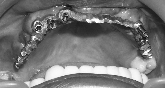
A 37-year-old male patient was referred to the Department of Prosthodontics, Madha dental college and Hospital, Chennai, for prosthetic rehabilitation. A local general practitioner had placed five EZ-Hitech implants in prosthetically unfavorable locations and angulations about 6 months earlier. Prosthetic rehabilitation became a challenge and the patient was referred. On examination his maxillary jaw was partially edentulous with 18 and 28 being the remaining natural teeth. Five healing abutments were visible in an otherwise healed maxillary ridge (Fig. 1). The healing abutments approximately corresponded to location of 11, 12, 13, 24 and 25. Orthopanographic radiograph confirmed five implants in the said locations.
Fig. 1.

Preoperative
Patient was given the option of an additional implant in the 15 or 16 region in order to reduce the cantilever length and to be able to fabricate a fixed prosthesis with favorable load distribution. Patient turned down the option and insisted on a fixed prosthesis supported by the existing five implants.
Healing abutments were removed and impression posts were attached and secured to the implants with abutment screw. An open tray implant level impression [7] was made. Impression was removed after loosening the abutment screws, Implant analogues were fixed and Die stone cast poured. Castable abutments were fixed to the implant analogues in the cast (Fig. 2). A single bar was waxed up connecting all the Castable abutments. Sprue formers were attached (Fig. 3) and the wax and was invested. Casting procedure was carried out and the bar was cast with Nickel Chromium alloy [8]. Divestment was done, casting recovered, cleaned, sand blasted and trimmed.
Fig. 2.

Castable abutments secured to the implant analogues show the unfavorable position and angulation
Fig. 3.

Wax-up ready for investing
Misfit of the casting due to casting shrinkage was anticipated and so the casting was sectioned into five units. Sections were made in the bar between 13 and 12 region, 12 and 11 region, 24 and 25 region and finally in the midline. The five sections were secured on to the implants in vivo with abutment screws and the precise seating of the abutment was verified with the help of radiographs. On confirming proper seating of the individual portions, the sectioned regions were united with pattern resin. This was then removed, invested and cast joined [9] to obtain a single bar. The passive fit of the bar was once again verified in vivo (Fig. 4).
Fig. 4.
Fit of abutment bar after ‘Cast joining’
Base plate wax was attached over the bar to fabricate an occlusal rim. Jaw relation was recorded after securing the bar to the maxillary implants and then the maxillary cast was oriented. The mandibular cast was then mounted on Hanau articulator. Teeth arrangement was completed and access holes were drilled in teeth 11, 12, 13, 24 and 25, in order to be able to insert the abutment screw to secure the bar to the implants. “Try in verification” was done after securing the bar to the implants. The access holes were then blocked with lab putty and the denture processed by compression molding technique in such a way that the bar was incorporated in the denture base. The processed denture was recovered, trimmed and polished (Figs. 5, 6).
Fig. 5.
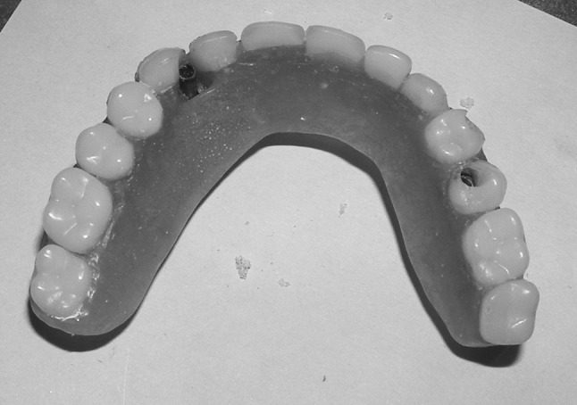
Occlusal view of prosthesis
Fig. 6.
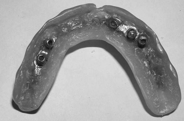
Intaglio surface of prosthesis
The labial flange of the denture was retained for aesthetics but the palatal flange was trimmed and modified to provide access for the patient to perform routine oral hygiene procedures. Denture was secured to the implants with abutment screws and occlusion checked (Fig. 7). The access holes were then sealed with composite resin and the patient was scheduled for recall on alternate days for the first 2 weeks to verify oral hygiene of the patient. Once the oral hygiene practice of the patient was found to be satisfactory and efficient, the patient was scheduled for a 6 monthly periodic recall appointment.
Fig. 7.
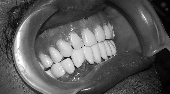
Occlusion of prosthesis
Discussion
Cement retained prosthesis was not an option because of the unfavorable position and angulation of the implants. Need for an economic screw retained prosthesis made an acrylic prosthesis as the most appropriate prosthesis in this situation. The cantilever length in the first quadrant is more than that in the second quadrant because of the absence of implant in the 15 or 16 regions. An acrylic prosthesis would flex in this region resulting in deleterious forces transferred to the implant in relation to 13. Fracture of acrylic prosthesis has also been reported [6] especially in case of presence of cantilever. Reinforcing acrylic provisional with a cast bar has been reported [10, 11] for use as provisional or as interim fixed denture in full mouth rehabitation, where fracture of provisional acrylic prosthesis is common. Reinforced provisional acrylic denture leuted to milled abutments with the help of resin cement has also been reported [12]. But the use of reinforced acrylic denture as long-term implant supported prosthesis has not been reported. Conventionally metal-ceramic restoration along with gingival porcelain is considered as a long-term restoration. But the incorporation of a cast bar in the acrylic prosthesis improves rigidity and strength of the prosthesis and so has been used here as a long term prosthesis.
Summary
Five implants placed in partially edentulous maxilla without prior planning became a challenge to prosthetically rehabilitate. Castable abutments were used and they were joined together in wax-up for a single bar. The bar was then incorporated in an acrylic denture that can be secured to the implants with abutment screws. Thus a fixed denture was processed and secured in position.
Contributor Information
P. R. Ganesh, Phone: +044-26421062, Phone: +919840148834, Email: drprg@rediffmail.com
G. Madhan, Email: drmadhanmds@gmail.com
References
- 1.Ribeiro FS, Pontes AE, Marcantonio E, Piattelli A, Neto RJ, Marcantonio E., Jr Success rate of immediate nonfunctional loaded single-tooth implants: immediate versus delayed implantation. Implant Dent. 2008;17(1):109–117. doi: 10.1097/ID.0b013e318166cb84. [DOI] [PubMed] [Google Scholar]
- 2.Tawse-Smith A, Payne AGT, Kumara R, Thomson WM. One-stage operative procedure using two different implant systems: a prospective study on implant overdentures in the edentulous mandible. Clin Implant Dent Relat Res. 2001;3–4:185–193. doi: 10.1111/j.1708-8208.2001.tb00140.x. [DOI] [PubMed] [Google Scholar]
- 3.Tanner T. Treatment planning for dental implants: considerations, indications, and contraindications. Dent Update. 1997;24(6):253–260. [PubMed] [Google Scholar]
- 4.Chan HL, Misch K, Wang HL. Dental imaging in implant treatment planning. Implant Dent. 2010;19(4):288–298. doi: 10.1097/ID.0b013e3181e59ebd. [DOI] [PubMed] [Google Scholar]
- 5.Spector L. Computer-aided dental implant planning. Dent Clin North Am. 2008;52(4):761–775. doi: 10.1016/j.cden.2008.05.004. [DOI] [PubMed] [Google Scholar]
- 6.Suarez-Feito JM, Sicilia A, Angulo J, Banerji S, Cuesta I, Millar B. Clinical performance of provisional screw-retained metal-free acrylic restorations in an immediate loading implant protocol: a 242 consecutive patients’ report. Clin Oral Implants Res. 2010;21(12):1360–1369. doi: 10.1111/j.1600-0501.2010.01956.x. [DOI] [PubMed] [Google Scholar]
- 7.Chaimattayompol N, Arbree NS, Wong SX. A simple method of making an implant-level impression when presented with limited space, unfavorable implant positions, or problematic implant angulations. J Prosthet Dent. 2002;87(6):684–687. doi: 10.1067/mpr.2002.126422. [DOI] [PubMed] [Google Scholar]
- 8.Barbosa GA, das Neves FD, de Mattos Mda G, Rodrigues RC, Ribeiro RF. Implant/abutment vertical misfit of one-piece cast frameworks made with different materials. Braz Dent J. 2010;21(6):515–519. doi: 10.1590/S0103-64402010000600006. [DOI] [PubMed] [Google Scholar]
- 9.Zoidis PC, Winkler S, Karellos ND. The effect of soldering, electrowelding, and cast-to procedures on the accuracy of fit of cast implant bars. Implant Dent. 1996;5(3):163–168. doi: 10.1097/00008505-199600530-00002. [DOI] [PubMed] [Google Scholar]
- 10.Saba Sebastian. Design of a cast bar reinforced provisional restoration for the management of the interim phase in implant dentistry. J Can Dent Assoc. 1999;65:160–162. [PubMed] [Google Scholar]
- 11.Degidi Marco, Gehrke Peter, Spanel Andre, Piattelli Adriano. Syncrystallization: a technique for temporization of immediately loaded implants with metal-reinforced acrylic resin restorations. Clin Implant Dent Relat Res. 2006;8(3):123–134. doi: 10.1111/j.1708-8208.2006.00011.x. [DOI] [PubMed] [Google Scholar]
- 12.Mirza RB, Gunaseelan R. Full-arch metal-resin cement-and screw-retained provisional restoration for immediately loaded implants. J Oral Implantol. 2010;36(3):219–223. doi: 10.1563/AAID-JOI-D-09-00048. [DOI] [PubMed] [Google Scholar]