Abstract
Objective: To explore the effects of Wnt5a and Wint7a on the differentiation of human embryonic stem cells (hESCs) into endometrium-like cells, and provide a basis for establishing endometrium-like cell models and a cell source for carrying out further endometrium-related experiments. Methods: The hESCs established by our center were differentiated into endometrium-like cells in 4 different media including Wnt5a (Group A), Wnt7a (Group B), secreted frizzled related protein (sFRP, an inhibitor of Wnt signal pathway, Group C) and medium alone (Group D). In the differentiated terminal cells, the expressions of cytokeratin (CK) and vimentin were detected with immunofluorescence, and the mRNA levels of CK18, epithelial cell adhesion molecule (EPCAM), estrogen receptor (ER) and progesterone receptor (PR) were determined with RT-PCR. At the same time, the differentiated terminal cells were incubated in medium containing medroxyprogesterone followed by determination of prolactin (PRL). Results: RT-PCR indicated that mRNA levels of CK18, EPCAM, ER and PR were significantly higher in Group A (Wnt5a) than in other groups (all P < 0.05), but were significantly lower in Group C (sFRP2) than in other groups (all P < 0.05). The changing trend of PRL mRNA was consistent with that of above genes in the 4 groups. Immunofluorescence displayed that the expression of cytokeratin was the strongest in Group A (Wnt5a), and the weakest in Group C (sFRP2) among the 4 groups. Conclusion: Wnt5a has promotive effects on the differentiation of hESCs into endometrium-like cells, but Wnt7a has no marked effects.
Keywords: Wnt5a, Wnt7a, human embryonic stem cells, endometrium-like cells
Introduction
In assisted reproductive technology, endometrial thickness is strongly correlated with the rates of embryo implantation and clinical pregnancy [1]. The thin endometrium can significantly reduce the rates of embryo implantation and clinical pregnancy. It has not been completely clear that how the endometrium develops and which signal pathways are involved in the endometrial development.
Although endometrial stem cells have been isolated from human endometrial tissue, their differentiation direction is not easily controlled because self-differentiation readily occurs during in vitro culture of adult stem cells. Human embryonic stem cells (hESCs) with self-renewal capacity and totipotency are obtained by isolation from cell mass of blastocysts and in vitro differentiated culture [2]. Observing the differentiation of hESCs into endometrium-like cells is conducive to understanding the endometrial development and critical signal pathways associated with endometrial regeneration, providing a basis for application of stem cells in clinical practice.
At present, cytokines are commonly used in the differentiation of stem cells. Epidermal growth factor (EGF), tumor growth factor (TGF-α) and platelet-derived growth factor (PDGF-BB) can promote the growth of endometrial stem cell clones [3], and are present in some epithelial tissues such as the skin, gastrointestinal tract and endometrium [4-6]. The uterus develops from Mullerian ducts in which the cells with Wnt7a expression give rise to epithelial cells of the fallopian tubes and uterus, and the cells with Wnt5a expression give rise to stromal cells of the uterus, cervix and vagina [7,8]. It is necessary to know whether Wnt5a and Wnt7a are involved in the differentiation of hESCs into endometrium-like cells and what roles they play.
In this study, 4 different media including Wnt5a, Wnt7a, secreted frizzled related protein (sFRP, an inhibitor of Wnt signal pathway) and medium alone were used in the differentiation of hESCs into endometrium-like cells to compare the differentiation efficiencies between the 4 media and establish more efficient differentiation scheme. This study provides a basis for establishing endometrium-like cell models and a cell source for carrying out further endometrium-related experiments.
Materials and methods
All study methods were approved by Institutional Review Board and Ethics Committee of the First Affiliated Hospital of Zhengzhou University.
Materials
hESCs were established by our center. Recombinant human Wnt5a, Wnt7a and sFRP2 were purchased from R&D Company (Emeryville, CA, USA). Recombinant human EGF, TGF-α and PDGF-BB were purchased from GIBCO Company (Grand island, NY, USA). Recombinant human 17β-E2 was purchased from Sigma (St. Louis, MO, USA). Primary antibodies of mouse anti-cytokeratin and rabbit anti-vimentin were purchased from Santa Cruz (L.A, California, USA). ALLPrep DNA/RNA extraction kit was purchased from Qiangen (Munich, Germany).
Culture and inductive differentiation of hESCs
The hESC, ZZU-hESC-2, established by our center was used in this study. After feeder layer was prepared and clones were thawed, the thawed hESCs were incubated in the prepared feeder layer at 37°C in an atmosphere of 5% CO2. Forty eight hours later, the adherent growth of clones was observed. hESC culture media mainly consisted of 80% KO-DMEM, 20% knockout serum replacement (KOSR), 1% non-essential amino acids (NEAA), 2mM L-glutamine, 0.1 mM β-mercaptoethanol and 8 ng/ml of basic fibroblast growth factor (bFGF). Medium was changed and the growth of clones was observed every day. One passage was performed with the mechanical method every 4-5 days.
The clones in good condition were used in inductive differentiation. The clones were respectively incubated in 4 different embryoid body-media containing 100 ng/mlof Wnt5a (Group A), 100 ng/mlof Wnt7a (Group B), 100 ng/mlof sFRP2 (Group C) and medium alone (Group D), respectively. Three days later, the embryoid bodies were placed in a dish covered by 0.1% of gelatin for inductive differentiation. Seven days later, these cells were continuously incubated in serum-free media instead of differentiated media for 14 days to obtain terminal cells. The differentiated media was prepared with DMEM containing 5% of serum, 1% NEAA, 10ng/ml PDGF-BB, 10ng/ml EGF, 10ng/ml transforming growth factor-α, transforming growth factor-α, 10-8mol/L 17-β estradiol.
Decidualization reaction
Some cells in each group were incubated in the media containing 10-6mol/L medroxyprogesterone for 10 days followed by determination of prolactin (PRL).
Immunofluorescence
The terminal cells were washed with PBS to get rid of residual media, fixed with 4% paraformaldehyde for 15 min, washed with PBS three times for 5 min of each time, punched with 0.1% Triton X-100 for 10 min, washed with PBS three times for 5 min of each time, blocked with 10% of pregnant mare serum for 30 min followed by addition of primary antibodies of mouse anti-cytokeratin (1:50) and rabbit anti-vimentin (1:50) at 4°C overnight. Samples were washed with PBS to get rid of residual media, and then secondary antibodies were added to incubate for one hour. After stained with DAPI and mounted, samples were observed.
RT-PCR
Total RNA was extracted using ALLPrep DNA/RNA extraction kit according to the instructions. cDNA was synthesized using reverse transcriptase kit. The reaction conditions were as follows: 50°C for 2 min, 95°C for 2 min; 95°C for 3s, 60°C for 30 s, 40 cycles. In this study, GAPDH was used as internal control and the levels of mRNA were calculated according to the following formula: ΔΔCt = (Ct of target gene-Ct of GAPDH) Sample A- (Ct of target gene-Ct of GAPDH) Sample B. Ct, a parameter without units, is the number of PCR cycles when the fluorescence reaches a threshold value. The design and synthesis of primers were performed by Takara Biological Engineering Co., Ltd (Table 1).
Table 1.
Sequences of specific primers
| Primer | Sequence of forward and reverse primers 5’-3 | GenBank accession no. | Annealing temperatuire (°C) |
|---|---|---|---|
| CK18 | GGAAGATGGCGAGGACTTTA | NM_199187. | 59 |
| AACTTTGGTGTCATTGGTCTC | 1 | ||
| EPCAM | TGCTGTTATTGTGGTTGTGGTG | NM_002354. | 61 |
| TACTTTGCCATTCTCTTCTTTCT | 2 | ||
| ER-a | TGCCAAGGAGACTCGCTA | NM_001122742.1 | 60 |
| TCAACATTCTCCCTCCTC | |||
| PR | ACACAAAACCTGACACCTCC | NM_001271161.2 | 60 |
| TACAGCATCTGCCCACTGAC | |||
| PRL | GGTGGCGACGACTCCTGGAGCCC | NM_000948.5 | 61 |
| GACACCAGACCAACTGGTAATG | |||
| GAPDH | AGAAGGCTGGGGCTCATTTG | NM_002046. | 59 |
| AGGGGCCATCCACAGTCTTC | 4 |
Notes: CK18: cytokeratin-18; EPCAM: epithelial cell adhesion molecule; ER-a: estrogen receptor; PR: progesterone receptor; PRL: prolactin.
Statistical analysis
Statistical treatment was performed with SPSS13.0 software. Measurement data were expressed as mean ± SD. One-factor analysis of variance was used in the comparison among groups. The comparison between two groups was performed with the least significant difference. Statistical significance was established at P < 0.05.
Results
Observation of terminal cells under light microscopy
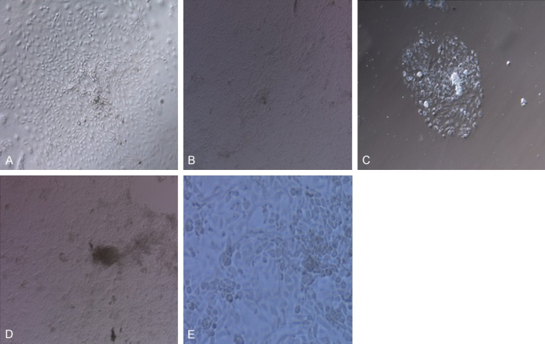
In Group D (medium alone), hESCs grew into embryoid bodies in early phase. With the prolongation of culture time, the embryoid bodies formed marked cysts. After incubated in differentiated media, monostratal adherent cells grew out towards periphery with the embryoid bodies as the center on the bottom of culture dishes. The terminal cells in Group B (Wnt7a) were similar to that in Group D (medium alone). In Group A (Wnt5a), the growth of monostratal adherent cells was denser than that in Group D (media alone). In Group C (sFRP2), the growth of monostratal adherent cells was markedly inhibited (Figure 1).
Figure 1.

Morphology of terminal cells and the morphology of cells after decidualization reaction under Leica inverted microscope × 200. A: In Group A (Wnt5a); B: In Group B (Wnt5a); C: In Group C (sFRP2), D: In Group D (medium alone) and E: Cells after decidualization reaction from Group A.
Decidualization reaction
After the terminal cells were incubated in the media containing 10-6 mol/L medroxyprogesterone for 10 days, cells became large, round and transparent with rich cytoplasm (Figure 1).
Expression of specific proteins in terminal cells of each group
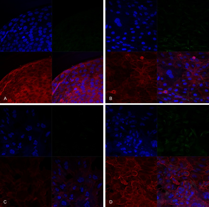
LSM710 confocal microscope showed that cytokeratin (CK) expression was present in 60% of cells and vimentin expression in 10% of cells in Group A (Wnt5a), suggesting that although most cells were epithelial cells after differentiation, there also were stromal cells. In groups B (Wnt7a) and D (medium alone), CK expression was positive in nearly 40% of cells. In Group C (sFRP2), only 20% of cells exhibited positive CK expression (Figure 2).
Figure 2.

Immunofluorescence shows the expressions of cytokeratin and vimentin in terminal cells under Zeiss confocal microscope LSM710 × 400. Notes: cytokeratin is red, vimentin green and cell nucleus blue. A: In Group A (Wnt5a); B: In Group B (Wnt7a); C: In Group C (sFRP2) and D: In Group D (medium alone).
Expression of specific genes in terminal cells of each group
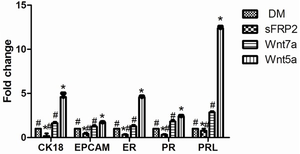
CK18 and epithelial cell adhesion molecule (EPCAM) are the specific makers of epithelial cells, and the presence of estrogen receptor (ER) and progesterone receptor (PR) suggests that the endometrial epithelial cells can be regulated by estrin and progestogen. RT-PCR indicated that mRNA expressions of CK, EPCAM, ER, PR and PRL were significantly higher in Group A (Wnt5a) than in control group (medium alone) (P < 0.05), but were significantly lower in Group C (sFRP2) than in control group (medium alone) (P < 0.05), and were similar between Group B (Wnt7a) and control group (medium alone). mRNA expressions of CK, EPCAM, ER, PR and PRL were the highest in Group A (Wnt5a) among the 4 groups (all P < 0.05), suggesting that cell functions were the most closed to that of endometrial epithelial cells and cells were the most sensitive to decidualization reaction in Group A (Wnt5a) among the 4 groups (Figure 3).
Figure 3.

mRNA expressions of specific genes in terminal cells. Notes: CK18: cytokeratin-18; EPCAM: epithelial cell adhesion molecule; ER: estrogen receptor; PR: progesterone receptor; PRL: prolactin; DM: medium alone; sFRP2: secreted frizzled related protein2. Data are expressed as mean ± SD, n = 3; *Indicates P < 0.05 as compared with control group (medium alone). #Indicates P < 0.05 as compared with Group A (Wnt5a).
Discussion
It has been an important issue to make hESCs differentiate into specific cell type. Exploring the differentiation of hESCs into endometrium-like cells can provide cell models and experimental basis for the researches about the early development of endometrium. It has been confirmed that the members of Wnt cytokine family play important roles in many developmental events [9-11], such as embryo development, cell proliferation, tissue regeneration and tumor formation.
So far, 20 kinds of secreted Wnt proteins have been found. They all can combine with the receptors of Frizzled family on the cell surface to activate three different signal pathways including Wnt/β-catenin classical pathway [12], and Wnt/cell polar [13] and Wnt/Ca2+ non-classical pathways [14]. At present, the mechanism about Wnt/β-catenin classical pathway is the clearest among the three signal pathways. The Wnt/β-catenin pathway is highly conserved during the evolution of species. It mainly regulates cell proliferation and differentiation, and plays a role in embryo development and tumor occurrence [15,16]. When Wnt signal pathway is activated, Wnt molecule combines with specific receptors on cell surface to regulate transcription and expression of downstream genes. It is reported that sFRP may block this pathway through competitive binding with receptors on the cell surface [17]. Wnt signal pathways attract more and more attention because they play an important role in self-renewal potentiality and differentiation of hESCs.
There have been different reports regarding the effect of Wnt signal pathways on hESCs. On one hand, Wnt signal pathway, an important factor in cell division, plays a role in self renewal of stem cells. It was reported that the activation of Wnt/β-catenin pathway activated proliferative signals of murine hematopoietic stem/progenitor cells in vitro [18,19]. On the other hand, there is no marked activation of Wnt signal pathways in quiescent stem cells. It was reported that the activation of Wnt/β-catenin pathway was found only in activated stem cells and progenitor cells derived from stem cells in vivo [20,21], and Wnt signal pathways were activated when primitive stem cells began differentiating [22-24]. However, in adult stem cells, self-renewal and differentiation are not mutually exclusive. During the regeneration and repair after tissue damage, only under the actions of activated Wnt signal pathways and proliferative signals, the quiescent stem/progenitor cells can produce offspring stem/progenitor cells, and finally form terminal cells [25,26].
In embryo development, during the process that primitive neural crest cells migrate to the skin, high expression of Wnt5a leads to changes in the morphology of developed cells; and when cells reach the target region, Wnt5a gene expression decrease [27]. Similarly, in endometrial stroma, there is Wnt5a expression which is necessary for the development of endometrial epithelial glands [28]. The results above suggest that Wnt5a is a main regulatory factor and plays an important role in cell growth and differentiation.
In this study, immunofluorescence indicated that cytokeratin expression was the highest in Group A (Wnt5a), the weakest in Group C (sFRP2), and similar between Group B (Wnt7a) and Group D (medium alone). RT-PCR displayed that mRNA expression of CK18, EPCAM, ER and PR in terminal cells was significantly increased in Group A (Wnt5a) as compared with other three groups; while in Group C (sFRP2), mRNA expression of CK18, EPCAM, ER and PR was significantly decreased. Our results suggest that Wnt5a has marked promotive effects on the differentiation of hESCs into endometrium-like cells, sFRP2 can decrease differentiation efficiency, and Wnt7a has no marked effects. It is reported that there are expressions of Wnt5a, Wnt7a and Wnt11 and their receptors including Fzd6, Fzd2 and accessory receptor LRP6, but sFRP2, Wnt receptor antagonist, is not found in the endometrial epithelial cells of neonatal sheep; with the development of endometrial glands, the expressions of Wnt7a and Wnt11 decrease, but sFRP2 expression increases and the level of sFRP2 is consistent with the density of endometrial glands. It may be speculated from this result that the formation of endometrial epithelial cells requires Wnt5a, Wnt7a and Wnt11, sERP2 inhibits the formation of endometrial epithelial cells but promotes the development of endometrial glands.
In summary, this study explored the effects of Wnt5a and Wnt7a on the differentiation of hESCs into endometrium-like cells in vitro for the first time, and found that 100 ng/ml of Wnt5a could significantly promote the differentiation of hESCs into endometrium-like cells, but Wnt7a had no marked effects. However, the functions of terminal cells differentiated with the scheme used in this study remains to be further investigated.
Acknowledgements
This study was supported by the National Natural Science Foundation of China (No. 31271605).
Disclosure of conflict of interest
None.
References
- 1.El-Toukhy T, Coomarasamy A, Khairy M, Sunkara K, Seed P, Khalaf Y, Braude P. The relationship between endometrial thickness and outcome of medicated frozen embryo replacement cycles. Fertil Steril. 2008;89:832–839. doi: 10.1016/j.fertnstert.2007.04.031. [DOI] [PubMed] [Google Scholar]
- 2.Thomson JA, Itskovitz-Eldor J, Shapiro SS, Waknitz MA, Swiergiel JJ, Marshall VS, Jones JM. Embryonic stem cell lines derived from human blastocysts. Science. 1998;282:1145–1147. doi: 10.1126/science.282.5391.1145. [DOI] [PubMed] [Google Scholar]
- 3.Gargett CE, Chan RW, Schwab KE. Hormone and growth factor signaling in endometrial renewal: role of stem/progenitor cells. Mol Cell Endocrinol. 2008;288:22–29. doi: 10.1016/j.mce.2008.02.026. [DOI] [PubMed] [Google Scholar]
- 4.Larroque-Cardoso P, Mucher E, Grazide MH, Josse G, Schmitt AM, Nadal-Wolbold F, Zarkovic K, Salvayre R, Negre-Salvayre A. 4-Hydroxynonenal impairs Transforming Growth Factor-beta1-induced elastin synthesis via Epidermal Growth Factor Receptor activation in human and murine fibroblasts. Free Radic Biol Med. 2014;17:427–436. doi: 10.1016/j.freeradbiomed.2014.02.015. [DOI] [PubMed] [Google Scholar]
- 5.Di Florio A, Sancho V, Moreno P, Delle Fave G, Jensen RT. Gastrointestinal hormones stimulate growth of Foregut Neuroendocrine Tumors by transactivating the EGF receptor. Biochim Biophys Acta. 2013;1833:573–582. doi: 10.1016/j.bbamcr.2012.11.021. [DOI] [PMC free article] [PubMed] [Google Scholar]
- 6.Schwenke M, Knofler M, Velicky P, Weimar CH, Kruse M, Samalecos A, Wolf A, Macklon NS, Bamberger AM, Gellersen B. Control of human endometrial stromal cell motility by PDGF-BB, HB-EGF and trophoblast-secreted factors. PLoS One. 2011;8:e54336. doi: 10.1371/journal.pone.0054336. [DOI] [PMC free article] [PubMed] [Google Scholar]
- 7.Miller C, Sassoon DA. Wnt-7a maintains appropriate uterine patterning during the development of the mouse female reproductive tract. Development. 1998;125:3201–3211. doi: 10.1242/dev.125.16.3201. [DOI] [PubMed] [Google Scholar]
- 8.Mericskay M, Kitajewski J, Sassoon D. Wnt5a is required for proper epithelial-mesenchymal interactions in the uterus. Development. 2004;131:2061–2072. doi: 10.1242/dev.01090. [DOI] [PubMed] [Google Scholar]
- 9.Nelson WJ, Nusse R. Convergence of Wnt, beta- catenin, and cadherin pathways. Science. 2004;303:1483–1487. doi: 10.1126/science.1094291. [DOI] [PMC free article] [PubMed] [Google Scholar]
- 10.Kleber M, Sommer L. Wnt signaling and the regulation of stem cell function. Curr Opin Cell Biol. 2004;16:681–687. doi: 10.1016/j.ceb.2004.08.006. [DOI] [PubMed] [Google Scholar]
- 11.Willert K, Brown JD, Danenberg E, Duncn AW, Weissman IL, Reya T, Yates JR 3rd, Nusse R. Wnt proteins are lipid-modified and can act as stem cell growth factors. Nature. 2003;423:448–452. doi: 10.1038/nature01611. [DOI] [PubMed] [Google Scholar]
- 12.Yang Y. Wnt signaling in development and disease. Cell Bioscie. 2012;2:14. doi: 10.1186/2045-3701-2-14. [DOI] [PMC free article] [PubMed] [Google Scholar]
- 13.Katoh M. WNT/PCP signaling pathway and human cancer (review) Oncol Rep. 2005;14:1583–1588. [PubMed] [Google Scholar]
- 14.Kohn AD, Moon RT. Wnt and calcium signaling: beta-catenin-independent pathways. Cell Calcium. 2005;38:439–446. doi: 10.1016/j.ceca.2005.06.022. [DOI] [PubMed] [Google Scholar]
- 15.Kalkman HO. A review of the evidence for the canonical Wnt pathway in autism spectrum disorders. Mol Autism. 2012;3:10–22. doi: 10.1186/2040-2392-3-10. [DOI] [PMC free article] [PubMed] [Google Scholar]
- 16.Van Amerongen R, Nusse R. Towards an integrated view of Wnt signaling in development. Development. 2009;136:3205–3214. doi: 10.1242/dev.033910. [DOI] [PubMed] [Google Scholar]
- 17.Weeraratna AT, Jiang Y, Hostetter G, Rosenblatt K, Duray P, Bittner M, Trent JM. Wnt5a signaling directly affects cell motility and invasion of metastatic melanoma. Cancer Cell. 2002;1:279–288. doi: 10.1016/s1535-6108(02)00045-4. [DOI] [PubMed] [Google Scholar]
- 18.Reya T, Duncan AW, Ailles L, Domen J, Scjerer DC, Willert K, Hintz L, Nusse R, Weissman IL. A role for Wnt signaling in self-renewal of haematopoietic stem cells. Nature. 2003;423:409–414. doi: 10.1038/nature01593. [DOI] [PubMed] [Google Scholar]
- 19.Alonso L, Fuchs E. Stem cells in the skin: waste not, Wnt not. Gene Dev. 2003;17:1189–1200. doi: 10.1101/gad.1086903. [DOI] [PubMed] [Google Scholar]
- 20.He XC, Zhang J, Tong WG, Tawfik O, Ross J, Scoville DH, Tian Q, Zeng X, He X, Wiedemann LM, Mishina Y, Li L. BMP signaling inhibits intestinal stem cell self-renewal through suppression of Wnt-beta-catenin signaling. Nat Genet. 2004;36:1117–1121. doi: 10.1038/ng1430. [DOI] [PubMed] [Google Scholar]
- 21.Otero JJ, Fu W, Kan L, Cuadra AE, Kessler JA. Beta-catenin signaling is required for neural differentiation of embryonic stem cells. Development. 2004;131:3545–3557. doi: 10.1242/dev.01218. [DOI] [PubMed] [Google Scholar]
- 22.Huelsken J, Vogel R, Brinkmann V, Erdmann B, Birchmeier C, Birchneier W. Requirement for beta-catenin in anterior-posterior axis formation in mice. J Cell Biol. 2000;148:567–578. doi: 10.1083/jcb.148.3.567. [DOI] [PMC free article] [PubMed] [Google Scholar]
- 23.Lee HY, Kleber M, Hari L, Brault V, Suter U, Taketo MM, Kemler R, Sommer L. Instructive role of Wnt/beta-catenin in sensory fate specification in neural crest stem cells. Science. 2004;303:1020–1023. doi: 10.1126/science.1091611. [DOI] [PubMed] [Google Scholar]
- 24.Scheller M, Huelsken J, Rosenbauer F, Taketo MM, Birchmeier W, Tenen DG, Leutz A. Hematopoietic stem cell and multilineage defects generated by constitutive beta-catenin activation. Nat Immunol. 2006;7:1037–1047. doi: 10.1038/ni1387. [DOI] [PubMed] [Google Scholar]
- 25.Kirstetter P, Anderson K, Porse BT, Jacobsen SE, Nerlov C. Activation of the canonical Wnt pathway leads to loss of hematopoietic stem cell repopulation and multilineage differentiation block. Nat Immunol. 2006;7:1048–1056. doi: 10.1038/ni1381. [DOI] [PubMed] [Google Scholar]
- 26.Trowbridge JJ, Moon RT, Bhatia M. Hematopoietic stem cell biology: too much of a Wnt thing. Nat Immunol. 2006;7:1021–1023. doi: 10.1038/ni1006-1021. [DOI] [PubMed] [Google Scholar]
- 27.Christiansen JH, Coles EG, Wilkinson DG. Molecular control of neural crest formation, migration and differentiation. Curr Opin Cell Biol. 2000;12:719–724. doi: 10.1016/s0955-0674(00)00158-7. [DOI] [PubMed] [Google Scholar]
- 28.Pukrop T, Binder C. The complex pathways of Wnt 5a in cancer progression. J Mol Med (Berl) 2008;86:259–266. doi: 10.1007/s00109-007-0266-2. [DOI] [PubMed] [Google Scholar]


