Abstract
Bovine herpesvirus 4 (BoHV-4) is increasingly considered as responsible for various problems of the reproductive tract. The virus infects mainly blood mononuclear cells and displays specific tropism for vascular endothelia, reproductive and fetal tissues. Epidemiological studies suggest its impact on reproductive performance, and its presence in various sites in the reproductive tract highlights its potential transmission in transfer-stage embryos. This work describes the biological and genetic characterization of BoHV-4 strains isolated from an in vitro bovine embryo production system. BoHV-4 strains were isolated in 2011 and 2013 from granulosa cells and bovine oocytes from ovary batches collected at a local abattoir, used as “starting material” for in vitro production of bovine embryos. Compatible BoHV-4-CPE was observed in the co-culture of granulosa cells and oocytes with MDBK cells. The identity of the isolates was confirmed by PCR assays targeting three ORFs of the viral genome. The phylogenetic analyses of the strains suggest that they were evolutionary unlinked. Therefore it is possible that BoHV-4 ovary infections occurred regularly along the evolution of the virus, at least in Argentina, which can have implications in the systems of in vitro embryo production. Thus, although BoHV-4 does not appear to be a frequent risk factor for in vitro embryo production, data are still limited. This study reveals the potential of BoHV-4 transmission via embryo transfer. Moreover, the high variability among the BoHV-4 strains isolated from aborted cows in Argentina highlights the importance of further research on the role of this virus as an agent with the potential to cause reproductive disease in cattle. The genetic characterization of the isolated strains provides data to better understand the pathogenesis of BoHV-4 infections. Furthermore, it will lead to fundamental insights into the molecular aspects of the virus and the means by which these strains circulate in the herds.
Introduction
Bovine herpesvirus 4 (BoHV-4) is a member of the Herpesviridae family, Gammaherpesvirinae subfamily, Rhadinovirus genus. BoHV-4 was isolated for the first time in 1963 from animals with respiratory and ocular signs in Europe [1]. Afterwards, virus isolates were classified into two groups by restriction endonuclease analysis [2–3]: Movar 33/63-like strains, isolated from Europe, and DN 599-like strains, isolated from North America [2–4]. In 2007, BoHV-4 was isolated in Argentina from vaginal secretions of aborted cows [5] and later from buffy coat fractions in association with bovine viral diarrhea virus [6]. More recently, the virus was isolated from bovine semen from an artificial insemination center [7].
Abortions associated with BoHV-4 are usually sporadic. Currently, the exact role of this virus in cattle diseases is unknown. However, several research groups [8–11] are studying its participation in the infection of the genital tract. Most reports of BoHV-4 infection in different countries associate BoHV-4 with the development of post-partum and chronic metritis, either alone or in combination with other pathogens [12–13]. Data on the relationship between BoHV-4 and infertility are limited [14]. However, a recent study has shown a positive correlation between infertility and BoHV-4 infection [15]. The presumptive implication of BoHV-4 in reproductive disorders is based on epidemiological observations, especially a higher seroprevalence in bovine females affected by metritis, abortion and/or infertility than in healthy bovine females [15–17].
Recently, Gur and Dogan [15] investigated the role of BoHV-4 infections in repeat breeding cows by serology and they found that BoHV-4 seropositivity is associated with increased risk of infertility: 69% of repeat breeder females (3–14 artificial inseminations) presented anti-BoHV-4 antibodies whereas only 44% of the cows that had received two inseminations or less were seropositive. However, such correlation was based only on epidemiological observations. In another recent study, the virus was isolated from 87% of the uteri from infertile cows obtained at slaughterhouses [18], indicating a high prevalence of BoHV-4 in reproductive tissues. These ‘materials of animal origin’ represent an important source of contamination when used in production and transfer of bovine embryos.
The virus has also been identified in aborted fetuses, mainly in spleen lymphocytes and monocytes and, occasionally from hepatic Kupffer cells and renal tubules [16–20]. It has also been evidenced in placental epithelium, where it replicates (especially within intraplacental inflammatory infiltrates) without causing any specific histological lesions. Because the virus has been isolated from various sites of the reproductive tract, it can be potentially transmitted by transfer-stage embryos [20]. Recent studies have shown other possible sources of infection such as spermatozoa, semen leukocyte fractions and colostral cells in which the viral genome was detected by PCR, indicating that reproductive cells might be a target for the virus [21].
The study of bovine herpesviruses in in vitro-produced embryos has been focused mainly on bovine herpesvirus-1 (BoHV-1) [22] and more recently, on bovine herpesvirus 5 (BoHV-5) [23–24]. However, there is little information on the infections by BoHV-4 in in vitro systems of embryo production. As detailed below, we successfully isolated BoHV-4 from granulosa cells and oocytes in 2011 and 2013, respectively. In the following years the virus was frequently isolated from samples of aborted cows, and hence we here describe a detailed characterization of the 2011 and 2013 strains.
Materials and Methods
Case History and Virological Findings
BoHV-4 strains were isolated in 2011 and 2013 from bovine granulosa cells and oocytes from batches of ovaries collected at a local abattoir located in Balcarce, Argentina (37° 50' 47” S, 58° 15' 19” W). Bovine ovaries were collected from a local slaughterhouse and transported in a thermic container to the Laboratory of in vitro Embryo Production (INTA), which is regulated by the principles established by the International Embryo Transfer (IETS) to assure quality and sanitary standards (that is, free of pathogens). Groups of 50 cumulus-oocyte complexes (COCs) corresponding to grade 1 and 2, were selected under a stereomicroscope and placed in four-well dishes (Nunc) in 400 μL of standard maturation medium (M199 plus 0.1 mg/mL L-glutamine and 2.2 mg/mL NaHCO3 was supplemented with 0.01 IU/mL rh-FSH [Gonal F-75, Serone, UK] and 10% estrous cow serum). After maturation, COCs were completely denuded of cumulus cells by pipetting in M199-HEPES containing 300 IU/mL hyaluronidase (H3506) for 2 min. Cumulus-free oocytes were washed five times in medium drops.
The material obtained from granulosa cells (30 ul), oocytes (n = 10) and washed medium drops (30ul) were co-cultured on confluent Madin-Darby bovine kidney (MDBK) cells grown in 96-well tissue culture plates. MDBK cells were incubated in minimum essential medium (MEM) supplemented with 10% fetal bovine serum. MDBK cells were provide by the ABAC (Argentinean Cell Bank, http://www.abac.org.ar/). They were BoHV-4-free, and certified as free of contaminating bacteria, mycoplasma and adventitious viruses.
Samples were subjected to three blind passages (48 h apart) at 37°C, 5% CO2 and observed daily for the presence of cytopathic effect (CPE). MDBK cells were mock-infected in parallel to provide a negative control. In addition, control procedures included routine PCR assessments of wash and culture media used for all the experiments.
Detection of Viral DNA by PCR
In all cell cultures DNA was purified from infected cells using the DNeasy Blood & Tissue Kit (Cat. 69504, Qiagen), according to the manufacturer's instructions. DNA concentration was determined by spectrophotometry at an absorbance of 260 nm. The presence of BoHV-4 DNA was evaluated by PCR assays targeting three ORFs of the viral genome: ORF8 (encoding glycoprotein B) [25], ORF22 (encoding glycoprotein H) [26] and ORF47 (encoding glycoprotein L) [27]. The BoHV-4 thymidine kinase (TK) gene was evaluated by a nested-PCR assay modified by [5]. The primers used for the amplifications are described in Table 1. DNA from mock-infected MDBK cells was used as negative control. The specificity of the reactions was evaluated by using DNA from reference strains of BoHV-1 and BoHV-5 as PCR template. The amplified products were separated by electrophoresis in a 2% agarose gel, and DNA bands were visualized by SYBR Safe DNA gel stain (Invitrogen).
Table 1. Primers used in this study.
| Gene | GeneBank accession no. | Orientation | Primer | Fragment expected (bp) |
|---|---|---|---|---|
| ORF22 | AF318573 | Forward | 5’-CCGGGTGAAACAAGTTCCTG-3’ | 563 |
| Reverse | 5’-GTCAGAGAACATATGATAACATC-3’ | |||
| ORF47 | AF318573 | Forward | 5’-CAGGCAAAAAGTGGCTTCTC-3’ | 138 |
| Reverse | 5’-TTGAGGGCCTGGATATTGTC-3’ | |||
| ORF8 | Wellenberg et al., 2001 | Forward | 5’-CCCTTCTTTACCACCACCTACA-3’ | 615 |
| Reverse | 5’-TGCCATAGCAGAGAAACAATGA-3’ | |||
| Orf3 | Verna et al., 2012 | Foward | 5’-GTTGGGCGTCCTGTATGGTAGC-3’ | 576 |
| Reverse | 5’-TGTATGCCCAAAACTTATAATATGACCAG-3’ | |||
| Orf3 | Verna et al., 2012 | Foward | 5’-TTGATAGTGCGTTGTTGGGATGTGGT-3’ | 216 |
| Reverse | 5’-CACTGCCCGGTGGGAAATAGCA-3’ |
* The pairs of primers used in this study were designed based on the sequences available in GenBank.
Sequencing and Evolutionary Analyses
The PCR amplicons obtained as described above were subjected to Sanger sequencing using PCR primers. Furthermore, the gB gene from 11 Genotype 1 strains (isolates 07_568, 08_433, 09_467, 09_759, 08_209, 10_154, 09_465, 08_362, DN_599, 08_476 and 08_263), two Genotype 2 strains (09_227, 07_435) and one strain (08_404) of uncertain parentage were also amplified and sequenced for their use in evolutionary analyses. The sequences described here were deposited in GenBank under accession numbers. The herein reported nucleotide sequences have been assigned GenBank accession numbers KP209014- KP209031.
For the evolutionary analyses, the TK sequences described here were combined with TK sequences of isolates 07_568, 08_433, 09_467, 09_759, 08_209, 10_154, 09_465, 08_362, DN_599, 08_476 and 08_263, 09_227, 07_435 and 08_404, described before [6]. We also determined the gB sequences of these isolates. After alignment and trimming of terminal alignment columns containing missing data, our dataset contained 435 positions of the gB gene and 212 from TK, of which 43 were informative. Sequence alignments were generated by the MAFFT multiple sequence alignment program, with default op and ep, and iterative refinement and weighted sum-of-pairs and consistency scores obtained from local alignments [28]. Numbers of informative sites were determined using the Phyloch package (http://www.christophheibl.de/Rpackages). Evolutionary models for the alignments obtained were selected with the MrAIC.pl. program distributed by the author [29]. These last analyses favored a JC69 model for both datasets.
Maximum Likelihood phylogenetic trees were inferred with the FastTree 2 program [30]. FastTree implements a Maximum Likelihood method of phylogenetic tree inference. The Maximum Likelihood approach evaluates a hypothesis in terms of the probability that a model and a hypothesized history would give rise to the observed sequences, which is to say the probability of the sequence alignment given the tree. It is assumed that the history (tree) associated with the higher data probability is preferred in comparison with a tree with a lower probability [31]. FastTree automatically scale search intensities according to dataset sizes by using up to 4xlog2(N) rounds of minimum-evolution nearest neighbour interchange, 2 rounds of subtree pruning and regraft moves and up to 2xlog(N) rounds of maximum-likelihood nearest neighbour interchanges, where N is the number of unique sequences in the dataset [30]. For the bootstrapped analyses, 100 resampled datasets were generated with the Seqboot component of the Phylip package [32], and subjected to FastTree 2 and the CompareToBootstrap.pl PERL script provided with the program.
Bayesian coalescent analyses were performed with BEAST [33]. BEAST is a program that implements a Bayesian framework for testing evolutionary hypotheses without conditioning on a single tree topology. This is done exploring the tree and parameter space by a Markov Chain Monte Carlo approach. Thus phylogenies and associated parameters are weighted in proportion to their posterior probabilities. A detailed description of the program and concepts implemented in the program can be found in [34]. The BEAST results were analyzed using Tracer v1.5 (available from: http://tree.bio.ed.ac.uk/software/tracer/). We performed two independent runs of 5E7 generations, which were inspected for convergence, likelihood; prior and posterior yielded an Effective Sample Size (ESS) greater than 200, and visually to check if there were any trends in the trace of each parameter. The runs were merged with LogCombiner (distributed with BEAST). The gB and TK datasets were combined into a single Bayesian coalescent analysis. However, the parameters of the corresponding site models, clocks and trees were unlinked, as preliminary phylogenetic analyses supported different evolutionary histories for the genes studied (please see also the following sections). Log-normal, relaxed molecular clocks were set, with uniform priors in the range of mutational rates known for herpesviruses and other double-stranded DNA viruses [35]. Although the isolation dates of the strains studied here are known, we considered the time interval encompassed by these dates to be too narrow in comparison with the evolutionary time scales of herpesviruses; i.e. codivergence with host species [36]. Thus, tip dates were not used to estimate the evolutionary rates and dates, but let them follow the prior. Convergence and adequate EES (Target 1000) were assessed with Tracer (available from http://beast.bio.ed.ac.uk/). The posterior sample of trees were summarized with TreeAnnotator [33].
Results
Characterization of Isolates
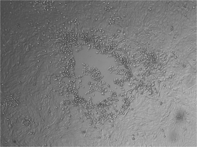
After oocyte maturation, granulosa cells were removed from oocytes and both samples were co-cultured with MDBK cells. After three blind passages, CPE resembling that of BoHV-4 was observed in the co-culture of granulosa cells and oocytes with MDBK cells but not in control ones (Fig 1). PCR and IF (FITC anti-BoHV-4 monoclonal antibody, Bio-X Diagnostics S.P.R.L.U) were used to confirm the presence of the virus in the inoculated cells.
Fig 1. Cytopathic effect induced by BoHV-4 strains from bovine ovary batches on MDBK cells.
Cytopathic effect associated with BoHV-4 infections. Light microscope (10X).
DNA was obtained from both samples and subjected to the PCR reactions described in Material and Methods (Table 1). A specific band for the amplified fragments was obtained from the DNA of oocytes and granulosa cells. As expected, BoHV-4 DNA was not detected in non-infected MDBK cells.
Evolutionary Analyses
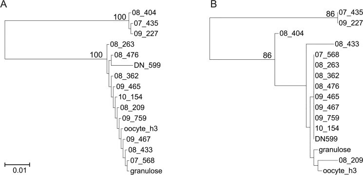
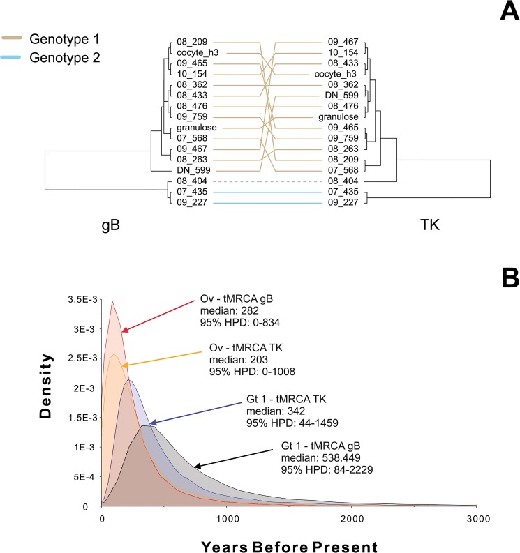
Phylogenetic comparisons with previously described BoHV-4 sequences indicated that the strains obtained from the bovine ovaries collected at slaughterhouse belonged to Genotype 1. However, there were conspicuous differences in the branch length and topology between the trees obtained from the genomic regions studied. For example, in the gB tree, the strain 08_263 was much closer to Genotype 1 viruses than in the TK phylogeny (Fig 2). Likewise, in the TK tree, a relatively large branch connected strain 08_433 to the base node of Genotype 1, whereas it displayed a shorter branch emerging from inside Genotype 1 clade in the gB tree (Fig 2). Fig 3A depicts in detail the topological incongruence observed for both genes using mirrored trees. The observed level of incongruence is consistent with the evolutionary scenarios observed for other herpesviruses [37]. As for the ovary sequences, they belonged to Genotype 1. However, they were unconnected to each other in both the TK and gB trees (Figs 2 and 3).
Fig 2. Phylogenetic analysis of the BoHV-4 strains studied here.
(A) Obtained from glycoprotein B gene sequence. (B) Obtained from thymidine kinase gene sequence. Branch lengths are proportional to genetic distances (the scale bar units are substitutions per aligned position). Numbers close to nodes correspond to bootstrap supports (n = 100); non-significant bootstrap supports are not shown. The trees were midpoint rooted.
Fig 3. Bayesian Evidence for the BoHV-4 sequences studied here.
(A) Mirrored maximum clade credibility trees. (B) Times to most recent common ancestor (tMRCA) posterior densities, medians, and 95% HPD intervals of the studied strains are shown. Branch lengths are proportional to median node heights. The links connecting terminal nodes are colored according to which genotype the corresponding strains belong to (tan Genotype 1; light blue Genotype 2). The link connecting terminals corresponding to strain 08_404 are dashed and gray colored to indicate uncertainty in genotype assignment. Ov strains isolated from ovaries (granulosa cells and oocyte_h3); Gt1 Genotype 1; gB glycoprotein B; TK thymidine kinase.
The Bayesian analyses revealed a mean time to most recent common ancestor (tMRCA) of 808.85 (95% Highest Posterior Density (HPD) interval 9.49–2234.49) and 279.50 (95% HPD interval 0.18–941.01) years for the gB and TK Genotype 1 sequences, respectively. The tMRCAs of the ovary sequences were 111.29 (95% HPD interval 0.00–393.08) and 154.54 (95% HPD 0.00–579.26) years for the gB and TK genes, respectively. The posterior densities for these data are provided in Fig 3B.
Discussion
Many studies have shown the effects of BoHV-4 on the reproductive tract. Nevertheless, there are no reports on the role of BoHV-4 in bovine infertility. In the present study, two BoHV-4 strains were isolated from granulosa cells and oocytes from ovaries obtained at slaughterhouse. The authenticity of the isolates was confirmed by PCR and sequencing of the TK locus as a genetic marker for the BoHV-4 genome, the 3' end of ORF1 (homologous to the EBV BVRF1 gene), ORF2 (homologous to the EBV BXRF1 gene), ORF3 (TK gene) and ORF22 (gH gene). This is the first report on the isolation and genomic characterization of BoHV-4 strains from ovaries obtained at slaughterhouse, which are routinely used as source of material for techniques of assisted reproduction in cattle.
The phylogenetic analyses of the strains isolated from bovine ovaries suggest that these strains were evolutionary unlinked. Therefore, it is possible that BoHV-4 ovary infections occurred regularly along the course of the evolution of the virus, at least in our country, which can have implications in the systems of in vitro embryo production, as discussed below. This phenomenon was also supported by the Bayesian analyses (Fig 3), which showed that the viruses isolated from the ovaries belong to lineages that have been circulating for long in our country, as can be inferred from the corresponding tMRCAs, which dates back to a period comprised among 0–834 (gB gene) or 0–1008 (TK gene) years before the isolation date. Both time periods widely overlap with the MRCA of genotype 1, strongly suggesting that all the strains from this genotype might be capable of producing ovary infections or at least be transmitted by ovary tissues. Furthermore, it should be noticed that cattle was introduced in Argentina around 400–500 years ago. Thus, based in the Bayesian tMRCAs, it is hypothesized that the lineages to which the ovary strains belongs to were introduced at that time. These data provide insight on the historical co-occurrence of ovary and "regular" infections. Both the chronograms obtained and the broad overlapping of the posterior density estimates of the tMRCAs are consistent with the recurrent infections of reproductive organs along the evolution of BoHV-4 in our country. These results suggest that BoHV-4 strains isolated from the reproductive tract belong to a viral lineage that has circulated for a relatively long period of time, contrary to the idea that the isolates correspond to a new viral variant with a specific tropism. Furthermore, these results support the concept that BoHV-4-related reproductive problems could be frequent and that, therefore, the virus represents a risk for the embryo production industry. In the case of BoHV-4, the information on this aspect is scarce, mainly because direct association between virus infection and disease has not been determined yet [35]. However, some studies have attempted to understand the interaction between the virus and in vitro-produced bovine embryos. Stringfellow et al. [20] demonstrated that BoHV-4 attaches to the intact zona pellucida of bovine embryos and that only trypsin treatment is able to remove the virus. On the other hand, in a preliminary study by using a green recombinant BoHV-4, it has been shown that the virus has low ability to infect in vitro-produced bovine embryos, which depends on the absence of zona pellucida, the amount of virus present and the stage of embryonic development [38]. In spite of these preliminary studies based on bovine embryos, there are no reports on the outcome of the interaction between BoHV-4 and gametes.
In the present work, viral isolation from oocytes with intact zona pellucida after several consecutive washes was consistent with the results obtained by [20]. This result has important implications about the effects of early BoHV-4 infections on vertical transmission. Recent studies have postulated that the virus is also able to cross the placental barrier and intensively replicate within the fetus [39]. However, it should be addressed that oocytes in this study were obtained from ovaries at slaughterhouse, which implies a possible natural infection of the host. It has been suggested that by placental cell destruction, induction of placental inflammation or alteration in the immune balance associated with pregnancy, the virus might alter placental function, leading eventually to delayed abortion [40].
Thus, although BoHV-4 does not appear to be a frequent risk factor for in vitro embryo production, data are still limited. However, this study reveals that the potential of BoHV-4 transmission via embryo transfer exists. Moreover, high variability among the BoHV-4 strains isolated from aborted cows in Argentina highlights the importance of further research on the role of this virus as an infectious agent able to cause reproductive disease in cattle [6]. The genetic characterization of the isolated strains can therefore provide data to better understand the pathogenesis of BoHV-4 infections. Furthermore, strain characterization will lead to fundamental insights into molecular aspects of the virus and the means by which these strains circulate in the herds.
Acknowledgments
We would like to thank M.Sc Germán Kaiser from the Laboratory of Biotechnology of Reproduction for his collaboration with oocytes manipulation and Dr. Maria Victoria Eusevi for language advice.
Data Availability
All relevant data are within the paper.
Funding Statement
Support was provided by Instituto Nacional de Tecnología Agropecuaria: Project PNSA1115053 and PNSA1115052; Agencia Nacional de Promoción Científica y Tecnológica: Project PICT 2011-0312. The funders had no role in study design, data collection and analysis, decision to publish, or preparation of the manuscript.
References
- 1. Bartha A, Juhasz M, Liebermann H (1966) Isolation of a bovine herpesvirus from calves with respiratory disease and keratoconjuntivistis. Acta Vet Acad Sci Hung 16: 357–358. [PubMed] [Google Scholar]
- 2. Bublot M, Lomonte P, Lequarre AS, Albrecht JC, Nicholas J, Fleckenstein B, et al. (1992) Genetic relationships between bovine herpesvirus 4 and the gammaherpesviruses Epstein-Barr virus and herpesvirus Saimiri. Virology 190: 654–665. [DOI] [PubMed] [Google Scholar]
- 3. Thiry E, Bublot M, Dubuisson J, Van Bressem MF, Lequarre AS, Lomonte P, et al. (1992) Molecular biology of bovine herpesvirus type 4. Vet Microbiol 33: 79–92. [DOI] [PubMed] [Google Scholar]
- 4. Henry BE, Ota R, Evermann JF (1986) Genetic relatedness of disease-associated field isolates bovid herpesvirus type 4. American Journal of Veterinary Research 47: 2242–2246. [PubMed] [Google Scholar]
- 5. Verna A, Leunda MR, Louge Uriarte E, Lomónaco M, Pereyra S, Odeón A (2008) Primera evidencia virológica de Herpesvirus bovino tipo 4 (BoHV-4) en Argentina. Rev Argent Microbiol 40: 130. [Google Scholar]
- 6. Verna AE, Manrique JM, Pérez SE, Leunda MR, Pereyra SB, Jones LR, et al. (2012) Genomic analysis of bovine herpesvirus type 4 (BoHV-4) from Argentina: High genetic variability and novel phylogenetic groups. Vet Microbiol 160: 1–8. 10.1016/j.vetmic.2012.04.039 [DOI] [PubMed] [Google Scholar]
- 7. Morán P, Favier P, Lomónaco M, Catena M, Chiapparrone M, Odeón A, et al. (2013) Search for the genome of bovine herpesvirus types 1, 4 and 5 in bovine semen. Open Veterinary Journal 3: 126–130. [PMC free article] [PubMed] [Google Scholar]
- 8. Egyed L, Ballagy-Pordany A, Bartha A, Belak S (1996) Studies of in vivo distribution of bovine herpesvirus type 4 in the natural host. Journal of Clinical Microbiology 34: 1091–1095. [DOI] [PMC free article] [PubMed] [Google Scholar]
- 9. Czaplicki G, Thiry E (1998) An association exists between bovine herpesvirus-4 seropositivity and abortion in cows. Prev Med Vet 33: 233–240. [DOI] [PubMed] [Google Scholar]
- 10. Frazier K, Pence M, Mauel MJ, Liggett A, Hines ME, Sangster L, et al. (2001) Endometritis in postparturient cattle associated with bovine herpesvirus-4 infection: 15 cases. J Vet Diagn Invest 13: 502–508. [DOI] [PubMed] [Google Scholar]
- 11. Kalman D, Janosi S, Egyed L (2004) Role of Herpesvirus 4 in bacterial bovine mastitis. Microbial Pathogenesis 37: 125–129. [DOI] [PubMed] [Google Scholar]
- 12. Wellemans G, Van Opdenbosch E, Mammerickx M (1986) Inoculation experimentale du virus LVR 140 (herpesvirus bovin IV) a des vaches gestantes et non-gestantes. Ann Rech Vet 17: 89–94. [PubMed] [Google Scholar]
- 13. Monge A, Elvira L, Gonzalez JV, Astiz S, Wellenberg GJ (2006) Bovine herpesvirus 4-associated postpartum metritis in a Spanish dairy herd. Res Vet Sci 80: 120–125. [DOI] [PubMed] [Google Scholar]
- 14. Nikolin VM, Donofrio G, Milosevic B, Taddei S, Radosavljevic V, Milicevic V (2007) First Serbian isolates of bovine herpesvirus 4 (BoHV 4) from a herd with a history of postpartum metritis. New Microbiol 30: 53–57. [PubMed] [Google Scholar]
- 15. Gur S, Dogan N (2010) The possible role of bovine herpesvirus type-4 infection in cow infertility. Anim Sci J 81: 304–308. 10.1111/j.1740-0929.2010.00743.x [DOI] [PubMed] [Google Scholar]
- 16. Naeem K, Goyal SM, Werdin RE (1989) Prevalence of bovid herpesvirus-4 and its antibody in cattle in Minnesota. Am J Vet Res 50: 1931–1935. [PubMed] [Google Scholar]
- 17. Bilge Dagalp S, Demir AB, Gungor E, Alkan F (2007) The seroprevalence of bovine herpesvirus type 4 (BoHV-4) infection in dairy herds in Turkey and possible interaction with reproductive disorders. Rev Med Vet 158: 201–205. [Google Scholar]
- 18. Fabian K, Makrai L, Sachse K, Szeredi L, Egyed L (2008) An investigation of aetiologial role of bovine herpesvirus 4 in bovine endometritis. Vet J 177: 289–292. [DOI] [PubMed] [Google Scholar]
- 19. Deim Z, Szeredi L, Tompo V, Egyed L (2006) Detection of bovine herpesvirus 4 in aborted bovine placentas. Microbiol Pathog 21: 144–148. [DOI] [PubMed] [Google Scholar]
- 20. Stringfellow DA, Lauerman LH, Nasti KB, Galik PK (1990) Trypsin treatment of bovine embryos after in vitro exposure to infectious bovine rhinotracheitis virus or bovine herpesvirus-4. Theriogenology 34: 427–434. [DOI] [PubMed] [Google Scholar]
- 21. Egyed L, Sassi G, Tibold J, Mádl I, Szenci O (2011) Symptomless intrauterine transmission of bovine herpesvirus 4 to bovine fetuses. Microb Pathog 50:322–5. 10.1016/j.micpath.2010.10.006 [DOI] [PubMed] [Google Scholar]
- 22. Bielanski A, Loewen KS, Del Campo MR, Sirard MA, Willadsen S (1993) Isolation of bovine herpesvirus-1 (BHV-1) and bovine viral diarrhea virus (BVDV) in association with the in vitro production of bovine embryos. Theriogenology 40: 531–8. [DOI] [PubMed] [Google Scholar]
- 23. Silva-Frade C, Gameiro R, Martins J Jr, Cardoso TC (2010) Apoptotic and development effects of bovine Herpesvirus type-5 infection on in vitro-produced bovine embryos. Theriogenology 74: 1296–1303. 10.1016/j.theriogenology.2010.06.001 [DOI] [PubMed] [Google Scholar]
- 24. Brenner MPC, Silva-Frade C, Ferrarezi MC, Gracia AF, Flores EF, Cardoso TC (2012) Evaluation of developmental changes in bovine in vitro produced embryos following exposure to bovine Herpesvirus type 5. Reprod Biol and Endocrinol 10: 53. [DOI] [PMC free article] [PubMed] [Google Scholar]
- 25. Wellenberg GJ, Verstraten ER, Belak S, Verschuren SB, Rijsewijk FA, Peshev R, et al. (2001) Detection of bovine herpesvirus 4 glycoprotein B and thymidine kinase DNA by PCR assay in bovine milk. J Virol Methods 97: 1001–1012. [DOI] [PubMed] [Google Scholar]
- 26. Gillet L, Minner F, Detry B, Farnir F, Willems L, Lambot M, et al. (2004) Investigation of the Susceptibility of Human Cell Lines to Bovine Herpesvirus 4 Infection: Demonstration that Human Cells Can Support a Nonpermissive Persistent Infection Which Protects Them against Tumor Necrosis Factor Alpha-Induced Apoptosis. J Virol 78(5): 2336–2347. [DOI] [PMC free article] [PubMed] [Google Scholar]
- 27. Lété C, Palmeira L, Leroy B, Mast J, Machiels B, Wattiez R, et al. (2012) Proteomic characterization of bovine herpesvirus 4 extracellular virions. J Virol 86: 11567–80. 10.1128/JVI.00456-12 [DOI] [PMC free article] [PubMed] [Google Scholar]
- 28. Katoh K, Toh H (2008) Recent developments in the MAFFT multiple sequence alignment program. Brief Bioinform 9: 286–98. 10.1093/bib/bbn013 [DOI] [PubMed] [Google Scholar]
- 29. Nylander JA, MrAIC.pl. (2004) Program distributed by the author Evolutionary Biology Centre, Uppsala University. [Google Scholar]
- 30. Price MN, Dehal PS, Arkin AP (2010) FastTree 2—approximately maximum-likelihood trees for large alignments. PLoS One 10;5: e9490 10.1371/journal.pone.0009490 [DOI] [PMC free article] [PubMed] [Google Scholar]
- 31. Felsenstein J (2004) Inferring Phylogenies; Sinauer Associates, Inc; ISBN: 978-0-87893-177-4. [Google Scholar]
- 32. Felsenstein J (1989) PHYLIP—Phylogeny Inference Package (Version 3.2). Cladistics 5: 164–166. [Google Scholar]
- 33. Drummond AJ, Rambaut A (2007) BEAST: Bayesian evolutionary analysis by sampling trees. BMC Evol Biol 7: 214 [DOI] [PMC free article] [PubMed] [Google Scholar]
- 34. Drummond AJ, Suchard MA, Xie D, Rambaut A (2012) Bayesian phylogenetics with BEAUti and the BEAST 1.7 Molecular Biology And Evolution 29: 1969–1973. 10.1093/molbev/mss075 [DOI] [PMC free article] [PubMed] [Google Scholar]
- 35. Duffy S, Sackleton LA, Holmes EC (2008) Rates of evolutionary change in viruses: patterns and determinants. Nature Reviews Genetics 9: 267–276. 10.1038/nrg2323 [DOI] [PubMed] [Google Scholar]
- 36. McGeoch DJ, Rixon FJ, Davison AJ (2006) Topics in herpesvirus genomics and evolution. Virus Research 117(1): 90–104. [DOI] [PubMed] [Google Scholar]
- 37. Shin YC, Jones LR, Manrique J, Lauer W, Carville A, Mansfield KG, et al. (2010) Glycoprotein gene sequence variation in rhesus monkey rhadinovirus. Virology 400: 175–186. 10.1016/j.virol.2010.01.030 [DOI] [PMC free article] [PubMed] [Google Scholar]
- 38. Donofrio G, Galli C, Lazzari G, van Santen VL, Cavirani S, Flammini C (2003) Interaction of a green recombinant bovine herpesvirus 4 with in vitro-produced bovine embryos. Vet Res Commun 27(5): 415–24. [DOI] [PubMed] [Google Scholar]
- 39.Chastant-Maillard S (2013) Impact of Bovine Herpesvirus 4 (BoHV-4) on Reproduction. Transboundary and Emerging Diseases 1–7. [DOI] [PubMed]
- 40. Deim Z, Szeredi L, Egyed L (2007) Detection of bovine herpesvirus 4 DNA in aborted bovine fetuses. Can J Vet Med 71: 226–229. [PMC free article] [PubMed] [Google Scholar]
Associated Data
This section collects any data citations, data availability statements, or supplementary materials included in this article.
Data Availability Statement
All relevant data are within the paper.


