Though developing biological markers for chronic pain has been a major goal of the field for decades, such biomarkers have not yet made their way into clinical practice. However, given the potential uses of biomarkers in multiple aspects of prevention and treatment—such as pain and risk factor assessment, diagnosis, prognosis, treatment selection, drug discovery, and more—efforts to discover new pain biomarkers have been expanding [5; 6; 8; 30].
Recent advances in human neuroimaging, including functional and structural Magnetic Resonance Imaging (fMRI/sMRI) combined with machine learning techniques, are bringing us closer to the goal of developing objective, brain-based markers of the neural functions and neuropathology that underlie chronic pain [2; 7; 25; 33]. These brain measures are particularly promising as biomarkers for chronic pain. Though pain is reliably induced by peripheral nociceptive input, many forms of chronic pain may arise from neuropathology in the supra-spinal circuits that govern the construction of pain experience and long-term motivation [1; 14; 26; 32].
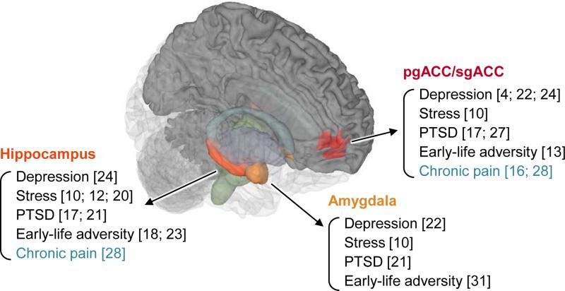
Particularly, structural neuroimaging measures could provide more stable markers of neuropathology of chronic pain, including stable features underlying pain risk and resilience [2; 3; 11; 19; 28; 29]. Gray-matter changes have also been associated with a number of conditions that are often co-morbid with chronic pain, including depression [4; 22; 24], stress [10; 12; 20], post-traumatic stress disorder [17; 21; 27], and early-life adversity [13; 18; 23; 31]. Therefore, structural measures may provide important clues about supra-spinal contributions to both pain and related risk factors (Fig. 1).
Figure 1.
Key common brain regions that show structural changes across different conditions related to chronic pain, including depression, stress, post-traumatic stress disorder (PTSD), and early-life adversity.
In this issue, Labus et al. [16] developed a new neuroimaging biomarker for irritable bowel syndrome (IBS) using structural MRI data, based on a relatively large sample of 80 IBS patients and 80 healthy controls. They used sparse Partial Least Squares-Discriminant Analysis (sPLS-DA), a method that allowed them to both develop a classification model based on brain structure and identify the regions that make the most important contributions to the classification. They subsequently tested the predictive model on a “holdout” sample of 26 IBS patients and 26 healthy controls. The model discriminated patients from controls with 70% accuracy (compared to a chance accuracy of 50%), providing a moderate but reliable morphological brain signature for IBS.
Rather than being the end of the story, this study serves as a starting point for biomarker discovery and validation. Like other brain ‘signatures’ [30], the signature they identified can become a ‘research product’ that can be tested on multiple samples from different laboratories, and validated or challenged in various ways. The more the marker for IBS status or IBS risk holds up to the scrutiny of being characterized across samples and populations, the more useful it will become.
Importantly, there is a set of desirable characteristics that a useful neuroimaging biomarker should demonstrate throughout the biomarker development process. We briefly describe several such characteristics (summarized in Table 1), and then relate them to the findings of Labus et al. [16].
Table 1.
Desirable characteristics of neuroimaging biomarkers
| Development Stages | Criteria | Definition | |
|---|---|---|---|
| Discovery | 1 | Diagnosticity | Sensitivity: Positive results when there is signal, effect size Specificity: Negative results when there is no signal |
| 2 | Interpretability | Neuroscientifically interpretable model | |
| Validation | 3 | Deployability | Precisely defined model and standardized testing procedure (well-described, clear and easy to deploy across research groups/clinics) |
| 4 | Generalizability | Generalizable results across different laboratories, scanners, populations, and variants of testing conditions. |
Criterion 1. Diagnosticity
Good biomarkers should produce high diagnostic performance in classification or prediction. Diagnostic performance can be evaluated by sensitivity and specificity. Sensitivity concerns whether a model can correctly detect signal when signal exists. Effect size is a closely related concept; larger effect sizes are related to higher sensitivity. Specificity concerns whether the model produces negative results when there is no signal. Specificity can be evaluated relative to a range of specific, alternative conditions that may be confusable with the condition of interest.
Criterion 2. Interpretability
Brain-based biomarkers should be meaningful and interpretable in terms of neuroscience, including prior neuroimaging studies and converging evidence from multiple sources (e.g., animal models, lesion studies, etc.). One potential pitfall in developing neuroimaging biomarkers is that classification or prediction models can capitalize on confounding variables that are not neuroscientifically meaningful or interesting at all (e.g., in-scanner head movement [9]). Therefore, neuroimaging biomarkers should be evaluated and interpreted in the light of existing neuroscientific.
Criterion 3. Deployability
Once the classification or outcome-prediction model has been developed as a neuroimaging biomarker, the model and the testing procedure should be precisely defined so that it can be prospectively applied to new data. Any flexibility in the testing procedures could introduce potential over-optimistic biases into test results, rendering them useless and potentially misleading. For example, “amygdala activity” cannot be a good neuroimaging biomarker without a precise definition of which ‘voxels’ in the amygdala should be activated and the relative expected intensity of activity across each voxel. A well-defined model and standardized testing procedure are crucial aspects of turning neuroimaging results into a ‘research product,’ a biomarker that can be shared and tested across laboratories.
Criterion 4. Generalizability
Clinically useful neuroimaging biomarkers aim to provide predictions about new individuals. Therefore, they should be validated through prospective testing to prove that their performance is generalizable across different laboratories, different scanners or scanning procedures, different populations, and variants of testing conditions (e.g., other types of chronic pain). Generalizability tests inherently require multi-study and multisite efforts. With precisely defined model and standardized testing procedure (Criterion 3), we can easily test the generalizability of biomarkers and define the boundary conditions under which they are valid and useful.
Evaluating the neuroimaging biomarker for IBS by Labus et al
We hope that more studies will use criteria such as those described above to evaluate existing and new biomarkers. Here, we apply our criteria to Labus et al.'s new neuroimaging biomarker for IBS, and in so doing point towards some opportunities for future development
Criterion 1
Labus et al.'s sMRI signature for IBS showed 68% sensitivity, 71% specificity, and 70% classification accuracy in holdout test data. While this accuracy level is similar to other brain structure-based tests (e.g., 73% accuracy in [2]), it is not high enough to be used as a biomarker for IBS, as Labus et al. acknowledged. However, the signature could be still useful as a marker for a potential risk factor for IBS, in combination with other measures, or as a probe for resilience given a brain propensity for IBS. There are avenues for potential improvement, including refinement of the algorithm, generation and selection of important brain features, data quality control, multi-modal assessment, and refined phenotyping (i.e., using multiple functional or symptomatic outcomes rather than diagnostic categories). Labus et al. tested healthy controls, but later studies could also evaluate specificity relative to other types of chronic pain or other mental health conditions that may share similar brain features (e.g. depression).
Criterion 2
Through stability analysis and variable importance in projection scores, Labus et al. tried to obtain an interpretable classification model and discussed the brain findings based on prior literature. However, we still need more evidence to fully understand the roles of these brain structures in IBS or chronic pain broadly, and to know which features are related to pain versus other co-morbid risk factors. Converging evidence from different approaches (e.g., fMRI, animal models) and across patient groups will be helpful.
Criterion 3
Labus et al. developed their model on 160 participants, and then they applied the a priori model on new holdout test data. They also reported precise model weights. These are strong features of the study. The research community could facilitate deployment across laboratories and patient groups using new innovations in technology. For example, Labus et al. could provide an online platform where researchers can upload structural images that they want to test the signature on, and signature scores can be sent to the researchers. In this way, we can maximize ease of deployment and minimize the number of choices researchers can make in applying Labus et al.'s results to their data.
Criterion 4
Like the vast majority of studies, Labus et al. used data only from one laboratory and one scanner. However, importantly, they used data obtained from six different acquisition sequences, which could help generalize their findings across different sequences. They included only female participants in this study, so the results cannot be generalized to males and/or different types of visceral pain disorders yet. Therefore, the next step could include testing their a priori signature on new data from different laboratories and different scanners, and also on male participants and patients with other chronic pain conditions (including other types of chronic visceral pain and other types of chronic pain, such as chronic low back pain).
Conclusion
Labus et al. [16] took an exciting step toward a neuroimaging biomarker for IBS, and more broadly, chronic visceral pain. Taking Labus et al. as a starting point, collaborative, multi-site efforts will help facilitate the biomarker development process, particularly focusing on the criteria above. We believe that Pain and Interoception Imaging Network (PAIN; painrepository.org [15]) repository will provide great resources for the biomarker discovery and validation process.
Acknowledgments
This work was funded by NIDA R01DA035484-01 (T.D.W, P.I.).
Footnotes
Conflict of interest statement
The authors have no conflicts of interest or financial relationships relevant to this commentary to disclose.
References
- 1.Apkarian AV, Baliki MN, Geha PY. Towards a theory of chronic pain. Progress in neurobiology. 2009;87(2):81–97. doi: 10.1016/j.pneurobio.2008.09.018. [DOI] [PMC free article] [PubMed] [Google Scholar]
- 2.Bagarinao E, Johnson KA, Martucci KT, Ichesco E, Farmer MA, Labus J, Ness TJ, Harris R, Deutsch G, Apkarian AV, Mayer EA, Clauw DJ, Mackey S. Preliminary structural MRI based brain classification of chronic pelvic pain: A MAPP network study. Pain. 2014;155(12):2502–2509. doi: 10.1016/j.pain.2014.09.002. [DOI] [PMC free article] [PubMed] [Google Scholar]
- 3.Baliki MN, Schnitzer TJ, Bauer WR, Apkarian AV. Brain morphological signatures for chronic pain. PloS one. 2011;6(10):e26010. doi: 10.1371/journal.pone.0026010. [DOI] [PMC free article] [PubMed] [Google Scholar]
- 4.Bora E, Fornito A, Pantelis C, Yucel M. Gray matter abnormalities in Major Depressive Disorder: a meta-analysis of voxel based morphometry studies. Journal of affective disorders. 2012;138(1-2):9–18. doi: 10.1016/j.jad.2011.03.049. [DOI] [PubMed] [Google Scholar]
- 5.Borsook D, Becerra L, Hargreaves R. Biomarkers for chronic pain and analgesia. Part 1: the need, reality, challenges, and solutions. Discovery medicine. 2011;11(58):197–207. [PubMed] [Google Scholar]
- 6.Borsook D, Becerra L, Hargreaves R. Biomarkers for chronic pain and analgesia. Part 2: how, where, and what to look for using functional imaging. Discovery medicine. 2011;11(58):209–219. [PubMed] [Google Scholar]
- 7.Callan D, Mills L, Nott C, England R, England S. A tool for classifying individuals with chronic back pain: using multivariate pattern analysis with functional magnetic resonance imaging data. PloS one. 2014;9(6):e98007. doi: 10.1371/journal.pone.0098007. [DOI] [PMC free article] [PubMed] [Google Scholar]
- 8.Duff EP, Vennart W, Wise RG, Howard MA, Harris RE, Lee M, Wartolowska K, Wanigasekera V, Wilson FJ, Whitlock M, Tracey I, Woolrich MW, Smith SM. Learning to identify CNS drug action and efficacy using multistudy fMRI data. Science translational medicine. 2015;7(274):274ra216. doi: 10.1126/scitranslmed.3008438. [DOI] [PubMed] [Google Scholar]
- 9.Eloyan A, Muschelli J, Nebel MB, Liu H, Han F, Zhao T, Barber AD, Joel S, Pekar JJ, Mostofsky SH, Caffo B. Automated diagnoses of attention deficit hyperactive disorder using magnetic resonance imaging. Frontiers in systems neuroscience. 2012;6:61. doi: 10.3389/fnsys.2012.00061. [DOI] [PMC free article] [PubMed] [Google Scholar]
- 10.Ganzel BL, Kim P, Glover GH, Temple E. Resilience after 9/11: multimodal neuroimaging evidence for stress-related change in the healthy adult brain. Neuroimage. 2008;40(2):788–795. doi: 10.1016/j.neuroimage.2007.12.010. [DOI] [PMC free article] [PubMed] [Google Scholar]
- 11.Geha PY, Baliki MN, Harden RN, Bauer WR, Parrish TB, Apkarian AV. The brain in chronic CRPS pain: abnormal gray-white matter interactions in emotional and autonomic regions. Neuron. 2008;60(4):570–581. doi: 10.1016/j.neuron.2008.08.022. [DOI] [PMC free article] [PubMed] [Google Scholar]
- 12.Gianaros PJ, Jennings JR, Sheu LK, Greer PJ, Kuller LH, Matthews KA. Prospective reports of chronic life stress predict decreased grey matter volume in the hippocampus. Neuroimage. 2007;35(2):795–803. doi: 10.1016/j.neuroimage.2006.10.045. [DOI] [PMC free article] [PubMed] [Google Scholar]
- 13.Kelly PA, Viding E, Wallace GL, Schaer M, De Brito SA, Robustelli B, McCrory EJ. Cortical thickness, surface area, and gyrification abnormalities in children exposed to maltreatment: neural markers of vulnerability? Biological psychiatry. 2013;74(11):845–852. doi: 10.1016/j.biopsych.2013.06.020. [DOI] [PubMed] [Google Scholar]
- 14.Klit H, Finnerup NB, Jensen TS. Central post-stroke pain: clinical characteristics, pathophysiology, and management. The Lancet Neurology. 2009;8(9):857–868. doi: 10.1016/S1474-4422(09)70176-0. [DOI] [PubMed] [Google Scholar]
- 15.Labus JS, Naliboff B, Kilpatrick L, Liu C, Ashe-McNalley C, Dos Santos IR, Alaverdyan M, Woodworth D, Gupta A, Ellingson BM, Tillisch K, Mayer EA. Pain and Interoception Imaging Network (PAIN): A multimodal, multisite, brain-imaging repository for chronic somatic and visceral pain disorders. Neuroimage. 2015 doi: 10.1016/j.neuroimage.2015.04.018. [DOI] [PMC free article] [PubMed] [Google Scholar]
- 16.Labus JS, Van Horn JD, Gupta A, Alaverdyan M, Torgerson C, Ashe-McNalley C, Irimia A, Hong J-y, Naliboff B, Tillisch K, Mayer EA. Multivariate morphological brain signatures predict chronic abdominal pain patients from healthy control subjects. Pain. 2015:XXX–XXX. doi: 10.1097/j.pain.0000000000000196. [DOI] [PMC free article] [PubMed] [Google Scholar]
- 17.Li L, Wu M, Liao Y, Ouyang L, Du M, Lei D, Chen L, Yao L, Huang X, Gong Q. Grey matter reduction associated with posttraumatic stress disorder and traumatic stress. Neuroscience and biobehavioral reviews. 2014;43:163–172. doi: 10.1016/j.neubiorev.2014.04.003. [DOI] [PubMed] [Google Scholar]
- 18.Luby J, Belden A, Botteron K, Marrus N, Harms MP, Babb C, Nishino T, Barch D. The effects of poverty on childhood brain development: the mediating effect of caregiving and stressful life events. JAMA pediatrics. 2013;167(12):1135–1142. doi: 10.1001/jamapediatrics.2013.3139. [DOI] [PMC free article] [PubMed] [Google Scholar]
- 19.May A. Chronic pain may change the structure of the brain. Pain. 2008;137(1):7–15. doi: 10.1016/j.pain.2008.02.034. [DOI] [PubMed] [Google Scholar]
- 20.McEwen BS, Gianaros PJ. Stress- and allostasis-induced brain plasticity. Annual review of medicine. 2011;62:431–445. doi: 10.1146/annurev-med-052209-100430. [DOI] [PMC free article] [PubMed] [Google Scholar]
- 21.Morey RA, Gold AL, LaBar KS, Beall SK, Brown VM, Haswell CC, Nasser JD, Wagner HR, McCarthy G, Mid-Atlantic MW. Amygdala volume changes in posttraumatic stress disorder in a large case-controlled veterans group. Archives of general psychiatry. 2012;69(11):1169–1178. doi: 10.1001/archgenpsychiatry.2012.50. [DOI] [PMC free article] [PubMed] [Google Scholar]
- 22.Pezawas L, Meyer-Lindenberg A, Drabant EM, Verchinski BA, Munoz KE, Kolachana BS, Egan MF, Mattay VS, Hariri AR, Weinberger DR. 5-HTTLPR polymorphism impacts human cingulate-amygdala interactions: a genetic susceptibility mechanism for depression. Nat Neurosci. 2005;8(6):828–834. doi: 10.1038/nn1463. [DOI] [PubMed] [Google Scholar]
- 23.Rao U, Chen LA, Bidesi AS, Shad MU, Thomas MA, Hammen CL. Hippocampal changes associated with early-life adversity and vulnerability to depression. Biological psychiatry. 2010;67(4):357–364. doi: 10.1016/j.biopsych.2009.10.017. [DOI] [PMC free article] [PubMed] [Google Scholar]
- 24.Redlich R, Almeida JJ, Grotegerd D, Opel N, Kugel H, Heindel W, Arolt V, Phillips ML, Dannlowski U. Brain morphometric biomarkers distinguishing unipolar and bipolar depression. A voxel-based morphometry-pattern classification approach. JAMA psychiatry. 2014;71(11):1222–1230. doi: 10.1001/jamapsychiatry.2014.1100. [DOI] [PMC free article] [PubMed] [Google Scholar]
- 25.Rosa MJ, Seymour B. Decoding the matrix: benefits and limitations of applying machine learning algorithms to pain neuroimaging. Pain. 2014;155(5):864–867. doi: 10.1016/j.pain.2014.02.013. [DOI] [PubMed] [Google Scholar]
- 26.Schwartz N, Temkin P, Jurado S, Lim BK, Heifets BD, Polepalli JS, Malenka RC. Chronic pain. Decreased motivation during chronic pain requires long-term depression in the nucleus accumbens. Science. 2014;345(6196):535–542. doi: 10.1126/science.1253994. [DOI] [PMC free article] [PubMed] [Google Scholar]
- 27.Sekiguchi A, Sugiura M, Taki Y, Kotozaki Y, Nouchi R, Takeuchi H, Araki T, Hanawa S, Nakagawa S, Miyauchi CM, Sakuma A, Kawashima R. Brain structural changes as vulnerability factors and acquired signs of post-earthquake stress. Mol Psychiatr. 2013;18(5):618–623. doi: 10.1038/mp.2012.51. [DOI] [PubMed] [Google Scholar]
- 28.Smallwood RF, Laird AR, Ramage AE, Parkinson AL, Lewis J, Clauw DJ, Williams DA, Schmidt-Wilcke T, Farrell MJ, Eickhoff SB, Robin DA. Structural brain anomalies and chronic pain: a quantitative meta-analysis of gray matter volume. The journal of pain : official journal of the American Pain Society. 2013;14(7):663–675. doi: 10.1016/j.jpain.2013.03.001. [DOI] [PMC free article] [PubMed] [Google Scholar]
- 29.Ung H, Brown JE, Johnson KA, Younger J, Hush J, Mackey S. Multivariate classification of structural MRI data detects chronic low back pain. Cerebral cortex. 2014;24(4):1037–1044. doi: 10.1093/cercor/bhs378. [DOI] [PMC free article] [PubMed] [Google Scholar]
- 30.Wager TD, Atlas LY, Lindquist MA, Roy M, Woo CW, Kross E. An fMRI-Based Neurologic Signature of Physical Pain. New Engl J Med. 2013;368(15):1388–1397. doi: 10.1056/NEJMoa1204471. [DOI] [PMC free article] [PubMed] [Google Scholar]
- 31.Yap MB, Whittle S, Yucel M, Sheeber L, Pantelis C, Simmons JG, Allen NB. Interaction of parenting experiences and brain structure in the prediction of depressive symptoms in adolescents. Archives of general psychiatry. 2008;65(12):1377–1385. doi: 10.1001/archpsyc.65.12.1377. [DOI] [PubMed] [Google Scholar]
- 32.Yarnitsky D. Conditioned pain modulation (the diffuse noxious inhibitory control-like effect): its relevance for acute and chronic pain states. Current opinion in anaesthesiology. 2010;23(5):611–615. doi: 10.1097/ACO.0b013e32833c348b. [DOI] [PubMed] [Google Scholar]
- 33.Zhang S, Seymour B. Technology for chronic pain. Current biology : CB. 2014;24(18):R930–935. doi: 10.1016/j.cub.2014.07.010. [DOI] [PubMed] [Google Scholar]


